J Korean Neurosurg Soc/ Volume 30/ April, 2001 514
KISEP Case Reports J Korean Neurosurg Soc 30::::514-517, 2001
악성형질변환으로 조기 재발한 측뇌실내 수막종
- 증 례 보 고 -
전남대학교 의과대학 신경외과학교실
주성필·정 신·이정길·김태선·김재휴·김수한·강삼석·이제혁
= Abstract =
Early Recurrence of a Lateral Ventricle Meningioma with Malignant Transformation
- --
- A Case Report --- -
Sung-Pil Joo, M.D., Shin Jung, M.D., Jung-Kil Lee, MD., Tae-Sun Kim, M.D., Jae-Hyoo Kim, M.D., Soo-Han Kim, M.D.,
Sam-Suk Kang, M.D., Je-Hyuk Lee, M.D.
Department of Neurosurgery, Chonnam National University Medical School, Kwang-Ju, Korea
lthough malignant transformation of meningiomas has been reported, it is extremely rare in meningiomas of ventricular system. Less than 2% of all meningiomas show malignant transformation from benign meningioma.
We report a case of meningioma with early recurrence and malignant transformation and investigated possible underlying factors using immunohistochemistry for PCNA and p53 protein expression.
KEY WORDS:Meningioma・Early recurrence・Malignant transformation.
서 론
악성수막종은 전체 뇌 수막종중 약 10% 이하의 발생빈 도를 보이고, 악성수막종의 약 29%는 양성수막종으로부터 악성형질변환되어 발생되는 것으로 보고되었다9). 뇌수막종 의 조직학적 악성도를 분류하는데 현재까지 WHO분류를 사용하여 왔는데 이는 그 정의 자체가 다양하게 해석될 수도 있고 재현성의 부족과 함께 그 신뢰도가 다소 떨어진다1). 뇌 수막종의 조기재발 및 악성형질변환과 관련된 조직병리학 적 특징은 논란의 여지가 있어 왔으나 최근 대두된 인자로 는 p53 면역반응, MIB-1 지수(labelling index, LI), apo- ptosis등이 제시되고 있고 종양의 악성형질변환을 예측할 수 있는 중요한 인자로는 짧은 기간을 가지면서 재발하며 PCNA-LI와 미세괴사의 증가, 세포외 기질의 증가, 클론 성 복잡 핵형으로의 점차적인 변환과 같은 조직학적 변화 등이다3)7). 본 교실에서는 측뇌실의 삼각부에 위치한 양성
수막종의 완전적출술 후 약 11개월만에 조기재발을 하였 고 조직학적으로 악성으로 형질변환된 증례를 경험하였기 에 이를 문헌 고찰과 함께 보고하는 바이다.
증 례
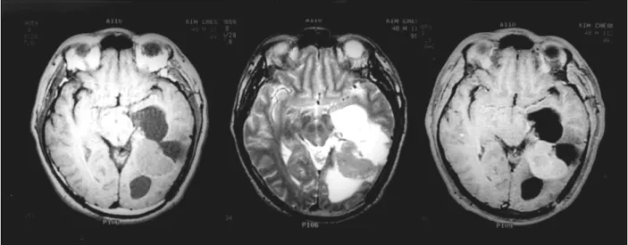
환자는 49세 남자환자로 내원 1일전에 경험한 전신 발 작을 주소로 내원 하였다. 과거력상 약 11개월 전에 두부 동통과 우측 측두부 반맹을 주소로 내원하여 실시한 자기 공명영상촬영상 7×7×6cm3크기의 비교적 경계가 좋은 종괴가 좌 측뇌실의 삼각부에 관찰되었다. 이 종괴는 T1강 조영상에서 중간정도의 신호강도을 보였고 T2강조영상상 비균질의 고 신호강도을 보이며 조영제 투여시 종괴는 비 균질성으로 조영증강이 되었으며 종양의 크기에 비해 주변 의 부종은 심하지 않았으며 우측으로 중심선의 전위가 관 찰되었다(Fig. 1A).
환자는 후 중간 측두이랑접근법으로 종양의 전 적출을 시
AAAA
주성필·정 신·이정길·김태선·김재휴·김수한·강삼석·이제혁
J Korean Neurosurg Soc/ Volume 30/ April, 2001 515
행받았고 조직학적으로 양성수막종으로 진단되었다(Fig. 2).
수술후 약 6개월에 시행한 추적 뇌 자기 공명영상에서 잔여 종양이나 종양의 재발은 보이지 않았다(Fig. 1B). 술후 11
개월 후전신 간대성 발작을 주소로 다시 내원하여 시행한 뇌 자기공명영상에서 좌 측뇌실에 4.2×3cm 크기의 재발 종양이 보여 전적출을 시행하였다(Fig. 3).
Fig. 1. Pre- and post-operative MRI. A:A large sized mass locating in the trigone of the left lateral ventricle with heterogenous enhancement. B:There is no evidence of the remaining tumor recurrence after surgery.
Fig. 2. Histology of the tumor removed from first operation. A:Photomicrograph demonstrating highly cellular tumor consisting of fibroblast and synctyial meningeal cells. Necrosis and mitosis are not evident(H & E, ×200). B:Immunoreactivity for PCNA is present in more than 10% of the tumor cells(immunohistochemistry for PCNA, ×200).
A A AA
BB BB
A AA
A BBBB
악성형질변환으로 조기 재발한 측뇌실내 수막종
J Korean Neurosurg Soc/ Volume 30/ April, 2001 516
수술은 전에 사용하였던 후 중간 측두이랑접근법에 의하 여 종양을 전 적출하였으며 기원한 측뇌실내의 맥락총에 대 한 연속 동결 절편 조직 검사를 하여 종양의 침윤이 없음을 확인하여 수술을 마쳤다. 병리조직학적으로 유사분열, 많은 병소에 괴사 및 주변뇌조직 침윤등의 소견이 보여 악성수 막종으로 진단되었고 PCNA지수(LI)는 40%이상이 양성 반응을 보였으며(Fig. 4) 또한 첫 번째 수술에서 얻은 조직 도 PCNA LI가 높은 양성반응(>10%)을 보였다(Fig. 2B), 술후 종양부위에 방사선 치료(5940cGy)를 시행받고 외래 추적 관찰중인 환자이다.
고 찰
측뇌실의 삼각부에 발생하는 수막종은 전체 뇌수막종의 0.5~4.5%를 차지하는 비교적 드문 병변이고 이 부위에 위
치하는 맥락총에서 기원하는 것으로 알려져 있다10). 수술 적 전적출후 거의 재발을 하지 않아 그 예후가 좋다는 것은 이미 알려진 사실이다. 본 증례에서와 같이 WHO 악성도의 분류법에 의하여 양성 수막종으로 조직학적 진단이 내려졌 던 환자가 수술후 11개월만에 악성수막종으로 형질변환되 어 조기에 재발이 된 경우는 극히 드물다.
조직학적으로 악성과 재발을 나타내는 지표로는 세포의 유사분열, 괴사, 병리학적 구조의 파괴, 혈관과다 및 혈철소 의 침착, 저명한 핵소체, 다형성 핵 등이 지금까지 대두되 어 왔지만 최근에 이것만으로 악성종양의 생물학적 지표로 서 사용되기에는 문제점이 많은 것으로 밝혀지고 있다1)5)6). 최근 Perry등의 보고에 따르면 뇌 수막종의 악성도를 분 류할 때 mitotic index를 많이 고려한 보다 쉬운 분류법을 제시하였다8).
Matsuno 등은 세포충실도(cellularity)는 술후 재발군과
Fig. 3. On MRI taken 11 months after surgey, a recurrent mass is noted in the previously operation site.
Fig. 4. Histology of the recurrent meningioma with malignant transformation. A:Mitoses are noted at least four in this field(H & E,
×200). B:High PCNA immunoreactivity is noted(immunohistochemistry for PCNA, ×200).
A A
AA BBBB
주성필·정 신·이정길·김태선·김재휴·김수한·강삼석·이제혁
J Korean Neurosurg Soc/ Volume 30/ April, 2001 517
비재발군사이에 유의한 차이가 없고 MIB-1이 3%이상인 경 우 임상경과중에 재발할 가능성이 높으며 Simpon’s grade 와 재발율 사이에도 연관관계가 있다고 하였다5). 그러나 양 성과 악성수막종에서 MIB-1 LI의 평균이 통계학적으로 유 의한 수준차이를 보였으나 MIB-1 LI만으로 수막종의 등 급을 나타내는 지표로 사용될 수는 없고 가장 좋은 접근은 MIB-1 LI와 조직학적인 특성이 같이 고려되어야 한다고 하였고 그 이유는 악성 수막종에서도 MIB-1 LI가 낮게 관 찰되는 경우도 있기 때문이다2).
뇌수막종의 재발에 있어 p53 면역반응과 MIB-1사이에 밀접한 관계가 있고 면역조직화학적 검사에 의한 p53 의 출현은 수막종의 번식능을 암시하고 향후 재발을 예견하는 중요한 인자가 될 수 있으며 p53 양성인 수막종은 활발한 유사분열능과 증가된 세포충실도, 세포의 다형성증, 고 핵 간 세포질의 비율을 보인다4).
조직학적으로 양성으로 진단되었다고 하더라도 종양의 성 장능의 표지자로 생각되는 MIB-1 positive indices 또는 PCNA가 2%이상인 경우 재발할 위험이 매우 높기 때문에 이러한 경우 조직학적으로 양성수막종인 경우일지라도 방 사선치료나 정위적 방사선수술를 고려해야한다고 보고하였 다3). 또한 수술중 얻은 수막종 조직의 DNA의 flow cyto- metic ananlysis는 조직학적으로 양성이고 육안적으로 완 전적출을 시행한 환자에서 재발을 예견하는데 중요하며 재 발군에서 proliferating index가 비재발군보다 높으며 PI가 20%이상인 경우 재발율이 매우 높다6). 그리고 apoptosis 또한 뇌수막종에서 관찰되기도 하며 비정형 및 악성변화에 연관관계가 있는 것으로 알려져있다7).
본 증례의 경우 후향적으로 일차 수술의 조직에 대한 PCNA와 p53에 대한 면역조직화학적 염색을 시행한 결과 PCNA에 강한 양성 반응을 보인 경우가 >10%으로 높게 나왔으며, 조기 재발후 이차 수술에서 얻은 조직은 10 high power fields상 20개 이상의 유사분열이 관찰되어 악성수 막종으로 진단되었다. 이에 대한 면역조직학적 염색에서도 비슷한 높은 PCNA LI와 p53에 음성반응을 보였다. 조직 학적 진단이 양성수막종일 지라도 종양 세포의 proliferative index가 높으면 수술후 방사선 치료등의 보조요법이 필요 할 것으로 사료된다. 환자는 술후 종양부위에 5940cGy의 방사선 조사를 받고 현재까지 외래 통원 치료중이다.
결 론
자들은 조기에 악성으로 형질 변환된 측뇌실의 삼각부에 발생한 양성수막종을 보고하며, 종양의 완전 적출을 시행
하고 조직학적으로 양성수막종일지라도 PCNA나 MIB-LI 과 같은 종양세포의 성장능이 높으면 술후 재발의 위험이 높을 것으로 사료되어 방사선치료를 고려해야 될 것으로 생각된다.
•논문접수일:2000년 4월 7일
•심사완료일:2000년 7월 7일
•책임저자:주 성 필
501-757 광주광역시 동구 학1동 8번지 전남대학교 의과대학 신경외과학교실
전화:062) 220-6606, 전송:062) 224-9865 E-mail:[email protected]
References
1) Abramovich CM, Prayson RA:Histopathologic features and MIB-1 labeling indices in recurrent and nonrecurrent mening- iomas. Arch Pathol Lab Med 123:793-800, 1999
2) Abramovich CM, Prayson RA:MIB-1 labeling indeces in benign, aggressive, and malignant meningiomas:a study of 90 tumors. Hum Pathol 29:1420-1427, 1998
3) Cerda-Nicolas M, Lopez-Gines C, Barcia-Salorio J, Llombart- Bosch A:Evolution to malignancy in a recurrent mening- ioma:morphological and cytogenetic findings. Clin Neurop- athol 17:210-215, 1998
4) Matsuno A, Magashima T, Matsuura R, Tanaka H, Hirakawa M, Tamura A, et al:Correlation between MIB-1 staining index and the immmunoreactivity of P53 protein in recurrent and non-recurrent meningiomas. Am J clin Pathol 106:776- 781, 1996
5) Matsuno A, Fujimaki T, Sasaki T, Nagashima T, Ide T, Asai A, et al:Clinical and histopathological analysis of proliferative potentials of recurrent and non-recurrent meningiomas, Acta Neuropathol 91:504-510, 1996
6) May PL, Broome JC, Lawry J, Buxton RA, Battersby RD:The prediction of recurrence in meningiomas, J Neurosurg 71: 347-351, 1989
7) Ng HK, Chen L:Apoptosis is associated with aypical or ma- lignant change in meningiomas. an in situ labelling and imm- unohistochemical study. Histopathology 33:64-70, 1998 8) Perry A, Scheithauer BW, Stafford SL, Lohse CM, Wollan
PC:“Malignancy” in meningiomas:a clinicopathologic study of 116 patients, with grading implications. Cancer 85: 2046-2056, 1999
9) Sutherland GR, Sima AA:Incidence and clinicopathologic features of meningioma., in Schimidek HH(ed):Meningiomas and their surgical menagement. Philadelphia, WB Saunders, 1991, pp10-21
10) Tukanowicz SA, Grant FC:The meningiomas of the lateral ventricles of the brain. J Neuropathol 17:367, 1958