152
basal values of pituitary hormone including the gonad, thyroid, and adrenal axes were normal on laboratory evaluation.
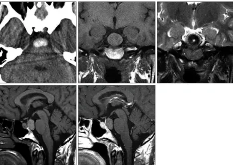
Computed tomography (CT) scan revealed a nonenhancing homogeneous hyperdense mass confined to the retrosellar re- gion. Mean Hounsfield unit (HU) for the central lesion was 105.1 HU. There was no bony abnormality in the sella turcica and the clivus. Magnetic resonance (MR) imaging showed an ovoid and well-demarcated mass without contrast enhancement displaying isointensity on T1-weighted images and hypointensity on T2- weighted (Fig. 1). This 25 mm-sized lesion was located just dor- sal to the pituitary gland and occupied the preopontine cistern.
Differential diagnosis included; craniopharyngioma, pituitary adenoma, neurenteric cyst, dermoid, and RCC.
A pterional craniotomy disclosed a gray-whitish tumor, which was not attached to the infundibulum, and extended up the third ventricle. On approaching the pituitary region, the cyst was observed between the optic nerve and carotid artery. The thin capsule was opened, yellow-whitish mucinous contents evacuated, and the cyst elevated with further dissection. There was no evidence of previous hemorrhage at surgery (Fig. 2A).
The tumor was gradually decompressed and totally excised through the space lateral to the carotid artery. The cyst was en- tirely located behind the dorsum sellae, there was no invasion into the diaphragm.
INTRODUCTION
Rathke’s cleft cysts (RCCs) are non-neoplastic congenital le- sions that develop from an epithelial remnant of Rathke’s pouch, a normal component of pituitary development that gives rise to the anterior lobe, pars intermedia, and pars tuberalis of the gland.
Topographically, RCCs usually occur in the sellar fossa with or without suprasellar involvement, but they can arise from every part of the craniopharyngeal duct, which is a part of Rathke’s pouch
11,14). Intracranial ectopic RCCs are exclusively found in the suprasellar region, likely a result of pouch remnants within the pars tuberalis above the diaphragma sellae
3). In rare instanc- es, RCCs may reside in the sphenoid sinus or clivus regions
8). In this report, the author presents a case of a RCC located in the retrosellar and prepontine area.
CASE REPORT
A 41-year-old woman presented with an acute-onset headache, which progressively worsened over the following 3 days. Her condition had been unresponsive to medical treatment. This pa- tient had signs of meningeal irritation with increased cerebrospi- nal fluid (CSF) cell number and protein levels. She did not show any endocrinological symptoms and signs on admission. The
A Case of Ectopic Rathke’s Cleft Cyst in the Prepontine Cistern
Ealmaan Kim, M.D., Ph.D.
Department of Neurosurgery, Keimyung University School of Medicine, Dongsan Medical Center, Daegu, Korea
A Rathke’s cleft cyst (RCC) is a benign pituitary cyst derived from the remnant of Rathke’s pouch, and usually presents as an intrasellar lesion with varying degrees of suprasellar extension. However, to date, a description of a primary prepontine RCC with no intrasellar component has not been reported. The author describes an exceptional case of a symptomatic RCC located behind the sella turcica in a 41-year-old woman who presented with severe headache. The author also provides an embryological hypothesis of the development of an ectopic RCC, with a special emphasis on ra- diologic characteristics.
Key Words : Prepontine cistern · Rathke’s pouch · Rathke’s cleft cyst · Sella turcica · Suprasellar cyst.
Case Report
•Received : January 28, 2012 •Revised : June 6, 2012 •Accepted : August 17, 2012
•Address for reprints : Ealmaan Kim, M.D., Ph.D.
Department of Neurosurgery, Keimyung University School of Medicine, 56 Dalseong-ro, Jung-gu, Daegu 700-712, Korea Tel : +82-53-250-7730, Fax : +82-53-250-7356, E-mail : [email protected]
•This is an Open Access article distributed under the terms of the Creative Commons Attribution Non-Commercial License (http://creativecommons.org/licenses/by-nc/3.0) which permits unrestricted non-commercial use, distribution, and reproduction in any medium, provided the original work is properly cited.
J Korean Neurosurg Soc 52 : 152-155, 2012
http://dx.doi.org/10.3340/jkns.2012.52.2.152
Copyright © 2012 The Korean Neurosurgical Society Print ISSN 2005-3711 On-line ISSN 1598-7876
www.jkns.or.kr
153
Ectopic Rathke’s Cleft Cyst | E Kim
HU density values, relatively acute hemorrhage (60-70 HU) or calcification (1000 HU) was thought unlikely and ruled out.
The major differential consideration for a hyperdense RCC in- cludes the classic colloid cyst and the rare “white” epidermoid cyst; because they may appear hyperattenuated on nonenhanced The patient recovered from the tran-
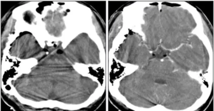
scranial surgery. Pathological examina- tion revealed the typical features of a RCC with columnar epitheliums and cystic contents (Fig. 2B-D). Postopera- tive CT scans showed no evidence of residual cyst in the prepontine cistern (Fig. 3). Her severe headache resolved postoperatively.
DISCUSSION
With the increased use and evolution of brain MR imaging technology more RCCs are being discovered; however, symptomatic cases are rare and accu- rate preoperative diagnoses can be dif- ficult. Excluding pituitary adenomas, RCCs are the most common lesion in the sellar and suprasellar region. These are developmental epithelial cysts filled with mucous, gelatinous or caseous content. RCCs are well encapsulated
and vary in size
1). Microscopically, they are typically composed of simple or pseudostratified columnar or cuboidal epithelium, often with ciliated or mucin-secreting goblet cells.
Neuroembryologically, the origin and pathogenensis of ecto- pic RCC is a subject of controversy, however, it is not surprising to assume that it is similar to the mechanism for ectopic occur- rence of the pituitary adenoma or craniopharyngioma
2,5). Ecto- pic locations include the nasopharynx, the sphenoid sinus, the clivus, and the posterior fossa
7,11,15). It has been proposed that RCCs originate from epithelial cell remnants of the obliterated craniopharyngeal duct along the path traversed by Rathke’s pouch and that location is determined by the embryological events of the sellar and/or suprasellar region. The pars tuberalis, while developing from the ventral portion of Rathke’s pouch, rotates to come in contact with the neuroectodermal layer of the developing ventral cerebral vesicle in the early embryonic period. As a result of different rates of rotation of the pars tube- ralis, Rathke’s pouch cells predominantly spread through sellar and suprasellar region, but few cells can be located outside the sella turcica
3,8). Failure of ectopic pouch remnants beyond the pituitary fossa to regress further during development can lead to cystic dilation and formation of a symptomatic RCC.
On CT scans, the majority of RCCs are homogenous, nonen- hancing, and of low-density, as are many other well-defined sellar and suprasellar lesions, including craniopharyngiomas, arach- noid cysts, and pituitary adenomas
13). However, cases of mixed density RCCs with iso-, hypo-, and hyperdense foci have been reported in the literature
15). The cyst in this report differed from ordinary RCCs because of its high density on CT, which was as- cribed to higher protein content
14). In this case, based on the CT
Fig. 2. Operative and pathology features of a Rathke’s cleft cyst in the pre- pontine cistern. Intraoperative view demonstrates resection of a cyst wall with thick mucoid and white-yellow substances (A). Photomicrographs of surgical specimens show the cystic contents and the waxy nodules (B and C). Histopathological examination of cyst wall illustrates ciliated co- lumnar epitheliums and inflammatory infiltrates (H&E, original magnifi- cation ×100) (D).
D B
C A
Fig. 1. Neuroimaging studies of an ectopic Rathke’s cleft cyst. Computed tomography scans show a homogenous oval-shaped high density in the prepontine cistern. Magnetic resonance images de- pict a nonenhancing retrosellar mass with an intracystic nodule that displays iso- to slight hyperinten- sity on T1-weighted sequence and hypointensity on T2-weighted sequence. The nodule (*) is sur- rounded by thin isointense cyst fluid (arrow).
154 J Korean Neurosurg Soc 52 | August 2012
For treating symptomatic sellar and suprasellar RCCs, the en- donasal transsphenoidal route including microscopic and en- doscopic is the procedure of choice. Results are reportedly good with low morbidity and recurrence rates
10). Although they are rare, the transcranial approach may be required for complex cysts or entirely supra- and retrosellar lesions, which was evi- dent in the present report. Regardless of the surgical approach chosen, treatment of retro- and suprasellar RCCs is more diffi- cult owing to their intimate proximity to the optic chiasm and pituitary stalk
3).
CONCLUSION
The author reports a rare case of a completely retrosellar type of RCC that was not attached to the pituitary fossa. The rarity of ectopic RCCs and their variable imaging features preclude pre- operative diagnosis. The unique MR findings of an intracystic nodule with low signal intensity on T2-weighted image may be an indicator in differentiating RCCs from other parasellar cys- tic lesions.
References
1. Aho CJ, Liu C, Zelman V, Couldwell WT, Weiss MH : Surgical out- comes in 118 patients with Rathke cleft cysts. J Neurosurg 102 : 189- 193, 2005
2. Anand VK, Osborne CM, Harkey HL 3rd : Infiltrative clival pituitary adenoma of ectopic origin. Otolaryngol Head Neck Surg 108 : 178-183, 1993
3. Barrow DL, Spector RH, Takei Y, Tindall GT : Symptomatic Rathke’s cleft cysts located entirely in the suprasellar region : review of diagnosis, management, and pathogenesis. Neurosurgery 16 : 766-772, 1985 4. Binning MJ, Gottfried ON, Osborn AG, Couldwell WT : Rathke cleft
cyst intracystic nodule : a characteristic magnetic resonance imaging finding. J Neurosurg 103 : 837-840, 2005
5. Suprasellar and infrasellar craniopharyngioma with a persistent cranio- pharyngeal canal : case report and review of the literature. Neuroradiol- ogy 43 : 760-762, 2001
6. Choi SH, Kwon BJ, Na DG, Kim JH, Han MH, Chang KH : Pituitary adenoma, craniopharyngioma, and Rathke cleft cyst involving both in- trasellar and suprasellar regions : differentiation using MRI. Clin Radiol 62 : 453-462, 2007
7. Chuang CC, Chen YL, Jung SM, Pai PC : A giant retroclival Rathke’s cleft cyst. J Clin Neurosci 17 : 1189-1191, 2010
8. Hanna E, Weissman J, Janecka IP : Sphenoclival Rathke’s cleft cysts : embryology, clinical appearance and management. Ear Nose Throat J 77 : 396-399, 1998
9. Kim JE, Kim JH, Kim OL, Paek SH, Kim DG, Chi JG, et al. : Surgical treatment of symptomatic Rathke cleft cysts : clinical features and re- sults with special attention to recurrence. J Neurosurg 100 : 33-40, 2004 10. Kleinschmidt-DeMasters BK, Lillehei KO, Stears JC : The pathologic,
surgical, and MR spectrum of Rathke cleft cysts. Surg Neurol 44 : 19- 26; discussion 26-27,1995
11. Megdiche-Bazarbacha H, Ben Hammouda K, Aicha AB, Sebai R, Bel- ghith L, Khaldi M, et al. : Intrasphenoidal rathke cleft cyst. AJNR Am J Neuroradiol 27 : 1098-1100, 2006
12. Nishioka H, Haraoka J, Izawa H, Ikeda Y : Magnetic resonance imaging, clinical manifestations, and management of Rathke’s cleft cyst. Clin En- docrinol (Oxf) 64 : 184-188, 2006
CT scans
6,16). However, colloid cysts should not be mistaken for RCCs since they typically occur only at the foramen of Monro.
Unlike RCCs, epidermoid cysts usually have an irregular con- figuration of the cyst wall and may calcify, and are usually locat- ed off midline.
Variable MR imaging characteristics of RCCs were presumed in part to be due to cholesterol contents, cell debris, mucopoly- saccharides, and rarely, hemorrhage within the cysts
10,16). Un- common RCCs like the this case that have high- and isointensi- ty on T1- and hypointensity on T2-weighted images are more likely to induce earlier symptoms, when the cysts are smaller than their CSF-like intensity counterparts
12,17). A RCC with low viscosity fluid may be confused with suprasellar arachnoid cysts, since they typically have the same signal intensity as CSF. It has been suggested that the presence of waxy nodules is the most reliable diagnostic indicator of RCCs
4). In the present case, the intracystic nodule was easily observed on T2-weighted images, but detection of the nodule on T1-weighted images was diffi- cult because of similarities in the cyst fluid and nodule intensi- ties. While T2 hypointensity is not observed in every RCC case, this is in contrast to pituitary adenomas where hypointensity on T2 would not be expected unless there were hemorrhages. RCC shows no enhancement or is limited to the cyst wall; conversely, craniopharyngiomas typically have nodular, globular, or rim enhancement on MR scanning
6). In the case of ectopic RCCs in the prepontine region, the major differential diagnosis is neur- enteric cyst or dermoid cyst because commonly they have a similar location
15). However, dermoid cysts have the same im- aging characteristics as fat because they contain liquid choles- terol. The best diagnostic clue for a neurenteric cyst is a lobulat- ed, nonenhancing, slightly hyperintense mass in front of the medulla on both T1-weighted and T2-weighted imagings
13).
RCCs may compress structures within and/or adjacent to the sella, which can elicit symptoms including visual impairment, pituitary endocrine dysfunction, and headache. Rarely, they also cause abscess formation, chronic inflammation, hyponatremia, cavernous sinus syndrome, diabetes insipidus, or meningitis
1). This patient presented with severe headache of acute-onset sug- gestive of leakage of the cystic contents. Acute symptoms that mimicked pituitary apoplexy were noted in up to 11.0% of re- ported cases
9).
Fig. 3. Postoperative computed tomography scans exhibit disappear- ance of the hyperdense cyst behind the pituitary fossa.
155
Ectopic Rathke’s Cleft Cyst | E Kim
16. Voelker JL, Campbell RL, Muller J : Clinical, radiographic, and patho- logical features of symptomatic Rathke’s cleft cysts. J Neurosurg 74 : 535-544, 1991
17. Zhou L, Luo L, Hui X, Chen H, Yu B, Guo G, et al. : Primary Rathke’s cleft cyst in the cerebellopontine angle associated with apoplexy. Childs Nerv Syst 26 : 1813-1817, 2010
13. Osborn AG, Preece MT : Intracranial cysts : radiologic-pathologic cor- relation and imaging approach. Radiology 239 : 650-664, 2006 14. Panagopoulos KP, Jolesz FA, el-Azouzi M, Black PM : Mucinous cysts of
the pituitary stalk. Report of two cases. J Neurosurg 71 : 276-278, 1989 15. Ross DA, Norman D, Wilson CB : Radiologic characteristics and results
of surgical management of Rathke’s cysts in 43 patients. Neurosurgery 30 : 173-178; discussion 178-179, 1992