• 검색 결과가 없습니다.

항암제와 방사선 치료에 반응이 없는 고형 가유두상 종양에서 Isotretionin 치료 1례

N/A
N/A
Protected

Academic year: 2021

Share "항암제와 방사선 치료에 반응이 없는 고형 가유두상 종양에서 Isotretionin 치료 1례"

Copied!
7
0
0

로드 중.... (전체 텍스트 보기)

전체 글

(1)

Abstract

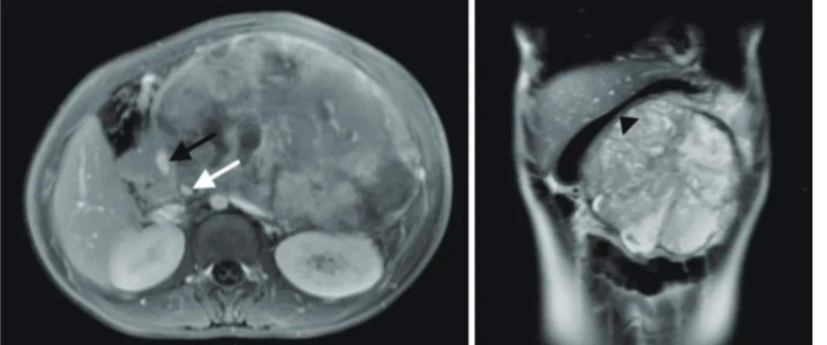
Solid pseudopapillary neoplasm (SPN) of the pancreas is an exocrine tumor with low malignant nature.

Although it’s treatment of choice is complete surgical resection, the patients who are not eligible for surgical therapy need other kinds of therapy and there are little reports. Vitamin A and its derivatives in some tumors are known to be prohibiting the tumors’ growth by reducing the manifestation of transcription factors. We present one case of SPN of the pancreas, which was not eligible for resection and was not responding to chemotherapy and radiation therapy, but, responded with the administration of Isotretionin (Roaccutan

). Therefore, for inoperable SPN of the pancreas, we consider Isotretionin therapy can be used as treatment and, in the future, new studies with more of Isotretionin treatment implemented cases should be conducted to identify their anticancer mechanism.

Key Words : Isotretionin, Solid-pseudopapillary neoplasm of the pancreas, Treatment

교신저자: 김흥식, 700-712 대구광역시 중구 달성로 56, 계명대학교 의과대학 소아청소년과학교실

Heung Sik Kim, M.D., Department of Pediatrics, Keimyung University School of Medicine 56 Dalseong-ro, Jung-gu, Daegu 700-712, Korea

Tel : +82-53-250-7516 E-mail : [email protected]

Departments of Pediatrics, Radiology

1

, Pathology

2

, Radiation Oncology

3

and Nuclear Medicine

4

, Keimyung University School of Medicine, Daegu, Korea

Yoon Jung Kim, M.D., Young Joon Ahn, M.D., Hee Jung Lee

1

, M.D., Hye Ra Jung

2

, M.D., Ok Bae Kim

3

, M.D., Kyoung Sook Won

4

, M.D., Ji Yoon Kim, M.D., Heung Sik Kim, M.D.

Effect of Isotretionin for Refractory Solid-pseudopapillary Tumor of the Pancreas which Previously did not Respond to Anticancer Drugs and Radiation Therapy

김윤정·안영준·이희정

1

·정혜라

2

·김옥배

3

·원경숙

4

·김지윤·김흥식

계명대학교 의과대학 소아청소년과학교실, 영상의학교실1, 병리학교실2, 방사선종양학교실3, 핵의학교실4

항암제와 방사선 치료에 반응이 없는 고형 가유두상 종양에서 Isotretionin 치료 1례

(2)

153

항암제와 방사선 치료에 반응이 없는 고형 가유두상 종양에서 Isotretionin 치료 1례

서 론

췌장의 고형 가유두상 종양은 낮은 악성도를 보이는 외분비성 종양이다. 병리학적으로 악성종양에 속하나 췌장에서 많이 발생하는 관선암 등과는 달리 주 변 조 직 으 로 의 침 습 이 나 국 소 림 프 절 침 범 , 원격전이 등이 매우 드물고 느리게 성장하는 특징이 있다[1]. 이 종양은 수술적 치료로 완치가 가능하며, 재발이 잘 되지 않고, 전이된 경우에도 완전 절제하면 장기 생존이 가능한 것으로 알려져 있다[2]. 또한 종양의 크기, 종양의 전이로 인해 절제가 불가능한 경우, 항암치료나 방사선 치료로 절제범위를 축소시킨 후 수술을 시행하여 완치된 예가 보고되고 있다[3-6].

레티노익산은 지난 수 십 년간 종양의 예방과 치료를 위해 연구되어 왔고[7], 소아 혈액종양 분 야 에 서 는 급 성 전 골 수 성 백 혈 병 과 고 위 험 신 경 모 세 포 종 의 치 료 에 성 공 적 으 로 사 용 되 고 있으나[8], 췌장의 고형 가유두상 종양에 사용하여 치료효과를 나타낸 예는 보고된 바가 없다.

저자들은 수술적 치료가 불가능하여 방사선 치료와 항암치료를 시행하였으나 종양의 크기에 변화를 보이지 않은 환아에게 레티노익산(isotretionin)을 사용한 후 종양의 크기가 감소한 1례를 경험하였기에 이를 보고하는 바이다.

증 례

12세 여자 환자로, 2주 동안의 간헐적인 명치부 복통을 주소로 다른 병원 방문하여 시행한 복부 전산화단층촬영(C T)에서 췌장 종괴가 발견되어 본원으로 전원되었다. 과거력 및 가족력에는 특이사항 없었고 특별한 약물복용력도 없었다. 전신소견상 명치부 복통을 호소하였고, 체중감소 및 식욕부진 등의 증상은 없었다. 전신진찰상 신장 151.5 cm, 체중 42.1 k g, 활력징후는 수축기혈압 100 m m H g, 이완기혈압 60 mmHg, 맥박 88/분, 호흡 20/분, 체온 36.9 ℃였으며, 황달은 없었으며 명치부에 압통을 동반한 4횡지 크기의 종괴가 촉지되었다. 전혈검사는 백혈구 7,140/µL, 혈색소 10.4 g/dL, 헤마토크리트

31.1%, 혈소판 354,000/µL이었고, 생화학적검사는 총단백 7.9 g/dL, 알부민 4.4 g/dL, 총 빌리루빈 0.4 mg/dL, ALT 13 IU/L, AST 30 IU/L, alkaline phosphatase 139 IU/L, amylase 107 U/L, lipase 14 U/L 이었다. CA 19-9, CEA, alpha fetoprotein 등의 종 양 표 지 자 검 사 는 모 두 정 상 범 위 였 다 . 복 부 자기공명영상검사 (MRI) 상 종괴는 췌장의 체부와 미부에 위치하여 비균질한 신호강도를 보이며 조영이 증강되는 157×101×133 m m의 크기로 경계가 명 확 하 며 섬 유 성 피 막 으 로 둘 러 싸 여 있 으 며 , 상 부 장 간 정 맥 , 복 강 동 맥 , 위 장 의 대 만 과 접해있었고(F i g. 1), 양전자단층촬영(PET) 상 고 형 부 분 에 중 등 도 의 불 균 질 한 F l u o r i n e - 1 8 Fluorodeoxyglucose (F-18 FDG) 섭취를 보이는 156×107×131 mm 크기의 큰 종괴가 관찰되었다(Fig. 2).

조직생검에서 고형 가성유두종양으로 진단되었으며(Fig. 3A), 면역조직학적 염색에서 α1-antitrypsin, vimentin, neuron specific enolase (NSE)에 양성반응을 보였다.

외과적 절제가 불가능한 상태로 판단되어 항암화학치료를 시행하기로 결정하였다. 먼저 Gemcitabine을 6개월간 6회 주기 투여 후 방사선 치료(180 cGy/fx, 5 fx/week, 4 ,5 0 0 c G y /5 w e e k s ) 를 시 행하 였 으나 , 복 부 전산화단층촬영(CT) 상 췌장 종괴의 크기에 변화가 없었고, 이후 vincristine (1.5 mg/m

2

/day)을 제 1병일, ifosfamide (1,800 mg/m

2

/day)를 제 1-4병일, etoposide (100 mg/m

2

/day)를 제 1-5병일, cisplatin (40 mg/m

2

/day)을 제 1-3병일에 투여하는 항암치료요법을 4주마다 3주기를 3개월간 지속하였으나 여전히 종양의 크 기 는 1 4 3 × 9 9 × 1 2 3 m m 로 크 게 감 소 하 지 않았으며(Fig. 4), 조직소견도 첫 번째 생검과 큰 차이를 보이지 않았다(Fig. 3-B). 이후 isotretionin (Roaccutan

) 100 mg/m

2

/day을 매일 투여 하였으며, 8 개 월 째 에 시 행 한 복 부 C T 상 종 괴 크 기 가 110×95×115 m m로 감소되고(F i g. 5), 조직 생검에서는 종괴의 일부가 남아있으나 뚜렷한 응고 괴사 및 기질의 심한 섬유화가 관찰되었다(Fig. 6).

투약을 지속한지 45개월째에 시행한 F-18 FDG PET/

CT 상 종괴 크기가 94×80×72 mm로 더욱 감소되고,

F-18 FDG 섭취 역시 감소한 소견 보여 (Fig. 7), 추후

절제수술을 시행할 예정이다.

(3)

고 찰

췌장의 고형 가성유두종양은 매우 드문 질환으로, 발생률은 췌장에서 발생하는 모든 고형 종양의 약 0.13~2.7%로 보고되고 있다[1]. 주로 20, 30대의 젊은 여 성 에 서 발 생 하 는 것 으 로 알 려 져 있 고 , 특 히

소아에서의 발생은 더욱 드물며[9], 국내에서는 2007년에 수술로 치료된 12세 남아의 증례가 처음 보고되었다[10].

췌장의 원발성 종양이 대부분 악성이며, 발견이 늦기 때문에 진단 당시 췌장 외 조직으로의 전이가 많아 예후가 불량한 반면, 이 종양은 주위 조직과의 Fig. 1. MRI T

1

-weighted axial image (left) and T

2

-weighted coronal image (right) reveal a large, well-demarcated solid mass (157 × 101 × 133 mm) with heterogenous signal intensity and heterogenous enhancement in the body and tail of the pancreas. The mass abuts the superior mesenteric vein (black arrow), celiac trunk (white arrow) and greater curvature of the stomach (arrow head).

Fig. 2. PET/CT scan of torso with F-18 FDG reveals a huge, mixed density mass (156 × 107 × 131 mm) in the

LUQ, with moderate FDG uptake in the solid portion (SUVmax 6.6) and no FDG uptake in the low density

portion containing irregular calcification, which is compressing the adjacent organs and vessels.

(4)

155

항암제와 방사선 치료에 반응이 없는 고형 가유두상 종양에서 Isotretionin 치료 1례

경계가 분명하고 전이나 재발을 거의 하지 않는 악성도가 낮은 종양으로, 외과적 수술로 대개 완치가 가능하다[1,2]. 국내에서도 췌장 고형 가성유두종양 환아들의 수술적 치료 후 추적 관찰기간 동안 재발 및 전이를 보이지 않고 무병 생존한 예가 보고되었다[11].

하지만 여러 예에서 전이, 재발, 인접 장기로의 침범 등을 보이는 병변이 확인되고 있으며[12], 수술적

절 제 만 으 로 완 치 가 불 가 능 한 경 우 추 가 적 으 로 시행하거나 대체할 수 있는 치료에 대한 여러 시도가 이루어지고 있다. 완전절제가 불가능하고 주위조직 및 장기와의 유착이 심한 경우에 방사선치료가 효과가 있다는 보고가 있으며[3], 수술적 절제가 불가능하여 gemcitabine으로 치료한 후 종양의 크기가 현저하게 감소하여 완전 절제 수술이 가능했던 1례가 보고되어 Fig. 3. A: No improvement after chemotherapy; pathologic findings of the solid papillary tumor of pancreas at diagnosis. B: Pathologic findings after chemotherapy.

Fig. 4. Follow up CT scan after 11 months with radiotherapy and chemotherapy reveals slightly decreased size of

the tumor mass in pancreas (143 × 99 × 123 mm).

(5)

있다[4]. 상장간막 정맥과 유착된 절제 불가능한 직경 15 cm의 췌장 두부 종양에서 6개월간 Cisplatin과 5-FU 병합 항암화학요법 후 종양 직경이 감소하여 성공적으로 종양의 절제를 시행한 1례가 있으며[5], C i s p l a t i n, i f o s f a m i d e, e t o p o s i d e를 이용한 항암화학요법에 반응이 없어 수술 후 방사선치료와 gemcitabine을 사용하여 관해를 보였던 1례 등이

보고되었다[6]. 그러나 수술적 절제가 불가능한 병변에 대해 항암 치료나 방사선 치료가 어느 정도의 효과가 있는지, 어떻게 시행해야 하는지에 대해 합의된 연구 결과는 아직 없다.

레티노이드는 배아의 발생, 시각, 생식, 뼈형성, 대사, 조혈, 분화, 증식, 세포자멸사 등 필수적인 생물학적인 과정에 생리적 조절인자로 이용된다[13]. 이는 Fig. 6. A: After retinoic acid treatment, the tumor mass shows mixed area of necrosis and fibrosis. B: Remaining tumor component with amorphous necrotic material. C: Marked stromal fibrosis. D: Coagulative necrosis.

Fig. 5. Follow up CT scan after 8 months with retinoic acid treatment reveals markedly decreased size of the tumor mass in pancreas (110 × 95 × 115 mm).

D

A B

B C

C D

(6)

157

항암제와 방사선 치료에 반응이 없는 고형 가유두상 종양에서 Isotretionin 치료 1례

종양세포의 핵수용체에 작용해 세포의 전사 과정을 조절하여 세포의 증식을 방해하고, 악성 세포의 분화를 촉진시키며, 종양 세포의 세포자멸사를 촉진하고, 종양에 의한 혈관 신생을 억제하여 종양 세포의 증식 억제 효과를 나타낸다[7]. 또한 동물실험을 통해 구강암, 피부암, 방광암, 폐암, 전립선암, 유방암에서 발암을 억제하는 것을 확인하였다[7,9]. 이러한 특성 때문에 레티노익산은 종양의 화학적 예방과 치료를 위해 오랫동안 연구되어 왔고[14,15], 특히 소아 혈액종양 분야에서는 급성 전골수성백혈병과 고위험 신경모세포종의 치료에 성공적으로 사용되고 있다[8].

본 증례에서 저자들은 수술적 치료가 불가능한 고 형 가 유 두 상 종 양 환 아 에 게 방 사 선 치 료 와 항암치료를 시행하였으나 종양의 크기가 크게 감소하지 않았고, 이 환아에게 레티노익산(isotretionin)을 투여한 후 종양 크기가 감소한 예를 경험하였다. 현재 레티노익산이 고형 가유두상 종양에 어떠한 영향을 미치는지에 대해 연구된 바가 없고, 치료효과를 나타낸 예도 보고되지 않고 있다. 이 증례 역시 어떤 기전으로 종양의 크기가 감소하였는지에 대한 실험적인 증명이 이루어지지 않은 상태로, 고형 가유두상 종양 세포의 체외 배양에서 레티노익산에 의한 증식 억제 효과가 나타나는지 확인하는 추가적인 연구가 필요할 것으로 생각된다.

요 약

췌장의 고형 가유두상 종양은 낮은 악성도를 보이는 외분비성 종양으로, 수술적 치료로 완치가 가능하며, 재발이 잘 되지 않고, 전이된 경우에도 완전 절제하면 장기 생존이 가능하며, 종양의 크기, 종양의 전이로 인해 절제가 불가능한 경우 항암치료나 방사선 치료로 종양의 크기를 감소시킨 후 절제하는 방법이 이용되고 있다. 본 증례에서 저자들은 방사선 치료와 항암치료에도 종양의 크기에 변화를 보이지 않은 췌장 고형 가유두상 종양 환아에게 레티노익산(isotretionin)을 사용한 후 종양의 크기가 감소한 1례를 경험하였기에 이를 보고하는 바이다.

Conflict of Interest

The authors report no conflict of interest in this work.

참 고 문 헌

1. Madan AK, Weldon CB, Long WP, Johnson D, Raafat

Fig. 7. Follow up F-18 FDG PET/CT scan after 45 months with retinoic acid treatment reveals decreased size and

metabolism of the mass in the pancreas (SUVmax 3.7, 94 × 80 × 72 mm) with decreased solid hypermetabolic

portion.

(7)

A. Solid and papillary epithelial neoplasm of the pancreas. J Surg Oncol 2004;85:193-8.

2. Horisawa M, Niinomi N, Sato T, Yokoi S, Oda K, Ichikawa M, et al. Frantz's tumor (solid and cystic tumor of the pancreas) with liver metastasis: successful treatment and long-term follow-up. J Pediatr Surg 1995;30:724-6.

3. Cappellari JO, Geisinger KR, Albertson DA, Wolfman NT, Kute TE. Malignant papillary cystic tumor of the pancreas. Cancer 1990;66:193-8.

4. Maffuz A, Bustamante Fde T, Silva JA, Torres-Vargas S. Preoperative gemcitabine for unresectable, solid pseudopapillary tumour of the pancreas. Lancet Oncol 2005;6:185-6.

5. Strauss JF, Hirsch VJ, Rubey CN, Pollock M. Resection of a solid and papillary epithelial neoplasm of the pancreas following treatment with cis-platinum and 5-fluorouracil: a case report. Med Pediatr Oncol 1993;21:365-7.

6. Shim YJ, Kang JH, Lee KS. A Case of Pediatric Solid Pseudopapillary Tumor of the Pancreas Treated with Gemcitabine. Clin Pediatr Hematol Oncol 2011;18:66- 9.

7. Lotan R. Retinoids and apoptosis: implications for cancer chemoprevention and therapy. J Natl Cancer Inst 1995;87:1655-7.

8. Smith MA, Parkinson DR, Cheson BD, Friedman MA.

Retinoids in cancer therapy. J Clin Oncol 1992;10:839-64.

9. Chao HC, Kong MS, Lin SJ, Lou CC, Lin PY. Papillary cystic neoplasm of the pancreas in children: report of three cases. Acta Paediatr Taiwan 2000;41:101-5.

10. Han CH, Moon JH, Cho YD, Jin SY, Lee JS, Lee MS, et al. A case of a solid pseudopapillary tumor in a male child. Korean J Med 2007;72:668-72.

11. Choi SH, Kim SM, Oh JT, Park JY, Seo JM, Lee SK.

Solid pseudopapillary tumor of the pancreas: a multicenter study of 23 pediatric cases. J Pediatr Surg 2006;41:1992-5.

12. P a p a v r a m i d i s T, P a p a v r a m i d i s S . S o l i d pseudopapillary tumors of the pancreas: review of 718 patients reported in English literature. J Am Coll Surg 2005;200:965-72.

13. Sun SY, Lotan R. Retinoids and their receptors in cancer development and chemoprevention. Crit Rev Oncol Hematol 2002;41:41-55.

14. Hansen LA, Sigman CC, Andreola F, Ross SA, K e l l o f f G J , D e L u c a L M . R e t i n o i d s i n chemoprevention and differentiation therapy.

Carcinogenesis 2000;21:1271-9.

15. Nagy L, Thomazy VA, Heyman RA, Davies PJ.

Retinoid-induced apoptosis in normal and neoplastic

tissues. Cell Death Differ 1998;5:11-9.

수치

Fig. 2. PET/CT scan of torso with F-18 FDG reveals a huge, mixed density mass (156 × 107 × 131 mm) in the  LUQ, with moderate FDG uptake in the solid portion (SUVmax 6.6) and no FDG uptake in the low density  portion containing irregular calcification, whi
Fig. 4. Follow up CT scan after 11 months with radiotherapy and chemotherapy reveals slightly decreased size of  the tumor mass in pancreas (143 × 99 × 123 mm).
Fig. 5. Follow up CT scan after 8 months with retinoic acid treatment reveals markedly decreased size of the  tumor mass in pancreas (110 × 95 × 115 mm).

참조

관련 문서

However, with the changes in the domestic and international environment after the reform and opening up, the new patriotism emerged in recognition of the

- 각종 지능정보기술은 그 자체로 의미가 있는 것이 아니라, 교육에 대한 방향성과 기술에 대한 이해를 바탕으로 학습자 요구와 수업 맥락 등 학습 환경에 맞게

co-treatment with hispidulin and TGF-β up-regulated the protein of expression E-cadherin and occludin against TGF-β-induced in MCF-7 and HCC38 cells.. The

First, after the Group Art Therapy, the scale of hopelessness depression in the test group decreased significantly in comparison with that before the group

(C-D) The hip anterioposterior and lateral radiograph shows bone ingrowth without subsidence or osteolysis after 62 months follow up after

Dombret H, Sutton L, Duarte M, Daniel MT, Leblond V, Castaigne S, Degos L: Combined therapy with all-trans retinoic acid and high-dose chemotherapy in

Utility of T1-and T2-weighted high-resolution vessel wall imaging for the diagnosis and follow up of isolated posterior inferior cerebellar artery dissection with

verify that USF2 interacts with BRCA1 in HeLa cells, and investigate changing interaction between.. BRCA1 and USF2 after treatment of ionizing radiation (IR), we