서 론
염증성 근병증은 병리학적으로 골격근내에 염증성 침윤 물( i n f i l t r a t e s )이 있다는 것이 특징이다. 그러나 다양한 종류의 근병증에서 병리학적 특징이 서로 비슷한 점이 있 음에도 불구하고 임상적 소견은 매우 다른 점이 많다. 이 런 염증성 근병증에는 여러 가지 근염이 있으나(Table 1) 그중 대표적인 것이 다발 근염(polymyositis), 피부 근염 (dermatomyositis), 및 봉입체 근염(inclusion body m y o s i t i s )이다. 그러나 봉입체 근염을 다발 근염이나 피 부 근염과 같은 군으로 분류하는 것에 대해서는 아직도 이견이 많이 있다. 비록 봉입체 근염이 임상적 및 병리학 적 소견상 다른 두가지 근염과 비슷한 점도 있으나 치료 에 대한 반응이 전혀 다르다는 점에서 상이한 병으로도 생각되어 진다.1 , 2
이 종설에서는 다발 근염, 피부 근염 및 봉입체 근염에 대한 일반적인 임상적 특징과 발병기전에 대해 정리해 보 고자 한다.
본 론
일반적인 임상 양상(Table 2)1 - 6
임상에서 가끔씩 보는 다발 근염, 피부 근염 및 봉입체 근염의 정확한 발생빈도는 모르나 대개 인구 1 0만명당 1
염증성 근질환의 발병기전
대구가톨릭대학교 의과대학 신경과학교실
이 동 국
Pathogenesis of Inflammatory Muscle Diseases
Dong Kuck Lee, M.D.
Department of Neurology, School of Medicine, Catholic University of Daegu
The inflammatory myopathies are divided into three major and distinct subsets as polymyositis(PM), dermatomyosi- tis(DM), and inclusion body myositis(IBM). This distinction is based on unique clinical, demographic, laboratory, his- tologic, therapeutic, prognostic, and immunopathologic criteria.
Although the causes of PM, DM, and IBM are unknown, autoimmune mechanisms are implicated, as supported by their association with other putative or definite autoimmune diseases or viruses, the evidence for a T cell-mediated myocytotoxicity or complement-mediated microangiopathy, the presence of various autoantibodies and their response to immunotherapies. But in IBM the immune-mediated process is weaker and IBM patients do not readily respond to immunotherapies, there are convincing immunopathological signs to suggest that a definite autoimmune component, similar to that seen in PM, also plays a role in the cause of IBM.
Key Words : Polymyositis, Dermatomyositis, Inclusion body myositis
Address for correspondence Dong-Kuck Lee, M.D., PhD.
Department of Neurology, School of Medicine, Catholic University of Daegu, 3056-6 Daemyung 4-dong, Nam-gu, Daegu 705-718, Korea Tel : +82-53-650-4267 Fax : +82-53-654-9786
E-mail : [email protected]
Table 1. The inflammatory myopathies1 Idiopathic
(1) Dermatomyositis (2) Polymyositis
(3) Inclusion body myositis
Associated with collagen vascular diseases (1) Systemic lupus erythematosus (2) Mixed connective tissue disease (3) Scleroderma
(4) Sjögren’s syndrome (5) Rheumatoid arthritis Infective
(1) Viral (2) Parasitic (3) Bacterial (4) Fungal Miscellaneous
(1) Eosinophilic myositis (2) Associated with vasculitis (3) Granulomatous
(4) Graft versus host disease (5) Macrophagic myofasciitis
명에서 발생한다고 생각된다. 그중 다발 근염은 성인에서 주로 발생하나 피부 근염은 소아와 성인 모두에서 발생한 다. 한편 봉입체 근염은 여성에 비해 남성에서 3배나 더 흔히 생기고 5 0세 이상에서 잘 생긴다.
이런 근육병들은 진행성이며 가끔은 대칭적인 근쇠약을 보인다. 그 결과 근위근의 운동을 필요로 하는 일어서기, 계단오르기, 머리 빗기나 머리감기 등이 힘들어 진다. 그 러나 원위근의 운동을 필요로 하는 단추끼우기, 바느질 하기나 글쓰기 같은 섬세한 손운동의 장애는 다발 근염과 피부 근염에서는 병의 말기가 되어야 겨우 생길 수 있으
나 봉입체 근염에서는 발병 초기에 생긴다. 그러므로 만 약 발병초기에 발의 신근과 손가락 굴근같은 원위근의 쇠 약이 있으면 봉입체 근염을 우선적으로 생각해야 한다.
또한 봉입체 근염에서는 발병 초기에 대퇴사두근( q u a - d r i c e p s )이 잘 침범되므로 넘어지는 증상이 흔히 생긴다.
그리고 염증성 근병증에서는 치료를 하지않거나 아주 진 행된 경우에도 안근은 침범되지 않는다. 만약 안근이 침 범되었다면 염증성 근병증보다는 다른 병을 우선 의심해 봐야 한다. 다발 근염과 피부 근염에서는 안면근이 침범 되지 않으나 봉입체 근염의 6 0 %에선 가벼운 안면근 침범 Table 2. Clinical and Laboratory Features Associated with Inflammatory Myopathies2,6,8
Characteristic Polymyositis Dermatomyositis Inclusion Body Myositis
Age of onset >18 yr Adulthood and childhood >50 yr
Sex Female>male Female>male Male>female
Development of muscle Subacute Acute Slowly
symptoms
Predominant involvement Proximal muscles Proximal muscles Proximal and distal muscles of muscle weakness
Muscle wasting Present in chronic forms Not prominent Nearly always pronounced in selected muscles
(triceps, finger flexors, quadriceps)
Myalgia Sometimes Often (especially in Never
acute cases)
Rash or calcinosis Absent Present Absent
Familial association No No Yes, in some casesa
Creatine kinase Increased (up to 50×normal) Increased (up to 50×normal) Normal or mildly increased (<10×normal)
Muscle biopsy Endomysial inflammation Perimysial and perivascular Endomysial inflammation;
inflammation; membrane rimmed vacuoles; amyloid attack complex, immunoglo- deposits; electron microscopy:
bulin, complement deposition 15- to 18-nm tubulofilaments on vessels
Cellular infiltrate CD8+T cells; macrophages CD4+T cells; B cells CD8+T cells; macrophages
Response to immunosupp- Yes Yes None or minimal
ressive therapy Associated conditions
Connective tissue diseases Yesa Scleroderma and mixed connec- Yes, in up to 20% of cases tive tissue disease
(overlap syndromes)
Other autoimmune diseasesb Frequent Infrequent Rare, but more frequently
recognized
Malignancy No Yes, in up to 15% of cases No
Viruses Yes, with HIV, HTLV-I,c Unproven Unproven
other viruses are uncertain (rare cases with HIV, HTLV-1)
Drugsd Yes Yes, rarely No
a Systemic lupus erythematosus, rheumatoid arthritis, Sjögren’s syndrome, systemic sclerosis, mixed connective tissue disease b Crohn’s disease, vasculitis, sarcoidosis, primary biliary cirrhosis, adult celiac disease, discoid lupus, ankylosing spondylitis, Behcet’s
syndrome, myasthenia gravis, acne fulminans, chronic graft-versus-host disease, dermatitis herpetiformis, psoriasis, Hashimoto’s dis- ease, granulomatous diseases, agammaglobulinemia, monoclonal gammopathy, hypereosinophilic syndrome, Lyme disease, Kawasaki disease, autoimmune thrombocytopenia, hypergammaglobulinemic purpura, hereditary complement deficiency, IgA defi- c i e n c y .
c HTLV-I, human T cell lymphotropic virus type I.
d Drugs include penicillamine(polymyositis and dermatomyositis), zidovudine(polymyositis), and contaminated tryptophan(dermato- myositis-like illness). Other myotoxic drugs may cause myopathy but not an inflammatory myopathy.
이 있다. 그리고 모든 형태의 염증성 근병증에서는 인두 근과 경부굴근이 자주 침범되어 연하곤란이나 목가누기가 힘들게 된다. 이 병이 진행된 경우나 드물게는 급성기에 호흡근이 침범되기도 한다. 만약 치료하지 않으면 심한 전신성 근쇠약과 근위축이 생긴다.
한편 염증성 근병증에서는 신경학적 진찰상 감각은 정 상이다. 심부 건반사는 대개 유지되나 만약 심한 근쇠약 과 근위축이 있거나 대퇴사두근과 원위근 위축이 생긴 봉 입체 근염에서는 심부 건반사가 없어지기도 한다. 근육통 과 근압통은 대개 발병초기에 일부 환자에서 생기고 팔에 잘 생기며 다발 근염보다는 피부 근염에서 더 자주 생긴 다. 다발 근염과 피부 근염에서는 수주일에서 수개월에 걸쳐 쇠약이 아급성으로 진행되나 드물게는 급성으로 진 행되기도 한다. 그러나 봉입체 근염에서는 마치 근이영양 증이나 운동신경원 질환과 비슷하게 수년간에 걸쳐 서서 히 병이 진행된다.
발병기전
염증성 근병증은 이 병이 다른 전신성 자가면역질환이 나 바이러스 또는 결체조직질환과 연관이 있다는 점, 다 양한 자가항체가 있다는 점, 조직적합유전자들과의 연관 성, T세포가 매개된 근세포독성이나 보체가 매개된 미세 혈관병증이 있는 점 및 면역치료에 대한 반응 등으로 미 루어 보아 자가면역기전의 이상에 의해 발병한다고 생각 된다. 그러나 아직도 근육이나 모세혈관에서 특수표적혈 관과 자가민감( s e l f - s e n s i t i z a t i o n )을 일으키는 작용물질 이 무엇인지는 규명되지 않았다.
1. 다발 근염(Fig. 1)1 , 4 , 6 - 1 6
다발 근염에서는 혈관보다는 괴사된 근섬유에서 염증반
응이 더 저명하며 피부 근염에서 보이는 것 같은 미세혈 관병증과 근허혈은 보이지 않는다. 또한 단일클론성 항체 검사상 T세포중 C D 8세포가 괴사된 근섬유주위에서 많은 비율을 차지하고 있다. 그 결과 왜 면역체계가 작동이 되 는지는 아직도 모르지만 CD8 T세포에 의해 매개되어 생 긴 항원과 M H C - I에 국한된 세포독성에 의해 다발 근염 이 발병한다고 생각되어지고 있다.
정상 근섬유의 근섬유초( s a r c o l e m m a )에서는 발현되지 않는 M H C - I이 다발 근염에서 발현되는 것은 활성화된 T 세포와 대식세포( m a c r o p h a g e )에 의해 분비된 c y t o k i n e 에 의해 이런 반응이 유도된 것으로 생각한다. 여러 가지 염증성 근병증에서 발견되는 T세포에서 유래된 c y t o k i n e (interleukin 2, 4, 5, interferon γ), 대식세포에서 유래 된 cytokine(interleukin 1, 6, TNF α), 백혈구 부착물 질(L-selectin, integrin LFA-1, VLA-4)과 내피세포 에 있는 연결물질(GlyCAM-1, ICAM-1, VCAM-1) 등 은 활성화된 T세포가 내피세포벽에 부착하여 이행 ( t r a n s m i g r a t i o n )이 되는 것을 촉진시킨다. 그리고 T세 포 metalloproteinase(MMP-2, MMP-9)도 T세포가 근육에 부착되도록 하여 세포독성을 증가시킨다.
그리고 염증성 침윤물은 근섬유속에 많으며 T 4보다는 주로 T 8으로 이루어진 T임파구가 B임파구보다 더 많이 보인다. 또한 CD8 T세포가 기저판(basal lamina)을 관 통하여 비괴사성 근섬유를 압박하고 있는 부분 침습 ( i n v a s i o n )을 보이는 것이 특징이다. 이 세포는 괴사를 유 발시키는 물질인 p e r f o r i n과 granzyme 등을 분비한다.
한편 다발 염증의 발병기전에 중요한 역할을 하는 항원 은 내인성 바이러스 펩티드보다는 내인성 근섬유초나 근 육내에서 만들어진 세포질 자가 단백이라고 생각되고 있 다. 왜냐하면 근섬유내에서 바이러스가 발견되지 않기 때 문이다.
2. 피부 근염1 , 3 , 4 , 6 , 7 , 1 0 - 1 2
피부 근염에서는 근내막침윤물에 B세포가 정상보다 많 고 C D 4+세포가 C D 8+세포보다 더 많으며 B세포와 대식 세포 가까이에 C D 4+세포가 있고 비괴사성 근섬유에 임파 구 침습이 없으며 보체가 활성화되어 있고 모세혈관에 membrane attack complex가 침착되어 있는 점 등으 로 보아 체액성(humoral) 면역에 의해 이 병이 발병한다 고 생각된다.
미세혈관 항체에 대해 면역과정이 생기는데 이것은 보 체 C5b-9 membranolytic attack complex에 의해 매 개된다. 그 결과 내피세포 괴사, 근내막 모세혈관 숫자의 감소, 허혈, 미세경색 비슷한 근섬유의 파괴 및 염증 등이 생긴다. 또한 근육내 큰 혈관도 같은 과정으로 침범받아 근경색이 생긴다. 그리고 주로 근다발주변에 저관류가 생 겨 근다발주위 위축이 생긴다. 그러나 이런 소견은 근육 에서만 생기는 것이 아니고 피부, 폐, 심장과 소화기에도 생긴다. 피부맥관구조에 이런 변화가 생기면 발진이 생긴 다. 병이 진행된 경우에는 전체 근육에서 근섬유괴사와 Figure 1. Schematic illustration of two major pathways of T-
cell-mediated cytotoxicity. One pathway depends on the secre- tion of perforin- and granzyme-containing cytotoxic granules (black triangles and dots, respectively)(bottom). The other pathway depends on ligation of the Fas death-receptor expressed on the target cell (top). Prior to activation of the cytotoxic pathways, the T-cell receptor (TCR) of the CD8+ cytotoxic T cell reacts with an antigenic peptide bound to a major histocompatibility complex (MHC) class I molecule on the target cell.9
퇴행이 생긴다.
또한 보체의 활성화는 proinflammatory cytokine의 분비를 유발시켜 내피세포에서 V C A M - 1과 I C A M - 1이 발현되게 하고 근내막과 근내막주위 공간으로 활성화된 림프양 세포의 이동을 촉진시킨다.
결과적으로 순환하는 면역복합체들이나 항체들은 혈관 손상을 일으키고 근육내 혈관에서 내피증식, 섬유소 혈전 과 모세혈관 폐색이 생기게 한다. 심지어는 광학현미경으 로는 정상인 경우에도 전자현미경검사에서는 모세혈관의 내벽에 미세소관(microtubular) 봉입과 공포가 나타나는 점으로 보아 모세혈관 내막이 면역반응의 표적이라고 생 각된다.
3. 봉입체 근염1 , 4 , 6 , 8 , 1 2 - 1 6
봉입체 근염은 다발 근염이나 피부 근염같은 일차성 염 증성 근병증으로 추정은 되나 자세한 병리기전은 아직도 모른다. 왜냐하면 봉입체 근염에서는 공포( v a c u o l e )가 있 으며 공포가 있는 일부 근섬유내에서는 아밀로이드-양성 침착물이 있고 사립체(mitochondria) DNA결손( d e l e- t i o n )을 가진 비정상적인 근사립체가 있으며 면역억제치 료에 대해 반응이 적은 점 등으로 보아 자가면역기전 외에 도 이차성 염증을 가지고 있는 근이영양증같은 퇴행성 근 병증의 기전도 함께 가지고 있는 것으로 보인다. 그러나 병리 소견상 비괴사성 근섬유의 부분 침범이 있으며 정상 으로 보이는 근섬유에서도 M H C - 1이 발현되고 염증성 침 윤이 있는 점 등은 다발 근염의 병리 소견과 비슷하다. 그 러므로 봉입체 근염은 저명한 염증반응이 있으며 면역조 직화학적 연구결과로 미루어 보아 주로 세포독성 T세포에 의해 매개되는 자가 면역질환이라고 생각되고 있다.
봉입체 근염의 특수한 병리소견으로는 근섬유 위축, 변 연성 공포 및 핵과 세포질 세사(filament) 등이 있다. 또 한 아밀로이드와 phosphorylated tau를 포함한 여러 가 지 단백질이 파괴된 근섬유에 축적되어 있다. 그러나 이 런 소견이 만성적인 손상 때문에 이차적으로 생긴 생긴 것인지 또는 병리기전에 일차적으로 작용하는 것인지에 대해선 아직도 이견이 많다.1 7
봉입체 근염의 병리소견은 알쯔하이머병의 뇌에 생긴 병리소견과 유사한 점이 많다. Askanas와 E n g e l1 8은 봉 입체 근염의 발병초기에 아밀로이드 β-전구단백이 과표현 된다고 했다. 또한 β-아밀로이드, 아밀로이드 전구단백, 프리온 단백, ubiquitin, α-antichymotrypsin, neuro- nal microtubule-associated protein 및tau 등에 대한 항체나 소식자( p r o b e )를 이용한 연구결과 이런 물질들은 근육의 공포와 연관이 있다는 것이 밝혀졌다. 그러므로 봉 입체 근염은 단백대사과정상의 이상 때문에 발병할 것이 라는 가능성도 제기되었다. 또한 β-아밀로이드 단백전구 유전자를 배양된 근세포에 직접 전달하면 봉입체 근염에 서 보이는 변화의 일부가 생긴다. 그러나 알쯔하이머병에 서 특징적으로 보이는 단백과 봉입체 근염과의 관계는 아 직도 모른다. 한편 B a n w e l l과 E n g e l1 9은 봉입체 근염의
비정상적인 근섬유에서 heat shock protein인 a l p h a B - c r y s t a l l i n이 저명하게 표현되어 있다고 했다.
한편 봉입체 근염에서는 사립체 D N A의 결손이 연령이 비슷한 정상인에서 보다 증가했다는 사실이 밝혀졌다. 이 런 사실은 근생검상 cytochrome oxidase-positive fiber 와 ragged-red fiber가 증가된 사실로도 알 수 있다. 그러 나 이런 소견이 봉입체 근염의 병리기전에 어떤 영향을 미 치는지는 아직도 모른다.2 0
비록 봉입체 근염의 조직학적인 연구가 주로 봉입체에 집중되어 있지만 이 병의 생검상 가장 저명한 특징은 C D 8+세포독성세포에 의한 근섬유의 침범이다. 결국 이것 은 면역학적으로 세포매개 세포독성이 있다는 것을 뜻한 다. 이런 사실은 침범된 근섬유에서 MHC-class 1 항원이 있다는 사실로도 알 수 있다. 그러나 프레드니손으로 치료 하면 세포반응은 호전되나 공포변화와 더불어 환자가 악 화된다는 사실은 세포반응이 이차적이라는 것을 뜻하므로 봉입체 근염의 병리기전은 아직도 자세히 모른다.
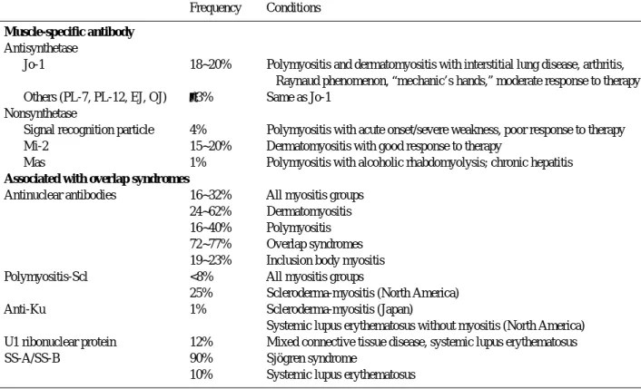
4. 염증성 근병증에서 발견되는 자가항체의 역할( T a b l e 3 )7 , 1 8 , 2 1
염증성 근병증의 2 0 %에서는 핵항원과 세포질항원에 대 한 여러 가지 자가항체가 있다. 다발 근염과 피부 근염의 10% 이하에서만 발견되는 세포질항원에 대한 항체는 세 포질 리보핵산 단백에 대한 것이다. 이 단백은 해독 ( t r a n s l a t i o n )과 단백생산을 담당한다. 또한 염증성 근병 증에서는 여러 가지 synthetase, 전이인자 및 정보를 인 식하는데 관여하는 단백에 대한 항체를 가지고 있다. 그런 자가항체중 a n t i - J o - 1이라고 불리는 h i s t i d y l - t r a n s f e r R N A에 대한 자가항체는 모든 항s y n t h e t a s e의 7 5 %를 차 지하고 있다. 임상적으로는 a n t i - J o - 1항체를 가진 사람 의 8 0 %이상이 간질성(interstitial) 폐질환을 가지고 있다 는 점이 중요하다.
그러나 이런 항체들은 근육에서만 생기는 특수한 것은 아니라고 생각된다. 왜냐하면 이런 항체들은 신체의 여기 저기에 있는 표적( t a r g e t )에 작용하며 병리기전상 의미가 없는 부수현상( e p i p h e n o m e n o n )을 나타내는지도 모르고 또한 임상적이나 면역병리학적으로 서로 다른 병이라고 생각되는 다발 근염, 피부 근염 및 봉입체 근염 모두에서 나타나며 거의 대부분에서 간질성 폐질환과 동반이 되기 때문이다. 때로는 활동성 근염을 동반하지 않은 간질성 폐질환에서도 이런 항체들이 나타나므로 이런 항체들은 근병증뿐만 아니라 간질성 폐질환에서도 중요한 지표가 될 것으로 생각된다. 피부 근염과 전신성 경화증이 중복 된 환자에서는 특수한 항핵 자가항체인 a n t i - P M / S c l이 생기는데 이것은 핵소체(nucleolar) 단백복합에 대한 항 체이다.
한편 a n t i - J o - 1항체를 가진 사람중 일부는 R a y n a u d 현상이나 비미란성(nonerosive) 관절염을 가지며 H L A 항원 D R 3와 D R w 5 2를 가진다. 다발 근염과 봉입체 근염 에서는 75% 이상에서 D R 3 ( D R B 1 0301, DQB1 0 2 0 1 )
의 h a p l o t y p e을 가지고 있는데 이것은 이런 대립유전자 ( a l l e l e )가 이 병을 일으키는 위험인자라는 것을 시사하는 것으로 생각되고 있다.
5. 바이러스 감염과 염증성 근병증과의 관련성4 , 6 - 8 coxsackievirus, paramyxovirus, 인플루엔자, 거대세 포바이러스, 볼거리 및 E p s t e i n - B a r r바이러스 등은 급성 과 만성 근염의 발병과 간접적인 관련이 있다. 그중 c o x- s a c k i e v i r u s는 molecular mimicry에 의해 유발된 기전 에 의해 자가면역성 근염을 일으킨다고 생각되고 있다. 왜 냐하면 Jo-1 항체의 공격목표가 되는 h i s t i d y l - t r a n s f e r RNA synthetase와 동물p i c o r n a v i r u s의 genomic RNA 는 구조가 서로 비슷하기 때문이다. 그러나 염증성 근병증 환자의 근생검을 아주 예민한 중합효소연쇄반응( P C R )으 로 조사해도 이런 바이러스를 발견할 수는 없었다.
다발 근염과 봉입체 근염이 바이러스와 관련성이 있다 는 것은 r e t r o v i r u s와의 관계를 보면 알 수 있다. 왜냐하 면 원숭이 면역결핍바이러스에 의해 감염된 원숭이와 인 간면역결핍바이러스( H I V )와 인간 T세포 림프영양성 바이 러스( H T L V )에 감염된 사람이 다발 근염을 일으키며 드물 게는 봉입체 근염도 일으키기 때문이다. HIV나 H T L V - 1 에 감염된 사람은 병의 초기 증상으로 염증성 근병증을 일으키기도 하고 병의 후기에 이런 근병증을 보이기도 한 다. 그러나 r e t r o v i r u s항원이 단지 근내막 대식세포에서 만 가끔 발견되고 근섬유에서는 발견되지 않는다는 것은 지속적인 감염이 있으며 바이러스 재생은 근육내에서는 일어나지 않는다는 것을 의미한다. HIV-1과 H T L V - 1감
염과 연관되어 생긴 다발 근염과 봉입체 근염의 조직소견 은 r e t r o v i r u s가 음성인 근염과 같다. 한편 HIV-1 양성 환자에서 생긴 다발 근염과 봉입체 근염은 z i d o b u d i n e으 로 장기간 치료함으로써 생긴 독성 근병증과는 감별해야 한다.
한편 항볼거리항체로 봉입체에 면역염색검사를 한 결과 를 보고 만성적으로 지속되는 볼거리가 봉입체 근염의 원 인이라고 의심하기도 하였으나 이런 가설은 그후 in situ h y b r i d i z a t i o n과 중합효소 연쇄반응검사상 볼거리 감염 이 확인되지 않음에 따라 폐기되었다. 또 소아마비후 증 후군환자의 조직이상소견이 봉입체 근염의 조직이상소견 과 비슷하다는 보고도 있다.
결 론
염증성 근병증에 대해선 아직도 의문점이 많다. 그중 가 장 기본적인 의문점이 병리기전과 치료에 대한 것이다. 일 반적으로 봉입체 근염은 여러 가지 특성상 다발 근염과 피 부 근염과는 다른 병으로 생각된다. 그러나 서로 다른 병리 기전에 의해 발병하는 것으로 생각되는 다발 근염과 피부 근염이 어떻게 MHC-1 발현과 항 J o - 1항체생산같은 공통 된 기전을 가지고 있을까? 봉입체 근염에서 보이는 단백축 적의 의미는 무엇일까? 봉입체 근염에서 보이는 염증성 변 화는 발병원인일까? 아니면 병의 결과로 생긴 것일까? 등 이런 여러 가지 의문은 앞으로 염증성 근병증에 대한 더 많 은 연구가 있는 후에야 해결될 것으로 생각된다.
Table 3. Autoantibodies in inflammatory myopathies8
Frequency Conditions Muscle-specific antibody
Antisynthetase
Jo-1 18~20% Polymyositis and dermatomyositis with interstitial lung disease, arthritis, Raynaud phenomenon, “mechanic’s hands,” moderate response to therapy Others (PL-7, PL-12, EJ, OJ) ≤3% Same as Jo-1
Nonsynthetase
Signal recognition particle 4% Polymyositis with acute onset/severe weakness, poor response to therapy
Mi-2 15~20% Dermatomyositis with good response to therapy
Mas 1% Polymyositis with alcoholic rhabdomyolysis; chronic hepatitis Associated with overlap syndromes
Antinuclear antibodies 16~32% All myositis groups 24~62% Dermatomyositis 16~40% Polymyositis 72~77% Overlap syndromes 19~23% Inclusion body myositis
Polymyositis-Scl <8% All myositis groups
25% Scleroderma-myositis (North America)
Anti-Ku 1% Scleroderma-myositis (Japan)
Systemic lupus erythematosus without myositis (North America) U1 ribonuclear protein 12% Mixed connective tissue disease, systemic lupus erythematosus
SS-A/SS-B 90% Sjögren syndrome
10% Systemic lupus erythematosus
REFERENCES
01. Hilton-Jones D. Inflammatory muscle diseases. Curr Opin Neurol 2001;14:591-596.
02. Pongratz D, Dalakas MC. Inflammatory myopathies. In:
Brant T, Caplan LR, Dichgans J, Christoph Diener H, Kennard C. Neurological disorders. 1st ed. New York:
Academic Press. 1996;965-969.
03. Bratt R, Shannon KM. Autoimmune and inflammatory disorders. In: Goetz CG, Pappert EJ. Textbook of clinical neurology. 1st ed. Philadelphia: W.B. Saunders Company.
1999;1026-1028.
04. Brooke MH. Disorders of skeletal muscle. In: Bradley WG, Daroff RB, Fenichel GM, Marsden CD. Neurology in clinical practice. 3rd ed. Vol. 2. Boston: Butterworth Heinemann. 2000;2223-2228.
05. Victor M, Ropper AH. Principles of neurology. 7th ed.
New York: McGraw-Hill. 2001;1482-1489.
06. Dalakas MC. Polymyositis, dermatomyositis, and inclu- sion body myositis. In: Brauwald E, Fauci AS, Kasper DL, Hauser SL, Longo DL, Jameson JL. Principles of internal medicine. 15th ed. Vol. 2. New York: McGraw-Hill. 2001;
2524-2529.
07. Dalakas MC. Immunopathogenesis of inflammatory myo- pathies. Ann Neurol 1995;37(S1):S74-S86.
08. Amato AA, Barohn RJ. Inflammatory myopathies: der- matomyositis, polymyositis, inclusion body myositis, and related diseases. In: Schapira AH, Griggs RC. Muscle dis- eases. 1st ed. Boston: Butterworth Heinemann. 1999;299- 317.
09. Behrens L, Bender A, Johnson MA, Hohlfeld R. Cytotoxic mechanisms in inflammatory myopathies: Co-expression of Fas and protective Bcl-2 in muscle fibers and inflam- matory cells. Brain 1997;120:929-938.
10. Hohlfeld R, Engel AG, Goebels N, Behrens L. Cellular immune mechanisms in inflammatory myopathies. Curr Opin Neurol 1997;9:520-526.
11. Nagaraju K, Raben N, Loeffler L. Conditional up-regula- tion of MHC class 1 in skeletal muscle leads to self-sus-
taining autoimmune myositis and myositis-specific autoantibodies. Proc Natl Acad Sci USA 2000;97:9209- 9214.
12. Arahatas K, Engel AG. Monoclonal antibody analysis of monoclonal cells in myopathies. I: quantitation of subsets according to diagnosis and sites of accumulation and de- monstration and counts of muscle fibers invaded by T cells.
Ann Neurol 1984;16:193-208.
13. Angel AG, Arahata K. Monoclonal antibody analysis of mononuclear cells in myopathies. II: phenotypes of autoin- vasive cells in polymyositis and inclusion body myositis.
Ann Neurol 1984;16:209-215.
14. Arahata K, Engel AG. Monoclonal antibody analysis of mononuclear cells in myopathies. III: immunoelectron microscopy aspects of cell-mediated muscle fiber injury.
Ann Neurol 1986;19:112-125.
15. Arahata K, Engel AG. Monoclonal antibody analysis of mononuclear cells in myopathies. IV: cell-mediated cyto- toxicity and muscle fiber necrosis. Ann Neurol 1988;23:
168-173.
16. Arahata K, Engel AG. Monoclonal antibody analysis of mononuclear cells in myopathies. V: identification and quantitation of T8+ cytotoxic and T8+suppressor cells.
Ann Neurol 1988;23:493-499.
17. Askanas V, Alvarez RV, Mirabella M, Engel WK. Use of anti-neurofilament antibody to identify paired-helical fila- ments in inclusion body myositis. Ann Neurol 1996;39:389- 3 9 1 .
18. Askanas V, Engel WK. Inclusion body myositis: newest concepts of pathogenesis and relation to aging and Alzhei- mer disease. J Neuropathol Exp Neurol 2001;60:1-14.
19. Banwell BL, Engel AG. AlphaB-crystallin immunolocal- ization yields new insights into inclusion body myositis.
Neurology 2000;54:1033-1041.
20. Moslemi AR, Lindberg C, Oldfors A. Analysis of multiple mitochonrial DNA deletions in inclusion body myositis.
Hum Mutat 1997;10:381-386.
21. Mozaffar T, Pestronk A. Myopathy with anti-Jo-1 antibod- ies: pathology in perimysium and neighbouring muscle fibres. J Neurol Neurosurg Psychiatry 2000;68:472-478.