Opuntia humifusa Supplementation Reduces Fat Weight by Increasing PPAR-
γand PGC-1α Protein Expression in the Skeletal Muscle of Rats
Daekeun Kwon, Junyong Kang, Jaeseung Kim and Youngju Song*
Laboratory of Sports Nutrition, Sunmoon University, Asan-si, Chung Nam, 336-708, Korea Received October 30, 2013 /Revised December 31, 2013 /Accepted January 22, 2014
This study was conducted to investigate the effects of supplementation withOpuntia humifusa on the expression of peroxisome proliferator-activated receptor-delta (PPAR-δ), peroxisome proliferator-acti- vated receptor-gamma (PPAR-γ) and peroxisome proliferator-activated receptor gamma coactivator- 1alpha (PGC-1α) in the skeletal muscle of rats fed a high-fat diet. Sixteen Sprague-Dawley male rats at 6 weeks of age were randomly divided into 2 groups: a control diet group (CG,n=8) and an ex- perimental diet group (EG, n=8). The rats were fed a high-fat diet (CG) or a high-fat diet supple- mented with 5%O. humifusa (EG) for 8 weeks. The results showed that the abdominal fat pad and epididymal fat pad weights were significantly lower in the EG than in the CG (p<0.01). In the blood, serum glucose, triglycerides, and total cholesterol in the EG group were lower than in the CG (p<0.01). The expression of PPAR-γ and PGC-1α protein in the skeletal muscle of the EG was increased compared with that of the CG (p<0.05). These results indicate that 8 weeks of O. humifusa supple- mentation lowers serum glucose and triglyceride levels and suppresses weight gain by reducing fat weight through an increase in the expression of PPAR-γ and PGC-1α in the muscle tissue of rats.
Key words : High-fat diet,Opuntia humifusa, PPAR-δ, PPAR-γ, PGC-1α
*Corresponding author
*Tel : +82-41-530-2239, Fax : +82-41-530-2810
*E-mail : [email protected].
This is an Open-Access article distributed under the terms of the Creative Commons Attribution Non-Commercial License (http://creativecommons.org/licenses/by-nc/3.0) which permits unrestricted non-commercial use, distribution, and reproduction in any medium, provided the original work is properly cited.
Journal of Life Science 2014 Vol. 24. No. 1. 67~73 DOI : http://dx.doi.org/10.5352/JLS.2014.24.1.67
Introduction
Peroxisome proliferator-activated receptors (PPARs), which are nuclear hormone receptors that activate lipid me- tabolism pathways such as lipogenesis and lipolysis, are well known to play an important role in maintaining cholesterol and lipid homeostasis. PPARs also regulate the genes in- volved in the metabolic regulation of lipoproteins, the activ- ity of insulin and the differentiation of adipocytes [6]. PPAR- δ is expressed in various tissues, particularly in muscle tis- sue, and is involved in the modulation of lipid catabolism and energy uncoupling in skeletal muscles [5]. In addition, PPAR-γ regulates fat storage and adipocyte metabolism [27], and it has been reported that PPAR-γ is involved in modulat- ing insulin sensitivity in the body [26]. Furthermore, PGC-1 α, which is a transcriptional coactivator associated with PPARs expression in tissues, is a regulator of mitochondria biogenesis and lipid metabolism activation [2, 12]. In partic- ular, it has been reported that PGC-1α is highly expressed
in brown adipose tissue, which is known to have a high capability for oxidation as well as activated transcription of PPAR-γ [20, 29].
In a previous study that related the reduction of fat weight and PPARs, Wang et al. [32] reported that activation of PPAR-δ decreased fat mass due to an increase in fatty acid oxidation and utilization in adipocytes and muscle tis- sues, and it has been reported that reduced expression of PPAR-γ protein in white adipose tissue affected body weight and reduced fat weight [30]. However, although the ex- pression of PPAR-γ is low in muscle, previous studies have suggested that lipid metabolism is related to PPAR-γ protein expression in muscle tissue. Norris et al. [22] reported that body weight significantly increased in PPAR-γ knockout mouse muscle tissue, in contrast with was observed in adipocytes. Nevertheless, although the protein expression of PPAR-γ and PGC-1α was increased in rat muscle tissue, body and fat weight was not affected. Therefore, PPAR pro- teins are expressed according to tissue-specific patterns.
Recently, many studies and development processes have focused on functional food, which can safely be ingested without toxicity as a long-term treatment, to activate lipid metabolism. Approximately 4,000 types of cacti exist in the world, and as a plant that is highly adaptable to semi-arid areas, some varieties have been used as a source of carbohy- drates and various vitamins. Particularly used in South
Table 1. Composition of freeze-dried O. humifusa
Ingredients Contents
Moisture (% w/w) Ash (% w/w)
Carbohydrate (g/100 g) Crude protein (g/100 g) Crude fat (g/100 g) Fiber (g/100 g) Fe2+ (mg/g) Ca2+ (mg/100 g) Mg2+ (mg/100 g) K+ (mg/100 g) Na+ (mg/100 g) P2+ (mg/100 g)
2.9 13.8 46.64.9 28.93.1 2931.35.8 1227.9 2155.5 30.9 653.2 Korea,O. humifusa, a tropical plants containing fruits on the
stems, is a member of Cactaceae family that has been culti- vated to grow in cold environment below -20℃ [8].O. humi- fusa contains not only minerals, such as calcium, magnesium, zinc, and ferrum but also soluble fiber, vitamin- C, vitamin- E, flavonoids, and polyphenols. It has been reported that these nutritional compounds play a predominant role in the physiological function of the body [10]. In a previous study, Dok-go et al. [4] described that supplementation withO. fi- cus-indica, which is repleted with flavonoids such as querce- tin that act as antioxidants, exhibited anti-inflammatory ef- fects and neuroprotective effects against oxidative injuries induced in cortical cell cultures in rats. It has been reported that cactus extract supplementation protects the gastric mu- cous membrane and suppresses carrageenan-induced edema and beta-glucuronidase and lysosomal enzyme secretion in neutrophils [24]. Although various research has been per- formed that reported thatO. humifusa supplementation has a positive effect on the physiological function to the body, there have not been any studies designed to evaluate the protein expression related to fatty acids regulation by O.
humifusa.
Therefore, in this study, we investigated the effects ofO.
humifusa supplementation on body weight and changes in fat weight as well as on PPAR-γ, δ and PGC-1α protein ex- pression, which plays an important role in transcriptional gene regulation of fatty acid metabolism in muscle tissue, in high-fat diet-induced rats.
Materials and Methods Experimental animals
All experimental protocols were approved by the Animal Study Committee of Sunmoon University. After the acclima- tization period during week one, sixteen 6-week-old male Sprague-Dawley rats (Samtaco Bio Korea, Hwaseong, Korea) were randomly divided into two groups: a control diet group (CG: high-fat diet group, n=8) and an experimental diet group (EG: 5%O. humifusa supplemented diet group, n=8), given free access to tap water and food for 8 weeks, and housed in groups of two per cage under controlled tem- perature (23±1°C) and relative humidity (50±5%). The light/dark cycle was automatically controlled (alternating 12-h periods), and lighting began at 8:00 pm. Food intake was measured daily and body weight was measured weekly.
At the end of the experimental period, the rats were anes-
thetized with diethyl ether after fasting for 12 h. Blood sam- ples were taken from the left ventricle and serum was ob- tained by centrifuging the blood at 700× g for 20 min at 4°C. Both hind limb muscles were dissected and immedi- ately immersed in liquid nitrogen. The serum samples and hind limb muscles were stored at -70°C until analyzed.
Preparation of experimental diet
O. humifusa, which was harvested in Asan, Chungnam, was cleaned and blended using a HMF-3150S blender (Hanil Electronics, Seoul, Korea). After blending, the O. humifusa was frozen in a freezer at a temperature of -70°C and then freeze-dried in a freeze dryer (Ilshin Co., Gyeonggi, Korea).
After freeze-drying, as shown in Table 1, a general compo- nent analysis of O. humifusa was performed using the Association of Official Analytical Chemists (AOAC) method for the following measurements: moisture, using an air-oven method; ash, using a dry-ashing method; carbohydrates, us- ing calculation; crude protein, using a Keldahl method;
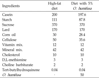
crude fat, using a Soxhlet extraction method; and fiber, using an enzymatic-chemical method. Mineral component analysis was performed using plasma atomic emission spectrometry (ICP-AES) to determine the composition of Fe2+, Ca2+, Mg2+, K+, Na+, and P2+. As shown in Table 2, the high-fat diet was composed of 20% protein, 48% carbohydrate and 20% fat and was modified from a previous study [18] and based on AIN-76G. The 5%O. humifusa diet was made by substituting a portion of carbohydrates, protein, fiber, and fat compo- nents of the high-fat diet. During the experimental period, the diet was prepared in batches sufficient for 3-5 days, and the experimental diets were stored at 4°C to maintain freshness.
Table 3. Comparison of body weight gain and fat tissue weights
CG EG
Body weight gain (g) Abdominal fat pad (g) Epididymal fat pad (g)
193.0±11.86 11.5±0.91 11.3±0.91
183.1±17.04 8.5±0.70* 8.5±0.78* Data are expressed as the mean ± SE; CG: Control diet group;
EG: Experimental diet group; *,p<0.05 compared with the CG.
Table 4. Changes in serum parameters
CG EG
Glucose (mg/dl) Triglyceride (mg/dl) HDLC (mg/dl) TC (mg/dl)
147.1±5.06 36.0±2.69 18.3±0.91 100.1±1.37
119.9±2.57**
18.0±3.07**
21.0±1.08 78.4±5.05 **
Data are expressed as the mean ± SE; CG: Control diet group;
EG: Experimental diet group; HDLC: high-density lipoprotein cholesterol; TC: total cholesterol; **,p<0.01 compared with the CG.
Table 2. Composition of the experimental diet (g/kg diet)
Ingredients High-fat
diet Diet with 5%
O. humifusa Casein
Starch Sucrose Lard Corn oil Cellulose Vitamin mix.
Mineral mix.
Cholesterol D,L-methionine Choline barbiturate Tert-butylhydroquinone O. humifusa
200111 370170 30 5012 4210 32 0.04
·
197.6 87.8 370170 28.4 35.612 4210
32 0.04
50
Serum analysis
Serum glucose, TG, TC, and HDLC levels were analyzed using enzymatic kits (Asan Pharmaceutical Co, Yongin, Korea).
Western blot analysis
For protein expression analysis, soleus muscle was homo- genized on ice with a polytron homogenizer in 20 mmol/l Tris-HCl buffer (pH 7.5) containing 5 mmol/l EDTA, 2 mmol/l PMSF, and 1:200 protease inhibitor cocktail (Sigma, St Louis, MO, USA). The protein concentrations were de- termined using Bradford reagent (Bio-Rad, Hercules, CA, USA), with bovine serum albumin as the standard. An ali- quot of tissue extract containing 20 μg of protein (for PPAR- δ, PPAR-γ and PGC-1α) was separated on a 10% SDS-PAGE gel. After electrophoresis, the proteins were transferred to a PVDF membrane (Millipore, Bedford, MA, USA) using a semi-dry blotting apparatus (Bio-Rad, Hercules, CA, USA).
After treating with blocking buffer (PBS containing 10%
skim milk) for 90 min, the membrane was incubated with primary polyclonal antibodies for 2 h (PPAR-δ, PPAR-γ and PGC-1α; Santa Cruz Biotechnology, CA, USA), followed by five 10 min. washes with PBS (5% Tween-20). The membrane was then incubated with HRP-conjugated anti-goat IgG or anti-rabbit IgG (Santa Cruz Biotechnology, CA, USA) for 1 h, followed by five 10 min. washes with PBS (5% Tween-20).
The target proteins were detected using an ECL kit (Amersham Pharmacia Biotech, Piscataway, NJ, USA). The films were photographed and the protein bands of interest were quantified using band analyzer software (Bio-Rad, Hercules, CA, USA).
Statistical analysis
All data were analyzed using SPSS software (version 16.0 for Windows). The data are expressed as the mean ± SE, and values were analyzed with the independent samplest test. Significance was defined as p<0.05.
Results Body weight gain and fat tissue weights
In order to determine the effect of O. humifusa supple- mentation on the body weight gain and fat tissue weights, both body weight and fat tissue weights were measured by using a digital electronic balance. As shown in Table 3, body weight gain was slightly lower in the EG compared with the CG after 8 weeks of O. humifusa supplementation.
However, the abdominal fat pad and epididymal fat pad were significantly smaller in the EG than in the CG (p<0.05).
Changes in serum parameters
The enzymatic kits were used to estimate the effect of O. humifusa supplementation on serum parameters such as glucose, TG, HDLC, and TC. After the 8 weeks of the experi- ment, as shown in Table 4, the glucose, TG and TC concen- trations of the EG were significantly lower than those of the CG (p<0.01). Additionally, the HDLC concentration of the EG tended to be higher than that of the CG, but the differ- ence was not significant.
Fig. 1. Effect ofO. humifusasupplementation on the expression levels of PPAR-δ and PPAR-γ proteins in skeletal muscle. The data are expressed as the mean ± SE. CG: Control diet group; EG: Experimental diet group; *, p<0.05; (A) PPAR-δ; (B) PPAR- γ.
Fig. 2. Effect ofO. humifusasupplementation on PGC-1α protein expressions in skeletal muscle. The data were expressed as the mean ± SE. CG: Control diet group; EG: Experim- ental diet group; *, p<0.05.
Skeletal muscle PPAR-δ, PPAR-γ, and PGC-1α protein expressions
Western blot analysis was performed to analyze the PPAR- δ, PPAR-γ, and PGC-1α protein expressions using ho- mogenated soleus muscles. As shown in Fig. 1A, the ex- pression of PPAR-δ protein in the EG tended to be higher than in the CG, but this difference was not significant.
However, PPAR-γ was significantly higher in the EG than in the CG (p<0.05, Fig. 1B). As shown in Fig. 2, PGC-1α pro- tein expression in the EG was significantly higher than in the CG (p<0.05).
Discussion
This study investigated the effect ofO. humifusa supple- mentation on body weight and changes in fat weight as well
as PPAR- δ, γ and PGC-1α protein expression on fatty acid metabolism in muscle tissue. In the present study, body weight was not significantly different between groups, whereas the PFT and EFT of the EG were significantly de- creased compared with the CG. Increases in calcium intake, one of the major components of O. humifusa, have been shown to improve fatty acid oxidation by suppressing fatty acid synthase and thus reducing the intra-adipocyte calcium level through decrease parathyroid hormone secretion and reduced fat accumulation as well as increased lipolysis [36].
In a previous study, Teegarden et al. [31] reported that the rate of fat oxidation was significantly increased after 12 weeks of a 1,400 mg/day calcium-supplemented diet in obese women aged 18-31 compared with controls. We ana- lyzed the nutritional composition of freeze-driedO. humifusa in the present study and found that, it contains 2,300 mg/100 g of calcium. In addition, we calculated that the ex- perimental diet group received approximately 1.3 times more calcium than the control group due to the 5%O. humi- fusa supplementation. In turn, fat oxidation of PFT and EFT in the EG was increased, which suppressed fat accumulation and increased lipolysis in the fat tissue of the EG, similar to what was observed in the previous study. Furthermore, we suggest that in the previous studies, increased expression of PPAR-δ, PPAR-γ, and PGC-1α proteins activated fatty acid metabolism [16, 32], suggesting that increased PPAR-γ and PGC-1α may have influenced fat weight reduction in the present study.
Serum analysis showed that serum glucose, TG, and TC of the EG were significantly decreased compared with the CG, and HDLC tended to increase in the EG compared with the Bwititi et al. [3] reported that orally humifusa extract
supplementation (20 mg/ 100 g body weight) lowers serum glucose levels through an insulin-independent mechanism in streptozotocin (STZ)-diabetic rats, and the glucose low- ering effect was due to an increase in intracellular glucose uptake through the renal reabsorption of Na+ after extract supplementation. In addition, it has been reported that in- sulin resistance was improved because TNF-α, which is a cytokine that is secreted from adipocytes and causes insulin resistance, is reduced by PPAR-γ agonists [21]. Therefore, increased PPAR-γ caused a reduction in glucose level in the present study. Meanwhile, it has been reported that in- creased lipoproteins such as TC, TG and LDLC cause hyper- lipidemia during high-fat diet supplementation in a rat mod- el [13, 35]. However, fiber, which is also a component of O. humifusa, improved hyperlipidemia through by suppress- ing endogenous cholesterol synthesis by hindering intestinal fat absorption, increasing bile acid synthesis, and forming fatty acids with a lower number of carbons, which are de- graded in the large intestine [1, 28]. Similar to our present data, Hahm et al. [11] found that glucose, TG, TC and LDLC were significantly decreased in STZ-induced diabetic rats af- ter oral supplementation with 250 mg/kg/dayO. humifusa extract. It has been reported that supplementation with 2.5 g /100 g diet prickly pear pectin, another cactus, has a pos- itive effect on lowering serum lipid profiles in hyper- lipidemia-induced guinea pigs [7], and serum LDLC, TG and TC levels are decreased byO. robusta administration in hy- perlipidemic middle-aged men [33]. As a result, we might assume that the reduction in serum TG and TC levels in the EG is caused by an improvement in hepatic cholesterol metabolism due to increased intake of fiber and pectin with the O. humifusa-supplemented diet.
Meanwhile, expression of PPAR-γ and PGC-1α in the muscle in the EG was significantly increased compared with the CG, and PPAR-δ in the EG tended to increase in muscle tissue compared with the control group. Although PPAR-δ protein is expressed in various tissues, its expression is high- est in muscle tissue [14], and it is involved in the catabolism of lipids as well as the regulation of energy uncoupling in skeletal cells [5]. Wang et al. reported that the activation of the PPAR-δ protein in adipocyte and skeletal muscle in- creased fat oxidation and utilization in an in vivo study us- ing obese Zucker rats. Therefore, we suggest thatO. humifusa supplementation partially affected fatty acid metabolism in muscles because an increasing tendency of PPAR-δ protein expression was observed afterO. humifusa supplementation.
Meanwhile, although PPAR-γ protein is highly expressed in adipose tissue, it is involved in fat oxidation-related gene expression in skeletal muscle tissue [19] and also plays an important role in lipid metabolism [9]. Similar to our present study, Kang et al. [17] reported that 8 weeks ofO. humifusa supplementation increased PPAR-γ protein expression in a rat model. We suggest that increased PPAR-γ protein ex- pression in the present study occurred through increased PGC-1α, which is a co-activator of PPAR-γ, due to intake of calcium-richO. humifusa. PGC-1α lead to PPAR expression as a co-activator and simulated mitochondrial biogenesis in muscle tissue as well as increased fatty acid oxidation through a transfer to slow twitch muscle fiber, which is fa- vorable for fatty acid oxidation metabolism [25]. Calcium, in whichO. humifusa is rich, plays an important role in the modulation of the intracellular signaling second messenger that leads to muscle contraction and gene expression.
Increased calcium intake is highly involved in improving lipid metabolism, oxidative phosphorylation, mitochondrial biogenesis, and PGC-1α protein expression due to an in- crease in the activation of AMP-activated protein kinase (AMPK) and calcium/calmodulin-dependent protein kinase (CAMK) [15, 23, 34]. As a result, we suggest that the intake of calcium-richO. humifusa increases the activation of intra- muscular AMPK and CAMK by increasing the serum cal- cium level, resulting in the expression of PPAR-γ and PGC-1 α, which are involved in lipid metabolism. Furthermore, as shown in a previous study, our results indicated that a re- duction in fat weight, an improvement in serum lipid pro- files, and a lowering of serum glucose level due to improve- ment of insulin sensitivity are caused by increased muscular PPAR-γ and PGC-1α protein expression, which lead to the activation of fatty acid metabolism.
These results show that supplementation withO. humifusa for 8 weeks plays a positive role in lowering serum glucose and TG levels and suppressing weight gain by reducing fat weight through increasing the expression levels of PPAR-γ and PGC-1α proteins in the muscle tissue of rats.
Acknowledgement
This research was supported by the Basic Science Research Program of the National Research Program through the National Research Foundation of Korea (NRF) funded by the Ministry of Education, Science and Technology (2011- 0011951).
References
1. Anderson, J. W. and Bridges, S. R. 1981. Plant fiber metabo- lites alter hepatic glucose and lipid metabolism.Diabetes30, 133-139.
2. Arany, I. 2008. When less is more: apoptosis during acute kidney injury. Kidney Int 74, 261-262.
3. Bwititi, P., Musabayane, C. T. and Nhachi, C. F. 2000. Effects ofOpuntia megacanthaon blood glucose and kidney function in streptozotocin diabetic rats.J Ethnopharmacol69, 247-252.
4. Dok-Go, H., Lee, K. H., Kim, H. J., Lee, E. H., Lee, J., Song, Y. S., Lee, Y. H., Jin, C., Lee, Y. S. and Cho, J. 2003. Neuro protective effects of antioxidative flavonoids, quercetin, (+)-dihydroquercetin and quercetin 3-methyl ether, isolated fromOpuntia ficus-indicavar. saboten.Brain Res965, 130-136.
5. Dressel, U., Allen, T. L., Pippal, J. B., Rohde, P. R., Lau, P. and Muscat, G. E. 2003. The peroxisome proliferator-acti- vated receptors β/δ agonist GW501516, regulates the ex- pression of genes involved in lipid catabolism and energy uncoupling in skeletal muscle cells. Mol Endocrinol 17, 2477-2493.
6. Evans, R. M., Barish, G. D. and Wang, Y. X. 2004. PPARs and the complex journey to obesity. Nat Med10, 355-361.
7. Fernandez, M. L., Lin, E. C. K., Trejo, A. and McNamara, D. J. 1994. Prickly pear (Opuntiasp.) pectin alters hepatic cholesterol metabolism without affecting cholesterol absorp- tion in guinea pigs fed a hypercholesterolemic diet.J Nutr 124, 817-824.
8. Goldstein, G. and Nobel, P. S. 1994. Water relations and low-temperature acclimation forcactus species varying in freezing tolerance. Plant Physiol 104, 675-681.
9. Gorla-Bajszczak, A., Siegrist-Kaiser, C., Boss, O., Burger, A.
G. and Meier, C. A. 2000. Expression of peroxisome pro- liferator-activated receptors in lean and obese Zucker rats.
Eur J Endocrinol 142, 71-78.
10. Gurrieri, S., Miceli, L., Lanza, C. M., Tomaselli, F., Bonomo, R. P. and Rizzarelli, E. 2000. Chemical characterization of sicilian prickly pear (Opuntia ficus indica) and perspectives for the storage of its juice.J Agric Food Chem48, 5424-5231.
11. Hahm, S. W., Park, J. and Son, Y. S. 2011.Opuntia humifusa stems lower blood glucose and cholesterol levels in strepto- zotocin-induced diabetic rats. Nutr Res31, 479-487.
12. Handschin, C. and Spiegelman, B. M. 2008. The role of ex- ercise and PGC1alpha in inflammation and chronic disease.
Nature454, 463-469.
13. Harrison, D., Griendling, K. K., Landmesser, U., Hornig, B.
and Drexler, H. 2003. Role of oxidative stress in atherosclerosis. Am J Cardiol91, 7A-11A.
14. Hatakeyama, Y. and Scarpace, P. J. 2001. Transcriptional regulation of uncoupling protein-2 gene expression in L6 myotubes. Int J Obes Relat Metab Disord25, 1619-1624.
15. He, Y. H., Li, S. T., Wang, Y. Y., Wang, G., He, Y., Liao, X. L., Sun, C. H. and Li, Y. 2012. Postweaning low-calcium diet promotes later-life obesity induced by a high-fat diet.
J Nutr Biochem23, 1238-1244.
16. Hu, S., Yao, J., Howe, A. A., Menke, B. M., Sivitz, W. I.,
Spector, A. A. and Norris, A. W. 2012. Peroxisome pro- liferator-activated receptor γ decouples fatty acid uptake from lipid inhibition of insulin signaling in skeletal muscle.
Mol Endocrinol 26, 977-988.
17. Kang, J. Y., Lee, J. H., Kwon, D. K. and Song, Y. J. 2013.
Effect of Opuntia humifusasupplementation and acute ex- ercise on insulin sensitivity and associations with PPAR-γ and PGC-1α protein expression in skeletal muscle of rats.
Int J Mol Sci 28, 7140-7154.
18. Kim, Y. J. and Park, T. S. 2008. Genes are differentially ex- pressed in the epididymal fat of rats rendered obese by a high-fat diet. Nutr Res 28, 414-422.
19. Lapsys, N. M., Kriketos, A. D., Lim-Fraser, M., Poynten, A.
M., Lowy, A., Furler, S. M., Chisholm, D. J. and Cooney, G. J. 2000. Expression of genes involved in lipid metabolism correlate with peroxisome proliferator-activated receptor gamma expression in human skeletal muscle. J Clin Endocrinol Metab85, 4293-4297.
20. Lin, J., Puigserver, P., Donovan, J., Tarr, P. and Spiegelman, B. M. 2002. Peroxisome proliferator-activated receptor gam- ma coactivator 1beta (PGC-1beta), a novel PGC-1-related transcription coactivator associated with host cell factor.J Biol Chem277, 1645-1648.
21. Miles, P. D., Romeo, O. M., Higo, K., Cohen, A., Rafaat, K. and Olefsky, J. M. 1997. TNF-alpha-induced insulin re- sistance in vivoand its prevention by troglitazone.Diabetes 46, 1678-1683.
22. Norris, A. W., Chen, L., Fisher, S. J., Szanto, I., Ristow, M., Jozsi, A. C., Hirshman, M. F., Rosen, E. D., Goodyear, L.
J., Gonzalez, F. J., Spiegelman, B. M. and Kahn, C. R. 2003.
Muscle-specific PPARγ-deficient mice develop increased adiposity and insulin resistance but respond to thiazolidine- diones. J Clin Invest112, 608-618.
23. Ojuka, E. O. 2004. Role of calcium and AMP kinase in the regulation of mitochondrial biogenesis and GLUT4 levels in muscle. Proc Nutr Soc63, 275-278.
24. Park, E. H., Kahng, J. H. and Paek, E. A. 1998. Studies on the pharmacological actions of cactus: identification of its anti-inflammatory effect. Arch Pharm Res 21, 30-34.
25. Petersen, K. F., Befroy, D., Dufour, S., Dziura, J., Ariyan, C., Rothman, D. L., DiPietro, L., Cline, G. W. and Shulman, G. I. 2003. Mitochondrial dysfunction in the elderly: possible role in insulin resistance. Science300, 1140-1142.
26. Russell, A. P., Hesselink, M. K., Lo, S. K. and Schrauwen, P. 2005. Regulation of metabolic transcriptional co-activators and transcription factors with acute exercise. FASEB J 19, 986-988.
27. Sears, I. B., MacGinnitie, M. A., Kovacs, L. G. and Graves, R. A. 1996. Differentiation-dependent expression of the brown adipocyte uncoupling protein gene: regulation by peroxisome proliferator-activated receptor gamma.Mol Cell Biol 16, 3410-3419.
28. Story, J. A. 1981. The role of dietary fiber in lipid metabolism.
Adv Lipid Res 18, 229-246.
29. St-Pierre, J., Lin, J., Krauss, S., Tarr, P. T., Yang, R., Newgard, C. B. and Spiegelman, B. M. 2003. Bioenergetic
초록:손바닥선인장 보충이 고지방식이 흰쥐 골격근의 PPAR-γ와 PGC-1α 단백질 발현 증가에 미치는 영향
권대근․강준용․김재승․송영주*
(선문대학교 스포츠영양학실)
본 연구는 안정 시 고지방식이 흰쥐의 골격근에서 PPAR-δ, PPAR-γ 그리고 PGC-1α 단백질 발현에 손바닥선인 장 보충이 미치는 효과에 대하여 연구하였다. SD계 수컷 흰쥐 16마리를 무작위로 대조군(CG, n=8)과 실험군(EG, n=8)으로 분류하였다. 8주 동안 대조군은 고지방식이를 부하하였으며, 실험군은 5% 손바닥선인장을 보충식이하 였다. 본 실험결과, 복부지방과 고환부 지방 중량은 EG군이 CG군에 비해 유의하게 낮게 나타났다(p<0.01). 또한 혈당, 중성지방, 총콜레스테롤의 농도도 EG군이 CG군에 비해 유의하게 낮게 나타났다(p<0.01). 한편, 골격근에서 PPAR-γ와 PGC-1α 단백질 발현은 EG군이 CG군에 비해 유의하게 높게 나타났다(p<0.05). 이상의 결과로부터 손 바닥선인장 보충이 고지방식이 흰쥐의 혈당과 중성지방 농도의 감소와 골격근에서 PPAR-γ와 PGC-1α 단백질 발 현을 증가시킴으로서 체지방을 감소시켜 체중증가 억제에 긍정적인 영향을 미치는 것으로 나타났다.
analysis of peroxisome proliferator-activated receptor gam- ma coactivators 1alpha and 1beta (PGC-1alpha and PGC-1beta) in muscle cells. J Biol Chem278, 26597-26603.
30. Sun, X. and Zemel, M. B. 2004. Calcium and dairy products inhibit weight and fat regain during ad libitum con- sumption following energy restriction in Ap2-agouti trans- genic mice. J Nutr 134, 3054-3060.
31. Teegarden, D., White, K. M., Lyle, R. M., Zemel, M. B., Van Loan, M. D., Matkovic, V., Craig, B. A. and Schoeller, D.
A. 2008. Calcium and dairy product modulation of lipid uti- lization and energy expenditure.Obesity (Silver Spring)16, 1566-1572.
32. Wang, Y. X., Lee, C. H., Tiep, S., Yu, R. T., Ham, J., Kang, H. and Evans, R. M. 2003. Peroxisome-proliferator-activated receptor delta activates fat metabolism to prevent obesity.
Cell 113, 159-170.
33. Wolfram, R. M., Kritz, H., Efthimiou, Y., Stomatopoulos, J.
and Sinzinger, H. 2002. Effect of prickly pear (Opuntia robus- ta) on glucose- and lipid-metabolism in non-diabetics with hyperlipidemia - a pilot study. Wien Klin Wochenschr 114, 840-846.
34. Wu, H., Kanatous, S. B., Thurmond, F. A., Gallardo, T., Isotani, E., Bassel-Duby, R. and Williams, R. S. 2002.
Regulation of mitochondrial biogenesis in skeletal muscle by CaMK. Science12, 349-352.
35. Yang, R., Le, G., Li, A., Zheng, J. and Shi, Y. 2006. Effect of antioxidant capacity on blood lipid metabolism and lip- oprotein lipase activity of rats fed a high-fat diet.Nutrition 22, 1185-1191.
36. Zemel, M. B., Shi, H., Greer, B., Dirienzo, D. and Zemel, P. C. 2000. Regulation of adiposity by dietary calcium.
FASEB J 14, 1132-1138.