Expression and Localization of 20α-Hydroxysteroid Dehydrogenase in Immature Pig Testis
Jeong-Soo Kim
1, Hun-Ki Seong
1, Munkhzaya Byambaragchaa
1, Bo-Woong Sim
2, Chang-Gi Her
3, Myung-Hwa Kang
4and Kwan-Sik Min
1*
1Animal Biotechnology, Graduate School of Future Convergence Technology, Department of Animal Resource Science, Institute of Genetic Engineering, Hankyong National University, Ansung 17579, Korea
2National Primate Research Center, Korea Research Institute of Bioscience and Biotechnology, Ochang 28116, Korea
3MediKinetics, PyeongTaek 17792, Korea
4Department of Food Science and Nutrition, Hoseo University, Asan 31499, Korea Received February 24, 2017 /Revised May 20, 2017 /Accepted July 20, 2017
In all mammalian species, progesterone is essential in the preparation for and maintenance of pregnancy. 20α-hydroxysteroid dehydrogenase (20α-HSD) predominantly converts progesterone into its biologically inactive form 20α-hydroxyprogesterone (20α-OHP), and plays a crucial role in the ter- mination of pregnancy and initiation of parturition. In this study, we characterized the expression and localization of 20α-HSDinthe testis of MediKinetics Micropigs
®. The testes were collected at days 6, 9, 12, 18, and 21 after birth. The 20α-HSD mRNA was found to be expressed in the testis at day 6 after birth by RT-PCR. The highest level of mRNA expression in the testis was detected on day 21 after birth. However, the mRNA was not detected in the placenta after parturition. Western blot for 20α-HSD reveal that the specific 37-kDa band was detected in immature pig testis. However, this band was not detected in testis tissue at day 6 after birth. In the immunohistochemical analysis of the testis, 20α-HSD was detected in the Sertoli cells and Leydig cells. Taken together, our study shows for the first time that the 20α-HSD mRNA and protein are expressed in pig testis after birth. Further inves- tigation is required to elucidate the functional mechanisms of 20α-HSD in pig testis after birth.
Key words : 20α-HSD, expression, IHC, miniature pig, testis
*Corresponding author
*Tel : +82-31-670-5421, Fax : +82-31-670-5417
*E-mail : [email protected]
This is an Open-Access article distributed under the terms of the Creative Commons Attribution Non-Commercial License (http://creativecommons.org/licenses/by-nc/3.0) which permits unrestricted non-commercial use, distribution, and reproduction in any medium, provided the original work is properly cited.
Journal of Life Science 2017 Vol. 27. No. 7. 739~745 DOI : https://doi.org/10.5352/JLS.2017.27.7.739
Introduction
The enzyme 20α-hydroxysteroid dehydrogenase (20α- HSD) catalyzes the conversion of progesterone into its in- active form, 20α-hydroxyprogesterone, and plays a critical role in the regulation of the luteal function in mammals by controlling progesterone activity [10].
In all mammalian species, progesterone is essential to pre- pare the body for pregnancy and for maintaining pregnancy, if it occurs [10]. Progesterone makes the endometrium ready for possible implantation and inhibits uterine contractions until birth.
Accordingly, 20α-HSD in the placenta may be involved in reducing the cytotoxic effects of progesterone in the devel-
oping fetus [15]. In rodents, 20α-HSD activity is suppressed in the corpus luteum (CL) by prolactin (PRL) or placental lactogens during pregnancy and abruptly increases before parturition, which is associated with a decrease in progester- one levels and a reciprocal increase in 20α-OHP levels [1, 22].
In previous studies, we evaluated 20α-HSD gene ex-
pression in the CL during the estrous cycle and determined
that it was highly expressed in the large luteal cells during
bovine estrous cycle [10], bovine early pregnancy [9], and
deer early pregnancy [11]. In the pig, we also reported that
20α-HSD was mainly localized in the trophoblast villus in
the placenta and was highly expressed in the glandular epi-
thelial cells of the endometrium and the luminal epithelial
cells of the uterus during early pregnancy and estrous cycle
[17, 21]. Moreover, we reported that 20α-HSD was detected
at high levels in macaque ovarian and placental tissues dur-
ing the pre-ovulation and pre-parturition phases, and that
it was mainly localized in the syncytiotrophoblasts of the
placenta and the isthmus cells of the oviduct [16]. Thus, 20α- HSD expression in the placenta plays a critical role during fetal development and/or the parturition process [25].
Recently, we also reported the presence of 20α-HSD mRNA and protein in the mouse testis after birth (Park et al., in- press). Additionally, the steroid hormones progesterone and estradiol were detected in the miniature pig during the es- trous cycle (Seong et al., inpress).
Moreover, prostaglandin F
2α (PGF
2α) is known to induce abortion in several species, including rodents [6], and ad- ministration of PGF
2α to pregnant rats increases luteal 20α- HSD activity [26]. The amount of 20α-HSD mRNA in cul- tured luteal cells increased with time and by treatment with the luteolytic agent PGF
2α. Immunofluorescence assays de- tected increased protein in cultured luteal cells [9]. However, little has been reported about the expression and localization of 20α-HSD in the pig testis after birth.
In the present study, we investigated the expression and localization of 20α-HSD in pig testis tissues. We anticipate that these results will provide useful information about male reproductive physiology, spermatogenesis and sexual matu- ration in the SPF MediKinetics Micropig
®.
Materials and Methods
Materials
The oligonucleotides used in this study were synthesized (Genotech, Daejon, Korea). The following reagents and mate- rials were also used: PCR Taq polymerase (Takara, Tokyo, Japan), Pro-Prep
TMprotein-extraction solution (Intron Bio- technology, Seoul, Korea), Lumi-Light western blot kit and Fast Start Essential DNA Green Master Kit (Roche, Basel, Switzerland), horse serum, goat serum, ABC detection kit, diaminobenzidine (DAB) staining kit, and hematoxylin (Vector Laboratories, Inc., Burlingame, CA. USA). The anti- body against bovine 20α-HSD was produced in our lab [10].
Xylene was purchased (Duksan Pure Chemical, Seoul, Korea). Permount was purchased (Thermo Fisher Scientific, Waltham, MA, USA). Slides and cover glasses were pur- chased (Paul Marienfeld GmbH & Co., Lauda-Königshofen, Germany). TRIzol reagent and First Strand Synthesis System Kit was purchased (Invitrogen, Carlsbad, CA, USA). All oth- er chemicals were obtained from local suppliers.
Testis Samples
Micropigs were obtained from MediKinetics (Pyeong Taek, Korea). Testis tissues were obtained by laparotomy of 10 pigs (two pigs per sample) under general anesthesia on days 6, 9, 12, 18, and 21 after birth (day 1 being the day after birth). After collection, testes were snap-frozen in liquid nitrogen and stored at -80℃. All animal experiments, includ- ing animal housing, were performed in accordance with the Hankyong National University Animal Care and Use Committee Guidelines (approval number: HKNU 2016-01).
Total RNA isolation
Total RNA was extracted from 100 mg of testis tissue with TRIzol
®reagent. 200 ml of chloroform per 1ml TRIzol
®re- agent was added and the sample was vortex for a few seconds. After incubating at room temperature (RT) for 3min, the tube was centrifuged at 14,000 rpm for 15 min at 4℃. Then, the supernatant was transferred to a new tube, 0.5 ml isopropyl alcohol was added, and the sample was gently mixed for a few seconds. After incubating for 10 min at RT, the tube was centrifuged at 14,000 rpm for 15 min at 4℃. The supernatant were carefully removed, the RNA pellet was rinsed with 1 ml 75% ethanol, and centrifuged at 7,500 rpm for 3 min at 4℃. Then, the supernatant was removed and the pellet was dried at RT. Finally, the total RNA pellet was dissolved in 50 ml diethyl pyrocarbonate (DEPC) water and incubated for 10min at 56℃. RNA concen- trations were measured using a Nano-Drop instrument.
RT-PCR analysis and qPCR-PCR for 20α-HSD expression in testis after birth
RT-PCR
cDNA was synthesized by First Strand Synthesis System
Kit with oligo (dT). One microgram of total RNA was mixed
with DEPC and brought up to a total volume of 8 ml. Then,
1 ml of oligo (dT) and 1 ml of dNTP were added and the
sample was incubated at 65°C for 5 min. Then, the tube was
placed on ice for 1 min. Next, 2 ml of 10× RT buffer, 4 ml
of MgCl
2, 2 ml of 0.1 M DTT, and 1 ml of RNase OUT was
added to the reaction solution. After centrifugation, the re-
action solution was incubated at 42℃ for 2 min. Then, 1
ml of SuperScript II RT was added to each tube and in-
cubated at 42℃ for 50 min. After termination of the reaction,
the sample was incubated at 70℃ for 15 min and then placed
on ice for 2 min. Finally, 1 ml of RNase H was added and
the sample was incubated for 20 min at 37℃.
After cDNA synthesis, the cDNA template was used for RT-PCR. The materials were as follows: The 50 ml reaction mixture contained 2 ml of cDNA, 1 ml of each primer, 4 ml of dNTP, 5 ml of 10× PCR buffer, 0.25 ml of EX Taq, and 35.25 ml of distilled water. The known PCR-amplified length of 20α-HSD is 572 bp. The PCR products were vi- sualized by electrophoresis on a 1% agarose gel with ethi- dium bromide staining. Pig 20α-HSD cDNA was cloned from ovary and placenta, as previously reported [21], and specific primers to amplify intended regions were designed.
The sequences of the synthetic oligosaccharides used in PCR were as follows: 20α-HSD (sense: 5ʹ-GTG AAG AGA GAA GAC ATA TTC TAC ACG-3ʹ; anti-sense: 5ʹ-ACG CTG CAG CTG GTA GCG AAG AGC-3ʹ) and GAPDH (sense:
5ʹ-ACC ACA GTC CAT GCC ATC AC-3ʹ; anti-sense: 5ʹ-TAC AGC AAC AGG GTG GTG GA-3ʹ). The following PCR am- plification protocol was used: 1 cycle of 2 min pre-denatura- tion at 94℃, 30 cycles of 1 min denaturation at 94℃, 40 s annealing at 59.8℃and 1 min extension at 72℃, and finally 8 min extension at 72℃.
qRT-PCR
The synthesized cDNA was used as template of qRT-PCR.
qRT-PCR was done using LifeCycler 96 and FastStart Essential DNA Green Master Kit. The reaction solution (20 ml) consisted of 2 ml of cDNA, 8 ml of PCR grade water, 1 ml of 10 pmol/ml of each primer, and 10 ml of 2× Master Mix. PCR was performed as follows: pre-incubation at 95℃
for 10 min and 45 cycles of 95℃ for 10 s, 60℃ for 10 s and 75℃ for 10 s. Reactions to generate melting curves were per- formed at 95℃ for 10 s, 65℃ for 60 s, and 97℃ for 1 s.
The results were analyzed by the relative Cq value of 20α- HSD to the Cq value of GAPDH.
Total protein extraction and western blotting Total proteins were extracted from 10-20 mg of testis sam- ple using Pro-Prep
TMprotein-extraction solution. Tissues were dissected out, transferred into tubes, homogenized in 600 μl of the Pro-Prep
TMsolution, and incubated for 30 min on ice, as reported previously [10]. The tubes were centri- fuged at 13,000 rpm at 4℃ for 5 min and the supernatants were transferred to new 1.5 ml tubes. Protein concentrations were measured using the Bradford method [4]. The obtained protein samples were subjected to SDS-PAGE and trans- ferred to PVDF membranes (0.2 μm) using a semi-dry elec- troblotting apparatus. The membranes were then washed with 1× TBST solution and blocked with a 5% skim-milk
blocking solution for 1 hr at RT. Membranes were incubated overnight at 4℃ with the primary antibody (1:5,000) against 20α-HSD diluted in a 1% blocking buffer, washed to remove unbound antibody, and then incubated with an anti-rabbit IgG-H&L secondary antibody (1:3,000) for 2 hr. Subsequently, membranes were incubated for 1 min with 2 ml of the Lumi-Light substrate solution and, after the substrate sol- ution was removed, the membranes were placed on Saran wrap, covered with a second piece of Saran wrap, and ex- posed to X-ray films for 1-10 min.
Immunohistochemistry
Immuohistochemical (IHC) staining of the testis samples was performed using a Vectastain ABC kit (Vector Laboratories), as reported previously [17]. The samples were fixed in 10% neutral buffered formalin at RT for 24 hr, wash- ed with phosphate-buffered-saline (PBS), rehydrated (twice for 1 hr each: 100% EtOH, 95% DEPC EtOH, and 70% DEPC EtOH), and then embedded in paraffin. The paraffin-em- bedded tissues were sectioned on a microtome at 5 μm thick- ness and mounted on poly-L-lysine-coated slides, which were then boiled in 10 mM sodium citrate for 1min and placed on ice for 20 min. For staining, the sections were washed with 3% hydrogen peroxide for 10 min, blocked for 1hr at RT, washed with PBS, incubated overnight at 4℃ with the primary antibodies produced in our lab [10] diluted in a 5% horse serum blocking buffer, washed, and then in- cubated for 2 hr with biotinylated secondary antibodies (polyclonal swine anti-rabbit IgG, 1:1,000). The sections were immunostained using the ABC detection kit and stained with 3,3¢-diaminobenzidine (DAB). Lastly, the sections were counterstained with 4ʹ,6-diamidino-2-phenylindole, mount- ed, and examined using a Nikon Eclipse TE-2000-E confocal microscope.
Results
Expression of 20α-HSD mRNA in the pig testis
Using specific primers for 20α-HSD and amplification by
RT-PCR, we detected the expression of 20α-HSD in the testis
after birth. Pig 20α-HSD mRNA was expressed on days 6,
9, 12, 18, and 21 after birth in the testis (Fig. 1). However,
it was not detected in the placenta after birth. Next, we de-
termined the mRNA expression by qRT-PCR to analyze the
quantity. As shown in Fig. 2, 20α-HSD mRNA was found
on day 6 after birth in the testis and its expression was the
Fig. 1. 20α-HSD mRNA expression in the testis after birth. Testes were collected on days 6, 9, 12, 18, and 21 after birth.
Total RNA was extracted and then subjected to RT-PCR.
The amplified products of the 20α-HSD and GAPDH genes were separated on an agarose gel and stained with ethidium bromide. Representative results are shown. M:
marker; NC: negative control.
Fig. 2. qRT-PCR results in the immature testis. Synthesized cDNA was used as a template for qRT-PCR. qRT-PCR was performed using LifeCycler 96 and FastStart Essential DNA Green Master Kit. The results were ana- lyzed by the relative Cq value of 20α-HSD to the Cq value level of GAPDH.
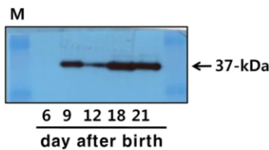
Fig. 3. Western blot analysis of 20α-HSD in the testis. Testes were obtained from 10 pigs and the extracted proteins were subjected to SDS-PAGE, transferred to PVDF membranes, and blotted with anti-20α-HSD primary an- tibodies and then with anti-mouse/rabbit IgG-POD sec- ondary antibodies. On the membranes, 37-kDa bands were detected.
highest on day 21. To our knowledge, this is the first study to report that 20α-HSD mRNA is expressed in the testis after birth.
Western blot analysis of 20α-HSDin the pig testis Next, we examined the expression of 20α-HSD in the pig testis using a specific anti-bovine 20α-HSD antibody devel- oped in our laboratory [10]. Western blotting revealed the expected 37-kDa band in the testes on days 6, 9, 12, 18, and 21 after birth (Fig. 3). The highest expression of the protein was detected on days 18 and 21 after birth. However, its
expression was not detected in the testis on day 6 after birth.
Comparison of the level of 20α-HSD protein with that of the mRNA in testis after birth showed a similar pattern. In this study, we identified an intense protein band by western blot of 20α-HSD in the testis on days 9 to 21 after birth.
Immunohistochemical localization of 20α-HSD in the testis after birth
To determine the cell types responsible for 20α-HSD ex- pression in the testis, we performed immunohistochemical analysis in the testis on days 6, 9, 12, 18, and 21 after birth.
As shown in Fig. 4, 20α-HSD was localized in Sertoli cells and Leydig cells. These results demonstrate that 20α-HSD is expressed at high levels in the immature testis after birth.
Thus, the protein expression of 20α-HSD in the immature testis may contribute to important functions, such as sper- matogenesis and regulation of maturation of the testis.
Discussion
In this study, we determined the expression and local- ization of 20α-HSD in the immature pig testis after birth.
Our results showed that 20α-HSD mRNA was expressed in the immature testis on days 6, 9, 12, 18, and 21 after birth.
Additionally, we also detected high levels of protein ex- pression in the testis after birth. IHC analysis revealed that 20α-HSD was localized in the Sertoli cells and Leydig cells of the immature testis
To the best of our knowledge, this is the first study that
demonstrated expression and localization of 20α-HSD in the
immature pig testis. We have studied the expression and
function of 20α-HSD in the ovary, placenta, and endome-
Fig. 4. Localization of 20α-HSD protein in the testis on days 6, 9, 12, 18, and 21 after birth. Proteins were detected usinganti-20α-HSD (right) antibodies. IHC analyses were performed using a Vectastain ABC kit. Sections of testes obtained from immature pigs are shown. The pre-immune serum was used as the staining control (left panel). Black arrows indicate Sertoli cells and Leydig cells.
trium during the estrous cycle and pregnancy [9, 10, 15-17, 21]. In porcine tissue, we reported that 20α-HSD mRNA and protein were coordinately expressed in the luteal cell of ova- ry throughout the estrous cycle and in the uterus on the day of pregnancy. Thus, the pig 20α-HSD might control im- portant mechanisms during the estrous cycle [21]. We also demonstrated that the pig 20α-HSD mRNA and protein are mainly localized in the trophoblast villus in the placenta on day 30 of pregnancy. The expression of the protein is also localized in the large luteal cells of the ovary. In addition, the protein is highly expressed in the glandular epithelial cells of the endometrium and the luminal epithelial cells of the uterus [17].
In the present study, we found that 20α-HSD mRNA and protein were highly expressed in the immature pig testis.
We also reported that 20α-HSD in the mouse is expressed in the seminiferous tubules of mature testis (Jeong et al., in- press). In a previous study of 3β-HSD, which is expressed in Leydig cells and converts testosterone into biologically active compounds, declining expression and activity were found [7]. Although testosterone at a low dose (50 μg/kg/
day) increased 3β-HSD mRNA expression, 3β-HSD ex-
pression was markedly decreased in rats treated with high doses of testosterone (200 or 1,000 μg/kg/day). Moreover, ketoconazole, diethylhexyl phthalate, and nonylphenol caused substantial downregulation of 3β-HSD mRNA in the testis at all doses tested [8].
During fish spermatogenesis, 11β-HSD activity was de- tected in the eel immature testis [18]. In catfish, IHC revealed the presence of the 20β-HSD protein predominantly in the interstitial cells and spermatogonia/spermatocytes [24]. 20β -HSD is expressed at a high level in the neonatal pig testis [13, 14], whereas avian 20β-HSD co-localizes with 11β-HSD and regulates the potency with which glucocorticoids stim- ulate intestinal Na
+absorption [5].
In bovine species, 20α-HSD from bull testis has been puri-
fied to homogeneity and characterized in terms of apparent
molecular weight, lack of subunit composition, substrate and
cofactor specificity, and certain kinetic parameters [19, 20,
27]. Bovine testicular 20α-HSD catalyzes transfer of the
4A-hydrogen from the dihydronicotinamide moiety of the
reduced cofactor [20]. We also reported the expression and
function of bovine 20α-HSD. 20α-HSD expression has been
detected in the ovary during the estrous cycle, and similarly
robust 20α-HSD protein expression has been identified on the cotyledon side of the placenta immediately before partu- rition [10]. 20α-HSD mRNA and protein are co-localized in large luteal cells, the placenta, and the endometrium during early pregnancy [9]. In monkey and deer, 20α-HSD is ex- pressed in the ovary at the pre-ovulation, and in the placenta at the pre-parturition stages [10, 11]. Thus, 20α-HSD ex- pression in both the ovary and the placenta is essential for the normal progression of the estrous cycle, pregnancy, and parturition.
20α-HSD activity in mice was downregulated by PRL [2]
and the inhibitory effect of PRL was found to cause a reduc- tion in 20α-HSD gene expression, with almost complete sup- pression of 20α-HSD expression in the CL [3]. The stim- ulation of cAMP generation by forskolin treatment was in- hibited 20α-HSD promoter activity in a dose-dependent manner [28]. In the mouse placenta, we also detected strong expression on days 16 and 18 of pregnancy (Jeong et al., inpress). The presence of 20α-HSD protein in the mouse pla- centa was reported in previous studies [22, 23].
This is the first study to examine the expression of 20α- HSD mRNA and protein in the immature pig testis. Our results suggest that 20α-HSD plays a pivotal role in the im- mature pig testis. Further studies on the functional sig- nificance of 20α-HSDare required to elucidate its role in sper- matogenesis and sex-maturation of male pigs.
Acknowledgment
This work was supported by a research grant from Hankyong National University in the year of 2016.
References
1. Akinola, L. A., Poutanen, M., Vihko, R. and Vihko, P. 1997.
Expression of 17β-hyxroxysteroid dehydrogenase type 1 and 2, P450 aromatase and 20α-hydroxysteroid dehydrogen- ase enzymes in immature, mature and pregnant rats.
Endocrinology 138, 2886-2892.
2. Albarracin, C. T. and Gibori, G. 1991. Prolactin action on luteal protein expression in the corpus luteum. Endocrinology 129, 1821-1830.
3. Albarracin, C. T., Parmer, T. G., Duan, W. R., Nelson, S.
E. and Gibori, G. 1994. Identification of a major prolactin- regulated protein as 20α-HSD:coordinate regulation of its activity, protein content, and messanger ribonucleic acid expression. Endocrinology 134, 2453-2460.
4. Bradford, M. M. 1976. A rapid and sensitive method for the quantitation of microgram quantities of protein utilizing
the principle of protein-dye binding. Anal. Biochem. 72, 248- 254.
5. Bryndova, J., Klusonova, P., Kucka, M., Mazancova- Vagnerova, K., Miksik, I. and Pacha, J. 2006. Cloning and expression of chicken 20α-HSD. J. Mol. Endocrinol. 37, 453-462.
6. Deis, R. P. 1971. Induction of lactogenesis and abortion by prostaglandin F2-alpha in pregnant rats. Nature 229, 568.
7. Ge, R. S. and Hardy, M. P. 1988. Variation in the end prod- ucts of androgen biosynthesis and metabolism during post- natal differentiation of rat Leydig cells. Endocrinology 139, 3787-3795.
8. Kim, H. H., Kwak, D. H., Yon, J. M., Baek, I. J., Lee, S.
R., Lee, J. E., Nahm, S. S., Jeong, J. H., Lee, B. J., Yun, Y.
W. and Nam, S. Y. 2007. Differential expression of 3I -hydroxysteroid dehydrogenase mRNA in rat testes ex- posed to endocrine disruptors. J. Reprod. Dev. 53, 465-471.
9. Kim, S. H., Shin, Y. S., Kang, M. H., Yoon, J. T. and Min, K. S. 2014. Gene expression and localization of 20α-HSD in reproductive tissues during early pregnancy of cattle. Anim.
Reprod. Sci. 147, 1-9.
10. Naidansuren, P., Park, C. W., Kim, S.H., Nanjidsuren, T, Park, J. J., Yun, S. J., Sim, B. W., Hwang, S. S., Kang, M.
H., Ryu, B. Y., Hwang, S. Y., Yoon, J. T., Yamanouchi, K.
and Min, K. S. 2011. Molecular characterization of bovine placental and ovarian 20α-HSD. Reproduction 142, 723-731.
11. Naidansuren, P., Park, C. W., Nanjidsuren, T., Park, J. J., Yun, S. J., Kang, M. H., Yamanouchi, K. and Min, K. S. 2012.
Ovarian and placental of 20α-HSD during pregnancy in deer. Anim. Reprod. Sci. 130, 63-73.
12. Nakajima, T., Yasuda, K., Nishizawa, M., Okada, H., Yoshi- mura, T., Ito, S. and Kanzaki, H. 2003. Expression of 20α- hydroxysteroid dehydrogenase mRNA in human endome- trium and decidua. Endocrine J. 50, 105-111.
13. Nakajin, S., Kawai, Y., Ohno, S. and Shinoda, M. 1989.
Purification and characterization of pig adrenal 20alpha-hy- droxysteroid dehydrogenase. J. Steroid Biochem. 33, 1181- 1189.
14. Nakajin, S., Ohno, S. and Shinoda, M. 1988. 20β-hydroxyste- roid dehydrogenase of neonatal pig testis: purification and some properties. J. Biochem. 104, 565-569.
15. Nanjidsuren, T. and Min, K. S. 2014. The transcription factor Ap-1 regulates monkey 20α-hydroxysteroid dehydrogenase promoter activity in CHO cells. BMC Biotechnology 14, 71.
16. Nanjidsuren, T., Naidansure, P., Park, C. W., Park, J. J., Yun, S. J., Sim, B. W., Kang, M. H., Lee, S. R., Chang, K. T. and Min, K. S. 2011. Expression and localization of the 20α-hy- droxysteroid dehydrogenase enzyme in the reproductive tissues of the cynomolgus monkey Macacafascicularis. J.
Steroid Biochem. Mol. Biol. 127, 337-344.
17. Nanjidsuren, T., Yun, S. J., Park, C. W., Kim, M. S., Kang, M. H. and Min, K. S. 2014. Expression and localization of 20α-HSD in porcine reproductive tissues during pregnancy.
Anim. Reprod. Sci. 148, 63-71.
18. Ozaki, Y., Higuchi, M., Miura, C., Yamaguchi, S., Tozawa, Y. and Miura, T. 2006. Roles of 11 beta-hydroxysteroid de-
초록:미니돼지 정소에서 20α-HSD의 발현 및 특성화
김정수
1․성훈기
1․밤바략차 뭉흐자야
1․심보웅
2․허창기
3․강명화
4․민관식
1*
(1한경대학교 미래융합기술대학원 동물생명공학전공, 동물생명환경과학과, 유전공학연구소, 2국가영장류센타,
3메디카이네틱스, 4호서대학교 식품영양학과)
모든 포유동물에 있어서 임신유지와 준비를 위해서는 프로게스테론 호르몬은 필수적이다. 20α-hydroxysteroid dehydrogenase (20α-HSD)는 프로게스테론을 생물학적으로 불활성인 20α-hydroxyprogesterone (20α-OHP)로 대 사시키는 중요한 효소이며, 임신말기와 분만개시에 중요한 역할을 하는 효소이다. 본 연구에서는 메디카이네틱스 미니돼지의 정소에서 20α-HSD의 발현에 대하여 분석하였다. 미니돼지 정소는 출생후 6, 9, 12, 18 및 21일에 채취 하였다. RT-PCR에 의한 mRNA 분석결과 분만 후 6일의 정소에서도 검출되었으며, 21일의 정소에서 가장 높은 발현을 하는 것으로 검출되었다. 그러나, 분만 후의 태반에서는 검출되지 않았다. 웨스턴블릇에 의한 단백질 분석 결과는 미니돼지 정소에서 특이적으로 37-kDa의 밴드가 검출되었다. 그러나 분만 후 6일의 정소에서는 단백질이 검출되지 않았다. 면역조직화학적 분석에 의하면 정소에서 20α-HSD는 Sertoli 세포와 Leydig 세포에서 특이적으 로 발현하였다. 따라서 이러한 연구결과는 분만 후 돼지 정소에서 20α-HSD 단백질의 발현에 대한 연구로서는 처음으로 밝혔으며, 향후 돼지 정소에서의 20α-HSD의 기능적인 역할에 대한 연구가 더욱 더 진행되어야 할 것으 로 사료된다.
hydrogenase in fish spermatogenesis. Endocrinology 147, 5139-5146.
19. Pineda, J. A., Murdock, G. L., Wason, R. J and Warren, J.
C. 1989. Stereospecificity of hydrogen transfer by bovine tes- ticular 20alpha-hydroxysteroid dehydrogenase. J. Steroid Biochem. 33, 1223-1228.
20. Pineda, J. A., Salinas, M. E. and Warren, J. C. 1985. Purifica- tion and characterization of 20alpha-hydroxysteroid de- hydrogenase from bull testis. J. Steroid Biochem. 23, 1001- 1006.
21. Seo, K. S., Naidansuren, P., Kim, S. H., Yun, S. J., Park, J.
J., Sim, B. W., Park, C. W., Nanjidsuren, T., Kang, M. H., Seo, H. W., Ka, H. K., Kim, N. H., Hwang, S. Y., Yoon, J. T., Yamanouchi, K. and Min, K. S. 2011. Expression of aldo-keto reductase family 1 member C1 (AKR1C1) gene in porcine ovary and uterine endometrium during the es- trous cycle and pregnancy. Reprod. Biol. Endocrinol. 9, 139.
22. Seong, H. H., Min, K. S., Kang, M. H., Yoon, J. T., Jin, H.
J., Chung, H. J., Chang, W. K., Yun, S. G. and Shiota, K.
2002. Change in ovarian and placental 20α-HSD activity during the pregnancy in the rat. Asian-Aust. J. Anim. Sci.
16, 342-347.
23. Shiota, K., Seong, H. H., Noda, K., Hattori, N., Ikeda, A.,
Ogura, A., Itagaki, S., Takahashi, M. and Ogawa, T. 1993.
20α-HSD activity in rat placenta. Endocrine J. 40, 673-681.
24. Sreenivasule, G., Senthilkumaran, B., Sridevi, P., Rajakumar, A. and Rasheeda, M. K. 2012. Expression and immunolocali- zation of 20α-hydroxysteroid dehydrogenase during tes- ticular cycle and after hCG induction, in vivo in the catfish, Clariasgariepinus. Gen. Comp. Endocrinol. 175, 48-54.
25. Sudeshna, T., Anand, K. and Medhamurthy, R. 2013. Anal- ysis of 20α-HSD expression in the corpus luteum of the buf- falo cow: effect of prostaglandin F2α treatment on circulating 20α-hydroxyprogesterone levels. Reprod. Biol. Endocrinol. 11, 111.
26. Telleria, C. M., Stocco, C. O., Stati, S. O. and Deis, R. P.
1999. Progesterone receptor is not required for progesterone action in the rat corpus luteum of pregnancy. Steroids 64, 760-766.
27. Warren, J. C., Murdock, G. L., Ma, Y., Goodman, S. R. and Zimmer, W. E. 1993. Molecular cloning of testicular 20α- HSD: identity with aldose reductase. Biochemistry 32, 1401- 1406.
28. Zhang, Y., Dufort, I., Rheault, P. and Luu-The, V. 2000.
Characterization of a human 20α-HSD. Mol. Endocrinol. 25, 221-228.