INTRODUCTION
Lipopolysaccharide (LPS) is a major component of the outer membranes of Gram-negative bacteria and has a dominant role in the host response to bacterial infection
1). Macrophages, which are the cells in the front line of host defense, play a pivotal role in the cellular response to LPS
2). Results from genetic and biochemical
studies showed that toll-like receptor 4 is the LPS recep- tor and mediates LPS-induced activation of downstream signaling pathways and expression of inflammatory tar- get genes
3). Stimulation of macrophages with LPS results in the production of various cytokines such as tumor necrosis factor-α, interleukin-1 and interleukin-6, and pro-inflammatory lipid mediators such as prostagla- ndins and leukotrienes
4,5). In addition, one of the key events in macrophage response to LPS stimuli is the expression of inducible nitric oxide synthase (iNOS) and the formation of NO, an important mediator involved in many host defense action in macrophages. These cytokines, lipid mediators, and NO participate in the innate immune response and may also lead to a harmful
* Corresponding author
Suk-Hwan Baek
Dept. of Biochemistry & Molecular Biology,
College of Medicine, Yeungnam Univ.
317-1 Daemyung-5 Dong, Nam-Gu, Daegu 705-717, South Korea
Tel: +82-53-620-4523 Fax: +82-53-623-8032
E-mail: [email protected]
Protein Kinase C-α αRegulates Toll-like Receptor 4-Mediated Inducible Nitric Oxide Synthase Expression
Jin-Gu Lee, Byung-Rho Chin*, Suk-Hwan Baek
Aging-associated Vascular Disease Research Center, Department of Biochemistry & Molecular Biology,
*Department of Dentistry, College of Medicine, Yeungnam University, Daegu, Korea
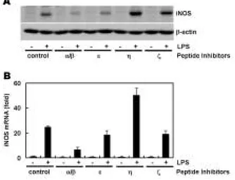
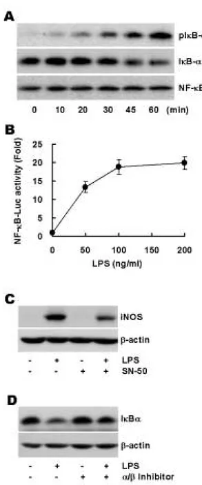
Purpose: The nitric oxide (NO) release by inducible nitric oxide synthase (iNOS) is the key events in macrophage response to lipopolysaccharide (LPS) which is suggested to be a crucial mediator for inflammatory and innate immune responses. NO is an impor- tant mediator involved in many host defense action and may also lead to a harmful host response to bacterial infection. However, given the importance of iNOS in a variety of pathophysiological conditions, control of its expression and signaling events in response to LPS has been the subject of considerable investigation. Materials and Methods: The Raw264.7 macrophage cell line was used to observe LPS-stimulated iNOS expression. The expression of iNOS is observed by Western blot analysis and real-time RT-PCR. Protein kinase C (PKC)-αoverexpressing Raw264.7 cells are established to determine the involvement of PKC-αin LPS-mediated iNOS expression. NF- κB activity is measured by IκBαdegradation and NF-κB luciferase activity assay. Results: We found that various PKC isozymes regulate LPS-induced iNOS expression at the transcriptional and translational levels. The involvement of PKC-αin LPS-mediated iNOS induc- tion was further confirmed by increased iNOS expression in PKC-αoverexpressing cells. NF-κB dependent transactivation by LPS was observed and PKC-αspecific inhibitory peptide abolished this activation, indicating that NF-κB activation is dependent on PKC-α.
Conclusion: Our data suggests that PKC-αis involved in LPS-mediated iNOS expression and that its downstream target is NF-κB.
Although PKC-αis a crucial mediator in the iNOS regulation, other PKC isozymes may contribute LPS-stimulated iNOS expression.
This finding is needed to be elucidated in further study.
Key words
Lipopolysaccharide, Inducible nitric oxide synthase, Protein kinase C, NF-κB Abstract
※ This work was supported by the Korean Science and Engineering Foundation via the Aging-associated Vascular
Research Center at Yeungnam University (R13-2005-005-02001-0 (2007)).