• 검색 결과가 없습니다.

The Therapeutic Effects of Optimal Dose of Mesenchymal Stem Cells in a Murine Model of an Elastase Induced-Emphysema

N/A
N/A
Protected

Academic year: 2021

Share "The Therapeutic Effects of Optimal Dose of Mesenchymal Stem Cells in a Murine Model of an Elastase Induced-Emphysema"

Copied!
7
0
0

로드 중.... (전체 텍스트 보기)

전체 글

(1)

The Therapeutic Effects of Optimal Dose of Mesenchymal Stem Cells in a Murine Model of an Elastase Induced-Emphysema

You-Sun Kim, Ph.D.

1,2

, Ji-Young Kim, M.S.

1

, Jin Won Huh, Ph.D.

3

, Sei Won Lee, Ph.D.

3

, Soo Jin Choi, Ph.D.

4

and Yeon-Mok Oh, M.D., Ph.D.

1,2,3

1

Asan Institute for Life Sciences, Seoul,

2

University of Ulsan College of Medicine, Seoul,

3

Departure of Pulmonary and Critical Care Medicine, Asan Medical Center, Seoul,

4

Biomedical Research Institute, MEDIPOST Co. Ltd., Seoul, Korea

Background: Chronic obstructive pulmonary disease is characterized by emphysema, chronic bronchitis, and small airway remodeling. The alveolar destruction associated with emphysema cannot be repaired by current clinical practices. Stem cell therapy has been successfully used in animal models of cigarette smoke- and elastase-induced emphysema. However, the optimal dose of mesenchymal stem cells (MSCs) for the most effective therapy has not yet been determined. It is vital to determine the optimal dose of MSCs for clinical application in emphysema cases.

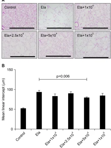
Methods: In the present study, we evaluated the therapeutic effects of various doses of MSCs on elastase-induced emphysema in mice. When 3 different doses of MSCs were intravenously injected into mice treated with elastase, only 5×10

4

MSCs showed a significant effect on the emphysematous mouse lung. We also identified action mechanisms of MSCs based on apoptosis, lung regeneration, and protease/antiprotease imbalance.

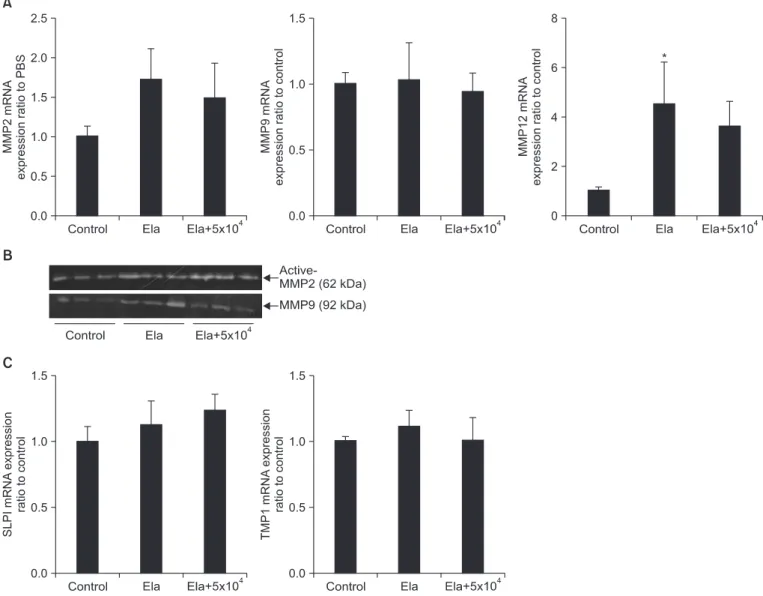
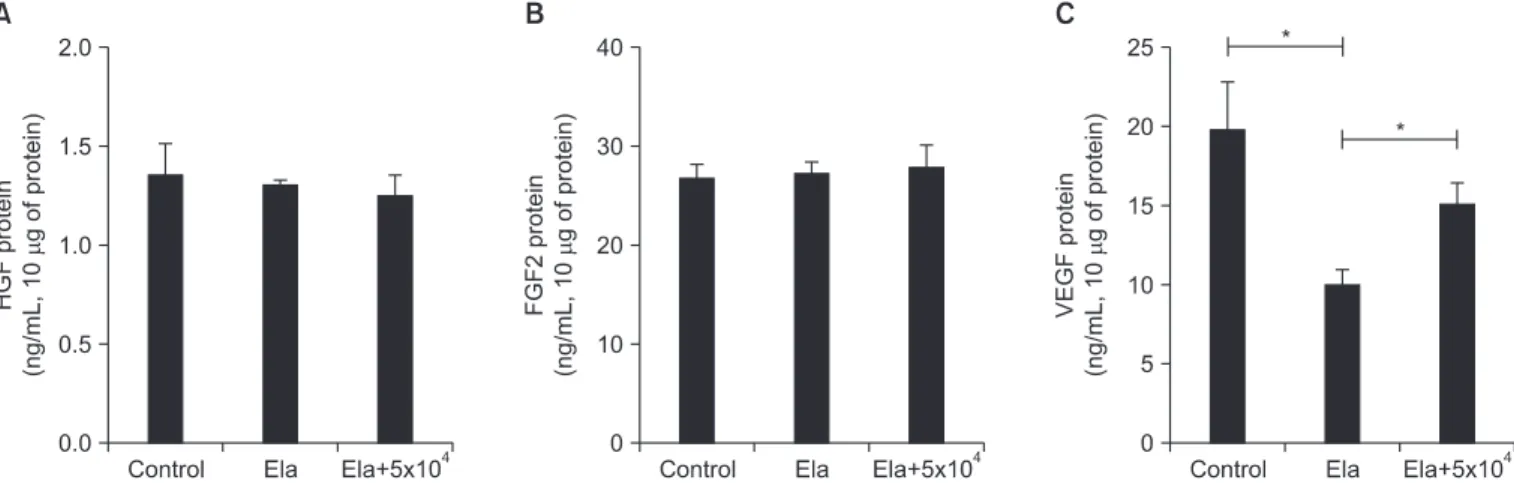
Results: The MSCs were not related with caspase-3/7 dependent apoptosis. But activity of matrix metalloproteinase 9 increased by emphysematous lung was decreased by intravenously injected MSCs. Vascular endothelial growth factor were also increased in lung from MSC injected mice, as compared to un-injected mice.

Conclusion: This is the first study on the optimal dose of MSCs as a therapeutic candidate. This data may provide important basic data for determining dosage in clinical application of MSCs in emphysema patients.

Keywords: Emphysema; Mesenchymal Stromal Cells; Therapy

Introduction

Chronic obstructive pulmonary disease (COPD) charac- terized by emphysema, chronic bronchitis, and small airway remodeling is one of the leading causes of death worldwide

1,2

. Cigarette smoking is a chief causative agent in the develop- ment of COPD characterized by progressive alveolar destruc- tion and persistent inflammation

3

. Emphysema is progres- sively marked by alveolar destruction and degradation of the extracellular matrix by protease-antiprotease imbalance and abnormal apoptosis and repair of resident lung cells

4,5

. Al- though some drugs exist that reduce airway obstruction in pa- tients with COPD, it is difficult to regenerate damaged alveolar structures or lung tissue

6,7

.

Our recent research has shown that bone marrow-derived mesenchymal stem cells (MSCs) are able to repair damage Copyright © 2015

The Korean Academy of Tuberculosis and Respiratory Diseases.

All rights reserved.

Address for correspondence: Yeon-Mok Oh, M.D., Ph.D.

Departure of Pulmonary and Critical Care Medicine, Asan Medical Center, 88 Olympic-ro 43-gil, Songpa-gu, Seoul 138-736, Korea Phone: 82-2-3010-3136, Fax: 82-2-3010-4650

E-mail: [email protected] Received: Sep. 16, 2014 Revised: Nov. 17, 2014 Accepted: Dec. 5, 2014

cc

It is identical to the Creative Commons Attribution Non-Commercial

License (http://creativecommons.org/licenses/by-nc/4.0/).

(2)

from cigarette smoke-induced emphysema

8

. Other research has also observed that MSCs from bone marrow and adi- pose tissue have a therapeutic effect in cigarette smoke- and elastase-induced emphysema

9,10

. Although most research studies to evaluate the therapeutic effect of MSCs in treating emphysema have applied intravenous injection, the optimal therapeutic dose of MSCs in a mouse model of emphysema remains unclear.

MSCs have been isolated from bone marrow, adipose tis- sue, umbilical cord blood, and placenta

11,12

. MSCs are consid- ered to have therapeutic capacities in a spectrum of diseases through paracrine, anti-inflammatory, immunomodulatory, and anti-apoptotic properties

13

. Recent reports have shown that different doses of MSCs exert different effects on healing or cytokine expression in a non-dose dependent manner

14

. However, another study reported that MSCs injected in the early period following acute myocardial infarction showed a significant effect and demonstrated dose dependence

15

.

Although it is important to determine the optimal doses of MSCs in treating emphysema, no such research has been done. Using an animal model is especially useful in determin- ing the optimal dose of MSCs for clinical application in em- physema cases. In the present study, we intravenously inject- ed different doses of cord blood MSCs into mice with elastase- induced emphysema and evaluated the therapeutic effects following each dose and the detailed action mechanisms of MSCs.

Materials and Methods

1. Animal model

C57BL/6J mice were purchased from Orientbio (Seongnam, Korea). Mice were bred in specific pathogen-free facilities at Asan Medical Center. All live mouse experiments were approved by the Institutional Animal Care and Use Commit- tee of Asan Medical Center (Korea). Female C57BL/6J mice (18–20 g, 7 weeks of age) were intratracheally injected with 0.4 U of porcine pancreatic elastase (Sigma-Aldrich, St. Louis, MO, USA) at day 0, were intravenously injected with different doses of MSCs at day 7, and were sacrificed at day 14.

2. Preparation of human umbilical cord blood MSCs Human umbilical cord blood-derived MSCs were provided by MEDIPOST (Seoul, Korea). The phenotype and differen- tiation ability of the MSCs were already confirmed by MEDI- POST. The MSCs were maintained with alpha-minimum es- sential medium (Gibco, Carlsbad, CA, USA), were replenished with fresh media twice a week, and were subcultured using 0.25% trypsin-EDTA (Gibco).

3. Histology and quantification of emphysema

The lungs were perfused with phosphate buffered saline and inflated by intratracheal infusion of 0.5% low-melting agar at 25 cm H

2

O, fixed in 4% paraformaldehyde, and embedded in paraffin. Lung sections of 4 μm thickness were stained with hematoxylin and eosin. The mean linear intercepts (MLI) were determined separately by two investigators in a blinded fashion

8

.

4. Measurement of caspase-3/7 activity

Proteins from lung tissue were prepared with a cell lysis buffer (Cell Signaling Technology, Danvers, MA, USA) and quantified based on the Bradford assay. The 10 μg of protein were incubated with caspase-3/7 substrate diluted with cas- pase-3/7 buffer in black multiwell plates (Promega, Madison, WI, USA). After 5 hours, the fluorescence of each sample was measured using a fluorometer.

5. Quantitative polymerase chain reaction

The mRNA from the lung tissue was prepared using the RNeasy Mini Kit (Qiagen, Duesseldorf, Germany) and quan- tified using spectrophotometer (NanoDrop Technologies, Rockland, DE, USA), and the cDNA was then synthesized using a cDNA synthesis kit (Thermo Scientific, Waltham, MA, USA). Quantitative polymerase chain reaction (PCR) analyses for matrix metalloproteinase (MMP) 2, MMP9, MMP12, SLIP, and tissue inhibitor of metalloproteinases-1 (TIMP1) were performed using a real-time LightCycler 480 PCR system (Roche Diagnostics, Basel, Switzerland) with LightCycler 480 SYBR Green I master (Roche Diagnostics). The primers used for MMP2, MMP9, MMP12, SLIP, and TIMP1 were displayed in Table 1.

6. Gelatin zymography

We measured MMP2 and MMP9 activity using gelatin zy- mography as previously described

16

. The lung homogenate

Table 1. Primer list

Gene Forward primer Reverse primer

Beta-actin cctcaccctcccaaaagc gtggactcagggcatgga

MMP2 acaagtggtccgcgtaaagt cggtcatcatcgtagttggtt

MMP9 aaccctgtgtgttcccgtt ggatgccgtctatgtcgtct

MMP12 tggtattcaaggagatgcacattt ggtttgtgccttgaaaacttttagt

SLPI ggccttttacctttcacggtg tacggcattgtggcttctcaa

TIMP1 gcaactcggacctggtcataa cggcccgtgatgagaaact

MMP: matrix metalloproteinase; TIMP1: tissue inhibitor of

metalloproteinases-1.

(3)

were prepared with cell lysis buffer without protease inhibitor.

The 10 μg of protein were loaded into sodium dodecyl sulfate polyacrylamide gel electrophoresis gel containing gelatin at a concentration 2.6 mg/mL. Gel were then incubated in 2.5% Triton X-100 and incubated for overnight in zymogaphy developing buffer at 37

o

C. And then gel were stained and de- stained with 0.5% comassie G250 in 30% ethanol/10% acetic acid and 30% ethanol/10% acetic acid, respectively.

7. Hepatocyte growth factor, fibroblast growth factor 2, and vascular endothelial growth factor measurement The 10 μg of protein from lung tissue were examined by

enzyme-linked immunosorbent assay (ELISA). The hepato- cyte growth factor (HGF), fibroblast growth factor 2 (FGF2), and vascular endothelial growth factor (VEGF) levels in the lung lysates were measured by ELISA according to the manu- facturer’s instructions (R&D Systems, Minneapolis, MN, USA).

8. Statistical analysis

The data are presented as the means±SE. The Mann- Whitney test was used to compare result between groups. A p- value of <0.05 was considered significant.

Results

1. Optimal dose of intravenously injected MSCs in a mouse model of elastase-induced emphysema To identify the optimal dose of MSCs for treatment of em- physema, C57BL/6J mice were treated intratracheally with 0.4 U of elastase on day 0 and were intravenously injected with 1×10

4

, 2.5×10

4

, 5×10

4

, or 1×10

5

of MSCs on day 7. Lung tissue was then collected on day 14. The mice treated with elastase only showed severe alveolar destruction compared to the control group, while the mice treated with MSCs showed less alveolar destruction (Figure 1A). The level of alveolar destruc- tion was quantified by measuring MLI. The mice treated with the 5×10

4

MSCs showed significantly lower MLI (78.15 μm) than the mice treated with elastase only (92.28 μm) (Figure 1B). The mice treated with the 1×10

4

MSCs, 2.5×10

4

, or 1×10

5

of MSCs also showed a pattern of decreased MLI (81.82 μm,

Figure 1. Optimal dose of mesenchymal stem cells (MSCs) to re- pair elastase induced emphysema in mice. C57BL/6J mice were administered 0.4 U of elastase on day 0 by intratracheal applica- tion and then intravenously injected with MSCs on day 7. Data are measures of the histological staining with hematoxylin and eosin in lung section on day 14. (A) Control (n=5), Ela (elastase only, n=15), Ela+1×10

4

(elastase+1×10

4

MSC, n=10), Ela+2.5×10

4

(elas- tase+2.5×10

4

MSC, n=10), Ela+5×10

4

(elastase+5×10

4

MSC, n=16), and Ela+1×10

5

(elastase+1×10

5

MSC, n=6) (×10). Scale bars=1.0 mm. (B) Morphometic analysis of the mean linear intercept. Values are presented as the mean±SEM.

Figure 2. The anti-apoptotic effects of mesenchymal stem cell

(MSC) in elastase induced emphysema. C57BL/6J mice were in-

tratracheally applied with 0.4 U of elastase on day 0 and then intra-

venously injected with MSCs on day 7. Lung tissue were collected

on day 14 (n=6–9 per group). Caspase 3/7 activity was measured

with fluorimetric emzymatic assay and normalized by protein

concentration in lung homogenates. Ela: elastase only; Ela+5×10

4

:

elastase+5×10

4

MSC.

(4)

88.75 μm, or 83.47 μm, respectively) compared to the mice treated with elastase only, but the difference was not signifi- cant (Figure 1B).

2. The mechanisms of MSCs in the elastase-induced emphysema model

To identify action mechanisms of MSCs in this model of elastase-induced emphysema, we obtained protein and mRNA in lung tissue from three of the mouse groups (control,

elastase-only, and elastase- and 5×10

4

MSC-treated groups).

The therapeutic mechanisms are divided in apoptosis, prote- ase/antiprotease and regeneration by growth factor. First, we test caspase 3/7 dependent apoptosis. Caspase 3/7 activity to detect cellular apoptosis is not significantly different among the three groups (Figure 2). Second, we performed an evalu- ation based on protease/antiprotease imbalance in the same three groups. The mRNA expression of MMP2, MMP9, and MMP12 were measured using quantitative PCR, and data were presented as a ratio of the control group (Figure 3A). In

Figure 3. The effects on protease and anti-protease in elastase induced emphysema with or without mesenchymal stem cells (MSCs).

C57BL/6J mice were intratracheally applied with 0.4 U of elastase on day 0 and then intravenously injected with MSCs on day 7. Lung tissues were collected on day 14 (n=6–9 per group). (A) Matrix metalloproteinase (MMP) 2, MMP9, and MMP12 mRNA expression were measured with quantitative polymerase chain reaction (PCR) using SYBR Green and normalized by β-actin expression and then displayed with ratio to control group. Values are presented as the mean±SEM. *p<0.05, as compared with control group. (B) Activity of MMP2 and 9 were measured with gelatin zymography. (C) The mRNA expression of SLPI (left) and tissue inhibitor of metalloproteinases-1 (TIMP1) (right) were mea- sured with quantitative PCR using SYBR Green and normalized by β-actin expression and then displayed with ratio to control group. PBS:

phosphate buffered saline; Ela: elastase only; Ela+5×10

4

: elastase+5×10

4

MSC.

(5)

the mouse group treated with elastase only, mRNA expression of MMP12 was higher than in the control group. The mouse group treated with both elastase and MSCs may be also higher than in the control group but there is not statistical difference.

Next, we performed gelatin zymography to observe the activ- ity of MMP2 and MMP9 (Figure 3B). The activity of MMP2 and MMP9 were induced in elastase only treated mouse group compared to the control group. In elastase and MSC treated mouse group, the MMP9 activity was decreased com- pared with the elastase only treated mouse group. The mRNA expression of SLPI and TIMP1 (representative antiprotease) were measured by quantitative PCR, and data were displayed as a ratio of the control group (Figure 3C). SLPI and TIMP1 ex- pression in lung is not significantly different among the three groups. HGF, FGF2, and VEGF production related to the lung regeneration was also measured in lung homogenate using ELISA. HGF and FGF2 were not significantly different among the three groups (Figure 4A, B). The VEGF in lung tissue were decreased in elastase only treated mouse group compared to control mouse group and were recovered in elastase and MSCs treated mouse group (Figure 4C).

Discussion

In this study, we identified the optimal dose of MSCs for therapy in a mouse model of elastase-induced emphysema.

A dose of 5×10

4

MSCs showed significant therapeutic effects in elastase-induced emphysema in C57BL/6J mice. A lower dose of 1×10

4

or 2.5×10

4

MSCs also showed slightly therapeu- tic effects but not to a significant degree. Moreover, we evalu- ated the action mechanisms of the dosage of 5×10

4

MSCs in elastase-induced emphysema. The effects of MSCs in emphy-

sematouse lung were reduction of MMP9 activity and induc- tion of VEGF production. Therefore, there is best therapeutic dose of MSCs in emphysema and these result may be become basic data to decide clinical application for patients with em- physema.

Some reports have shown that it is possible to apply stem cells as an effective therapy to regenerate destroyed alveo- lar structures

8-10

. Our previous report has shown that bone marrow cells (6×10

6

) and MSCs (6×10

5

) have the capability of repairing cigarette smoke-induced emphysema in rats exposed to cigarette smoke for 6 months

8

. Other researchers have observed positive effects in mice from the injection of adipose-derived stem cells (3×10

5

) every other week during 3 or 4 months of cigarette smoke exposure

10

. In the mouse model of elastase-induced emphysema, 2.8×10

6

bone marrow mononuclear cells can repair destroyed alveolar structures

9

. There is no standard therapeutic dose of stem cells in the ani- mal model of emphysema/COPD. These results represent the first report on the optimal dose of MSCs for the treatment of emphysema.

A recent report showed that when different doses of MSCs were injected into the injured ligaments of two groups of rats, the higher dose group exhibited increased expression of pro- inflammatory cytokines, including interleukin (IL)-1β, inter- feron γ, IL-2, while the lower dose group showed enhanced functional healing compared to the higher dose group

14

. It is noteworthy that in most clinical trials of MSCs, the amount of MSCs injected depends on the patient’s body weight

17-19

. Thus, patients with higher body weights will require more MSCs and incur greater costs. Determining the optimal dose of MSCs is vital because of a variety of factors, such as immune response, inflammatory cytokines, therapeutic effects, and financial burden.

Figure 4. The production of growth factors in elastase induced emphysema with or without mesenchymal stem cells (MSCs). C57BL/6J mice were intratracheally applied with 0.4 U of elastase on day 0 and then intravenously injected with MSCs on day 7. Lung tissue were collected on day14 (n=5–9 per group). The growth factors were measured with enzyme-linked immunosorbent assay for hepatocyte growth factor (HGF) (A), fibroblast growth factor 2 (FGF2) (B), and vascular endothelial growth factor (VEGF) (C). Values are presented as the mean±SEM.

*p<0.05 was statistical significance of the comparison between 2 groups. Ela: elastase only; Ela+5×10

4

: elastase+5×10

4

MSC.

(6)

Animal models for COPD have been developed through the agents of cigarette smoke and elastase. An elastase-induced emphysema model is widely applied, and supports the hy- pothesized role of protease/antiprotease imbalance in the development of emphysema. Although a mouse model of elastase-induced emphysema is not perfectly representative of human COPD/emphysema, this model focuses on alveo- lar destruction based on protease/antiprotease imbalance.

Therefore, we observed cellular apoptosis as measured by caspase 3/7 enzyme activity, and found that it was not higher in the elastase only and elastase and MSCs treated mouse group compared to the control mouse group. The induction of MMP12 mRNA expression was observed in the elastase only treated mouse group. MMPs is more important their activity than mRNA expression. Especially, it was well known about MMP2 and MMP9 activity linked with asthmatic and COPD patients

16

. These results are shown in decrease of MMP9 ac- tivity because of MSCs therapeutic effects.

Many other reports suggest that the paracrine properties of MSCs enable a key mechanism of action in the treatment of emphysema. In fact, it is well known that MSCs produce many cytokines and growth factors

13,20

. These include some candidate molecules in the categories of growth factors (HGF, keratinocyte growth factor, FGF2, and VEGF) and cytokines (IL-10). Although inflammation is a very important factor in the development of emphysema/COPD, we applied MSCs following complete recovery from inflammation. Therefore, this study did not confirm the anti-inflammatory effect of IL- 10. HGF is a viable candidate growth factor for the repair of lung destruction, as intranasally administered HGF has been shown to ameliorate the effects of elastase-induced emphy- sema in mice

21

. The other report demonstrated that growth factors derived from human MSCs were decreased in SLPI expression in mouse lung epithelial cells

22

. FGF2 is an impor- tant component of the regulation of lung development and need to proliferation and migration of alveolar epithelial cell

23

. Therefore, FGF2 is also critical to repair of lung injury. In this study, although MSCs showed a therapeutic effect on elastase- induced emphysema, MSCs were not found to increase HGF and FGF2 protein levels.

VEGF is important role during airway growth especially, development and maintenance of the pulmonary vascula- ture

23

. Moreover, inhibition of VEGF receptor in animal leads to emphysema features including alveolar space enlargement and alveolar cell apoptosis

24

. Recent report was shown that MSCs protect cigarette smoked lung damage partly via VEGF- VEGF receptors

25

. In this report, they observed that VEGF from MSCs was crucial for protection of lung damage. MSCs can induce the expression of VEGF and their receptors and enhanced VEGF signal prevent lung damage by cigarette smoking.

In clinical trial, MSCs were infused four monthly 100×10

6

cells into patient with COPD or control

26

. Systemic injec-

tion of MSCs was not significant improved in pulmonary function test or quality of life. Clinical trial for other diseases, some researchers applied with MSCs depending on patient’s weight such as (2–10)×10

6

cells/kg and (5.7–7.5)×10

8

cells/

kg for Hurler syndrome and Osteogenesis imperfecta, respec- tively

27,28

. Although additional researches need to determine MSCs dose for clinical trial, we suggest that optimal dose of MSCs in preclinical emphysema model be able to apply to clinical trial ratio to weight.

In conclusion, we report here for the first time that there is an optimal dose of MSCs for the treatment of elastase-induced emphysema in mice. And the therapeutic effects of MSCs are related with decrease of MMP9 activity and increase of VEGF production. Our report will provide important basic data for deciding on dosage in clinical application of MSCs.

Conflicts of Interest

No potential conflict of interest relevant to this article was reported.

Acknowledgements

The authors thank the members of the Asan Medical Cen- ter animal facility for their technical expertise. This study was supported by grants from the Korean Health Technology R&D Project, Ministry of Health & Welfare, Republic of Korea (no.

HI12C0169).

References

1. Senior RM, Anthonisen NR. Chronic obstructive pulmonary disease (COPD). Am J Respir Crit Care Med 1998;157(4 Pt 2):

S139-47.

2. Sutherland ER, Cherniack RM. Management of chronic ob- structive pulmonary disease. N Engl J Med 2004;350:2689-97.

3. Churg A, Cosio M, Wright JL. Mechanisms of cigarette smoke- induced COPD: insights from animal models. Am J Physiol Lung Cell Mol Physiol 2008;294:L612-31.

4. Segura-Valdez L, Pardo A, Gaxiola M, Uhal BD, Becerril C, Sel- man M. Upregulation of gelatinases A and B, collagenases 1 and 2, and increased parenchymal cell death in COPD. Chest 2000;117:684-94.

5. Yokohori N, Aoshiba K, Nagai A; Respiratory Failure Research Group in Japan. Increased levels of cell death and prolifera- tion in alveolar wall cells in patients with pulmonary emphy- sema. Chest 2004;125:626-32.

6. Aaron SD, Vandemheen KL, Fergusson D, Maltais F, Bour-

beau J, Goldstein R, et al. Tiotropium in combination with

placebo, salmeterol, or fluticasone-salmeterol for treatment

(7)

of chronic obstructive pulmonary disease: a randomized trial.

Ann Intern Med 2007;146:545-55.

7. Calverley PM, Anderson JA, Celli B, Ferguson GT, Jenkins C, Jones PW, et al. Salmeterol and fluticasone propionate and survival in chronic obstructive pulmonary disease. N Engl J Med 2007;356:775-89.

8. Huh JW, Kim SY, Lee JH, Lee JS, Van Ta Q, Kim M, et al. Bone marrow cells repair cigarette smoke-induced emphysema in rats. Am J Physiol Lung Cell Mol Physiol 2011;301:L255-66.

9. Longhini-Dos-Santos N, Barbosa-de-Oliveira VA, Kozma RH, Faria CA, Stessuk T, Frei F, et al. Cell therapy with bone mar- row mononuclear cells in elastase-induced pulmonary em- physema. Stem Cell Rev 2013;9:210-8.

10. Schweitzer KS, Johnstone BH, Garrison J, Rush NI, Cooper S, Traktuev DO, et al. Adipose stem cell treatment in mice atten- uates lung and systemic injury induced by cigarette smoking.

Am J Respir Crit Care Med 2011;183:215-25.

11. Kern S, Eichler H, Stoeve J, Kluter H, Bieback K. Comparative analysis of mesenchymal stem cells from bone marrow, um- bilical cord blood, or adipose tissue. Stem Cells 2006;24:1294- 301.

12. Wexler SA, Donaldson C, Denning-Kendall P, Rice C, Bradley B, Hows JM. Adult bone marrow is a rich source of human mesenchymal ‘stem’ cells but umbilical cord and mobilized adult blood are not. Br J Haematol 2003;121:368-74.

13. Murphy MB, Moncivais K, Caplan AI. Mesenchymal stem cells: environmentally responsive therapeutics for regenera- tive medicine. Exp Mol Med 2013;45:e54.

14. Saether EE, Chamberlain CS, Leiferman EM, Kondratko- Mittnacht JR, Li WJ, Brickson SL, et al. Enhanced medial collateral ligament healing using mesenchymal stem cells:

dosage effects on cellular response and cytokine profile. Stem Cell Rev 2014;10:86-96.

15. Richardson JD, Bertaso AG, Psaltis PJ, Frost L, Carbone A, Pa- ton S, et al. Impact of timing and dose of mesenchymal stro- mal cell therapy in a preclinical model of acute myocardial infarction. J Card Fail 2013;19:342-53.

16. Cataldo D, Munaut C, Noel A, Frankenne F, Bartsch P, Foidart JM, et al. MMP-2- and MMP-9-linked gelatinolytic activity in the sputum from patients with asthma and chronic obstruc- tive pulmonary disease. Int Arch Allergy Immunol 2000;123:

259-67.

17. Chang YS, Ahn SY, Yoo HS, Sung SI, Choi SJ, Oh WI, et al. Mes- enchymal stem cells for bronchopulmonary dysplasia: phase

1 dose-escalation clinical trial. J Pediatr 2014;164:966-72.e6.

18. Hare JM, Traverse JH, Henry TD, Dib N, Strumpf RK, Schul- man SP, et al. A randomized, double-blind, placebo-con- trolled, dose-escalation study of intravenous adult human mesenchymal stem cells (prochymal) after acute myocardial infarction. J Am Coll Cardiol 2009;54:2277-86.

19. Liang J, Zhang H, Hua B, Wang H, Lu L, Shi S, et al. Allogenic mesenchymal stem cells transplantation in refractory sys- temic lupus erythematosus: a pilot clinical study. Ann Rheum Dis 2010;69:1423-9.

20. Haynesworth SE, Baber MA, Caplan AI. Cytokine expression by human marrow-derived mesenchymal progenitor cells in vitro : effects of dexamethasone and IL-1 alpha. J Cell Physiol 1996;166:585-92.

21. Hegab AE, Kubo H, Yamaya M, Asada M, He M, Fujino N, et al.

Intranasal HGF administration ameliorates the physiologic and morphologic changes in lung emphysema. Mol Ther 2008;16:1417-26.

22. Katsha AM, Ohkouchi S, Xin H, Kanehira M, Sun R, Nukiwa T, et al. Paracrine factors of multipotent stromal cells ameliorate lung injury in an elastase-induced emphysema model. Mol Ther 2011;19:196-203.

23. Muyal JP, Muyal V, Kotnala S, Kumar D, Bhardwaj H. Thera- peutic potential of growth factors in pulmonary emphysema- tous condition. Lung 2013;191:147-63.

24. Kasahara Y, Tuder RM, Taraseviciene-Stewart L, Le Cras TD, Abman S, Hirth PK, et al. Inhibition of VEGF receptors causes lung cell apoptosis and emphysema. J Clin Invest 2000;106:

1311-9.

25. Guan XJ, Song L, Han FF, Cui ZL, Chen X, Guo XJ, et al. Mes- enchymal stem cells protect cigarette smoke-damaged lung and pulmonary function partly via VEGF-VEGF receptors. J Cell Biochem 2013;114:323-35.

26. Weiss DJ, Casaburi R, Flannery R, LeRoux-Williams M, Tash- kin DP. A placebo-controlled, randomized trial of mesenchy- mal stem cells in COPD. Chest 2013;143:1590-8.

27. Koc ON, Day J, Nieder M, Gerson SL, Lazarus HM, Krivit W.

Allogeneic mesenchymal stem cell infusion for treatment of metachromatic leukodystrophy (MLD) and Hurler syndrome (MPS-IH). Bone Marrow Transplant 2002;30:215-22.

28. Horwitz EM, Prockop DJ, Fitzpatrick LA, Koo WW, Gordon

PL, Neel M, et al. Transplantability and therapeutic effects of

bone marrow-derived mesenchymal cells in children with

osteogenesis imperfecta. Nat Med 1999;5:309-13.

수치

Table 1. Primer list
Figure 1. Optimal dose of mesenchymal stem cells (MSCs) to re- re-pair elastase induced emphysema in mice
Figure 3. The effects on protease and anti-protease in elastase induced emphysema with or without mesenchymal stem cells (MSCs)
Figure 4. The production of growth factors in elastase induced emphysema with or without mesenchymal stem cells (MSCs)

참조

관련 문서

After first field tests, we expect electric passenger drones or eVTOL aircraft (short for electric vertical take-off and landing) to start providing commercial mobility

1 John Owen, Justification by Faith Alone, in The Works of John Owen, ed. John Bolt, trans. Scott Clark, &#34;Do This and Live: Christ's Active Obedience as the

In addition, the expression of REDD1 was increased when Huh7 cells were treated with PA, and overexpression of REDD1 affected cell survival and lipid

In this study, two experiments were conducted to understand the effects of additional charge on the detailed growth mechanism of Alq 3 and to determine the effect of

We investigated the effects of BIX01294 on cellular senescence in human BM-MSCs (hBM-MSCs), and identified that an optimal treatment of BIX01294 leads to attenuated

In the present study we investigated the proliferation effect of human oral cancer KB cell treated with pulsatilla koreana extract.. We analyzed the effects of this

In particular, a recent study showed that intrathecal SOG administration has an antinociceptive effect in a formalin test of rat model and shows the possibility of

The main effect according to the results of the repeated measures ANOVA on physical fitness based on test time showed a significant difference in grip