• 검색 결과가 없습니다.

Empyema Necessitatis in a Patient on Peritoneal Dialysis

N/A
N/A
Protected

Academic year: 2021

Share "Empyema Necessitatis in a Patient on Peritoneal Dialysis"

Copied!
4
0
0

로드 중.... (전체 텍스트 보기)

전체 글

(1)

Empyema Necessitatis in a Patient on Peritoneal Dialysis

In Ho Moh, M.D.

1

, Young-Ki Lee, M.D.

1

, Hee Joon Kim, M.D.

1

, Hyun Yon Jung, M.D.

1

, Jae Hyun Park, M.D.

1

, Hye-Kyung Ahn, M.D.

2

and Jung-Woo Noh, M.D.

1

1

Department of Internal Medicine, Hallym Kidney Research Institute,

2

Department of Pathology, Hallym University College of Medicine, Seoul, Korea

Empyema necessitatis is a rare complication of an empyema. Although the incidence is thought to be decreased in the post-antibiotic era, immunocompromised patients such as patients with chronic kidney disease on dialysis are still at a higher risk. A 56-year-old woman on peritoneal dialysis presented with an enlarging mass on the right anterior chest wall.

The chest computed tomography scan revealed an empyema necessitatis and the histopathologic findings revealed a granulomatous inflammation with caseation necrosis. The patient was treated with anti-tuberculous medication.

Keywords: Empyema; Mycobacterium tuberculosis; Peritoneal Dialysis

Case Report

A 56-year-old woman presented with an enlarging soft mass on right anterior chest wall with pain lasting for 2 months.

There were no symptoms of fever, dyspnea, or cough. She denied recent chest trauma or exposure to tuberculosis. She was diagnosed with chronic kidney disease seven years ago and has been on peritoneal dialysis for four years. Her medical history was remarkable of hypertension, diabetes mellitus and hypothyroidism on medication. Vital signs on admission were blood pressure 134/68 mm Hg, temperature 36.9

o

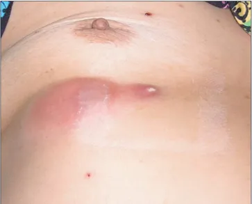
C, pulse rate 95 beats per minute, and respiratory rate 20 breaths per min- ute. Physical examination revealed a pink erythematous, cold, soft, tender, subcutaneous mass of 6×3 cm on the right chest wall between the fifth and seventh ribs (Figure 1).

Laboratory values revealed a white blood cell count of 17,270/mm

3

with 89.8% neutrophils. Hemoglobin was 7.2 g/dL and platelets were 416,000/mm

3

. Blood urea nitrogen was 47.7 mg/dL, creatinine was 8.15 mg/dL, sodium was 133 mEq/L and potassium was 3.7 mEq/L. Liver function tests were within normal limits and C-reactive protein was 72.4 mg/L. Chest X-ray revealed pleural thickening in the right lung (Figure 2).

Computed tomography (CT) scan of the chest revealed calci- fied pleural thickening with loculated fluid collection in the right lower anterior hemithorax suggestive of empyema, with multiple cystic extrapleural masses in the right anterior chest wall consistent with empyema necessitatis (Figure 3).

Fine needle aspiration biopsy (FNAB) was performed for Copyright © 2014

The Korean Academy of Tuberculosis and Respiratory Diseases.

All rights reserved.

Introduction

Empyema necessitatis is an abscess on the thoracic wall by extension of purulent pleural liquid through adjacent tissues

1

. Tuberculous empyema necessitatis is a rare complication of tuberculosis. However, immunocompromised patients such as patients with chronic kidney disease on dialysis are at a higher risk

1,2

.

Herein, we present a case of tuberculous empyema neces- sitatis in a patient with chronic kidney disease on peritoneal dialysis.

CASE REPORT

http://dx.doi.org/10.4046/trd.2014.77.2.94

ISSN: 1738-3536(Print)/2005-6184(Online) • Tuberc Respir Dis 2014;77:94-97

94

Address for correspondence: Young-Ki Lee, M.D.

Department of Internal Medicine, Kangnam Sacred Heart Hospital, Hallym University College of Medicine, 1 Singil-ro, Yeongdeungpo-gu, Seoul 150-950, Korea

Phone: 82-2-829-5114, Fax: 82-2-848-9821 E-mail: km2071@unitel.co.kr

Received: Jan. 29, 2014 Revised: May 20, 2014 Accepted: May 30, 2014

cc
It is identical to the Creative Commons Attribution Non-Commercial

License (http://creativecommons.org/licenses/by-nc/3.0/).

(2)

Empyema necessitatis in peritoneal dialysis

http://dx.doi.org/10.4046/trd.2014.77.2.94 95

www.e-trd.org

diagnosis. The mass was filled with pus-like materials and his- topathologic findings revealed granulomatous inflammation with caseation necrosis (Figure 4). Polymerase chain reaction for Mycobacterium tuberculosis was positive.

The patient was diagnosed with tuberculous empyema with tuberculous empyema necessitatis. An anti-tuberculous med- ication was started (with dose adjustment for chronic kidney disease; isoniazid 400 mg per day, rifampin 600 mg per day, pyrazinamide 1,500 mg 3 times per week, ethambutol 800 mg

three times per week). No surgical intervention was made to the chest wall. After 5 months, CT scan revealed reduced size of the multiple cystic extrapleural masses (Figure 3) and anti- tuberculous medication was continued.

Discussion

Empyema necessitatis is a collection of inflammatory tissue Figure 2. The initial chest X-ray reveals a pleural thickening in the right lung.

Figure 1. Skin lesions. Pink erythematous, cold, soft, tender sub- cutaneous mass of 6×3 cm size are noted on the right chest wall between the fifth and seventh ribs.

Figure 3. (A, B) On admission, the chest

computed tomography (CT) reveals a

calcified pleural thickening with loculated

fluid collection in the right lower anterior

hemithorax (arrowhead) and with mul-

tiple cystic extrapleural masses in right

anterior chest wall (arrows). (C, D) After

5 months of therapy, the chest CT reveals

a reduced loculated fluid collection in the

right lower anterior hemithorax (arrow-

head) and a reduced size of the multiple

cystic extrapleural masses in the right

anterior chest wall (arrows).

(3)

IH Moh et al.

96 Tuberc Respir Dis 2014;77:94-97 www.e-trd.org

that usually extends directly from the pleural cavity into the thoracic wall, forming a mass in the extrapleural soft tissues of the chest

1

. It is characterized by the extension of suppura- tion from the pleural space, typically through the thoracic wall, forming subcutaneous abscess. Clinically, patients show symptoms of enlarging, tender, erythematous mass, cough, chest pain and dyspnea

3

. The most common site is the ante- rior chest wall between the midclavicular and anterior axillary line and between the second and sixth intercostal spaces

4

. Less commonly, it can involve the bronchus, vertebral column, diaphragm, breast, mediastinum, retroperitoneum, esopha- gus, pericardium, flank or groin

5

. It was first described in 1640 by Baillon, who reported a woman with left sided pulsating tumor associated with pleurisy. The most common etiology is Mycobacterium tuberculosis and cases of Actinomycosis species, Streptococcus pneumonia, Staphylococcus aureus, Blastomycosis species, and neoplasms have been reported.

Three reports have reviewed cases of empyema necessitatis.

In 1940, Sindel

4

reviewed 115 cases. Mycobacterium tuber- culosis was the most common etiology with 84 (73%) cases.

In 2004, Freeman et al.

5

reviewed 26 cases from 1966 to 2004 and 13 (50%) cases was caused by Mycobacterium tuber- culosis. Recently, in 2010, Llamas-Velasco et al.

3

reviewed 20 cases from 2004 and Mycobacterium tuberculosis was less common (7 cases, 35%) than the previous reports but still the most common infectious agent. There were no data concern- ing the prevalence of chronic kidney disease or patients on dialysis in these reports.

Empyema necessitatis is thought as an unusual complica- tion of empyema with decreasing incidence, while most of its patients were described in the preantibiotic era. However, im- munocompromised patients including patients with chronic kidney disease on dialysis are at a higher risk of empyema ne-

cessitatis

1,2

. Cellular and humoral immunity are both reduced in patients with chronic kidney disease on dialysis and risk of tuberculosis infection is 6.9- to 62.5-fold higher than the gen- eral population

6

. Additionally, extra pulmonary involvements are more frequent than isolated pulmonary disease in these patients, leading to delayed diagnosis

7

.

Mortality is significantly decreasing with antibiotic therapy, from 66% in the preantibiotic era to less than 5% nowadays

3,4

. Mortality from recent cases were also associated with reduced immune defence such as old age

8

, neoplastic

9

, or cystic fibro- sis

10

. Although there has been no recent case of mortality re- ported with patients on dialysis, higher mortality is expected.

The differential diagnosis of empyema necessitatis includes neoplasms, Wegener’s granulomatosis, sarcoidosis, infective endocarditis and septic embolization

11

. CT scan and biopsy are important for accurate diagnosis. CT is helpful in detection and characterization of empyema necessitatis while tissue and fluid for cytological diagnosis and culture can be aspirated by FNAB

12,13

.

Treatment of empyema necessitatis includes antimicrobial therapy and surgical drainage. While standard antituberculo- sis regimen can be used for treatment

14

, dose adjustments for patients with chronic kidney disease are necessary.

In summary, empyema necessitatis should be considered in any patient with an enlarging soft tissue mass on the chest wall especially in immunocompromised patients such as pa- tients with chronic renal failure on dialysis, even though his- tory or symptoms of infection are absent. Early diagnosis and proper management can be associated with the best outcome.

Conflicts of Interest

No potential conflict of interest relevant to this article was reported.

References

1. Jover F, Andreu L, Cuadrado JM, Montagud J, Merino J. Tu- berculous empyema necessitatis in a man infected with the human immunodeficiency virus. South Med J 2002;95:751-2.

2. Aylk S, Qakan A, Aslankara N, Ozsoz A. Tuberculous abscess on the chest wall. Monaldi Arch Chest Dis 2009;71:39-42.

3. Llamas-Velasco M, Dominguez I, Ovejero E, Perez-Gala S, Garcia-Diez A. Empyema necessitatis revisited. Eur J Derma- tol 2010;20:115-9.

4. Sindel EA. Empyema necessitates. Q Bull Sea View Hosp 1940;6:1-49.

5. Freeman AF, Ben-Ami T, Shulman ST. Streptococcus pneu- moniae empyema necessitatis. Pediatr Infect Dis J 2004;23:

177-9.

6. Hussein MM, Mooij JM, Roujouleh H. Tuberculosis and Figure 4. A needle biopsy of the right anterior chest wall shows a

caseogranulomatous inflammation, consistent with tuberculosis

(H&E stain, ×200).

(4)

Empyema necessitatis in peritoneal dialysis

http://dx.doi.org/10.4046/trd.2014.77.2.94 97

www.e-trd.org

chronic renal disease. Semin Dial 2003;16:38-44.

7. el-Shahawy MA, Gadallah MF, Campese VM. Tuberculosis of the spine (Pott’s disease) in patients with end-stage renal disease. Am J Nephrol 1994;14:55-9.

8. Akgul AG, Orki A, Orki T, Yuksel M, Arman B. Approach to empyema necessitatis. World J Surg 2011;35:981-4.

9. Reyes CV. Cutaneous tumefaction in empyema necessitatis.

Int J Dermatol 2007;46:1294-7.

10. Noyes BE, Michaels MG, Kurland G, Armitage JM, Orenstein DM. Pseudomonas cepacia empyema necessitatis after lung transplantation in two patients with cystic fibrosis. Chest

1994;105:1888-91.

11. Tonna I, Conlon CP, Davies RJ. A case of empyema necessita- tis. Eur J Intern Med 2007;18:441-2.

12. Reyes CV, Thompson KS, Jensen J. Fine needle aspiration bi- opsy of mastitis secondary to empyema necessitatis: a report of two cases. Acta Cytol 1999;43:873-6.

13. Glicklich M, Mendelson DS, Gendal ES, Teirstein AS. Tuber- culous empyema necessitatis. Computed tomography find- ings. Clin Imaging 1990;14:23-5.

14. Chaiyasate K, Hramiec J. Images in clinical medicine. Tuber-

culosis empyema necessitatis. N Engl J Med 2005;352:e8.

수치

Figure 1. Skin lesions. Pink erythematous, cold, soft, tender sub- sub-cutaneous mass of 6×3 cm size are noted on the right chest wall  between the fifth and seventh ribs.

참조

관련 문서

The index is calculated with the latest 5-year auction data of 400 selected Classic, Modern, and Contemporary Chinese painting artists from major auction houses..

SigmaNEST 펀칭 파워팩 제품 관리, 자동 다이나믹™ 배 열 및 펀칭 툴 관리를 갖춘 터렛 펀칭 프로그래밍을 위한 가장 완벽하고 최적화된

1 John Owen, Justification by Faith Alone, in The Works of John Owen, ed. John Bolt, trans. Scott Clark, "Do This and Live: Christ's Active Obedience as the

Preoperative evaluation of pelvic lateral lymph node of patients with lower rectal cancer: comparison study of MR imaging and CT in 53 patients?.

Forces exerted by a flowing fluid on a pipe bend, enlargement, or contraction in a pipeline may be computed by a application of the impulse-momentum principle...

 In the present study, as a preliminary study, a LACC-based Compton CT was developed to estimate the activity of the spot inside the radioactive waste drum. To

• In any flow of a barotropic inviscid fluid, the circulation about any closed path does not vary with time if the contour is imagined to move with the fluid, that is, always to

n The magnitude of the buoyant force acting on a floating body in the fluid is equal to the weight of the fluid which is displaced by