ORIGINAL ARTICLE
산겨릅나무 줄기 추출물의 세포독성 억제 및 라디칼 소거 활성
임태진*
상지대학교 동물생명자원학부 동물생명공학전공
Anticytotoxic and Radical Scavenging Activities of Acer tegmentosum Maxim Stem Extracts
Tae-Jin Rhim*
Department of Animal Biotechnology in Division of Animal and Life Resources, Sangji University, Wonju 220-702, Korea
Abstract
The objective of this study was to investigate anticytotoxic and antioxidatative capacities of ethanol extracts from Acer tegmentosum Maxim (A. tegmentosum) stem in vitro. The extract at concentration of 200 ug/mL inhibited 10 and 20 ug/mL arsenic trioxide-induced cytotoxicity of HepG2 cells by 79.3 and 57.5%, respectively. The extract at concentration of 200 ug/mL inhibited 0.2 and 0.5 mM t-BHP-induced cytotoxicity of HepG2 cells by 66.3 and 35.7%, respectively. Antioxidative effects of the extract were examined via measurement of ABTS, superoxide, and peroxyl radical scavenging activities. ABTS radical scavenging activity of the extract was higher than that of α-tocopherol. Superoxide scavenging activity of the extract was higher than that of catechin. Oxygen radical absorbance capacity of the extract was higher than that of ascorbic acid.
Cupric reducing antioxidant capacity of the extract was higher than that of α-tocopherol. The extract at concentrations of 100 and 500 μg/mL inhibited 10 mM t-BHP-induced lipid peroxidation of HepG2 cells by 38.2 and 80.7%, respectively. The extract prevented supercoiled DNA strand breakage induced by hydroxyl or peroxyl radical. Total phenolic and flavonoid contents of the extract at concentration of 100 μg/mL were 71.3 nmol/mL gallic acid and 18.8 nmol/mL catechin equivalents, respectively. Thus, strong cytoprotective and antioxidant effects of A. tegmentosum stem extract seem to be due to, at least in part, the prevention from free radicals-induced oxidation as well as high levels in polyphenolic contents.
Key words : Acer tegmentosum Maxim, Anticytotoxic, Radical scavenging
1. 서론1)
비소는 자연계에 널리 분포하는 준금속 오염원이며, 무기와 유기 형태로 분류된다. 유기 비소에 비해 무기 비 소가 독성이 더 강하다고 알려져 있으며, 비소는 다양한 경로를 통해 인체에 흡입되는데, 일반적으로 음용수와 식품을 통한 경로가 비소의 주요한 노출 경로이다
(Gebel, 1999). 세계보건기구는 음용수 중 비소의 규제 농도를 0.01 mg/L로 설정하고 있고(WHO, 2011), 우리 나라도 먹는 물의 수질 기준과 관련하여 비소의 건강상 유해영향 무기물질에 관한 기준은 0.01 mg/L를 넘지 아 니할 것으로 정하고 있다. 비소화합물에 장기간 노출되 면 암, 동맥경화증, 당뇨 등의 질병이 유발된다고 보고된 바 있다(Wang et al., 2007). 동물세포 실험에서 비소화
Received 26 July, 2015; Revised 25 August, 2015;
Accepted 28 August, 2015
*Corresponding author : Tae-Jin Rhim, Department of Animal Biotechnology in Division of Animal and Life Resources, Sangji University, Wonju 220-702, Korea
Phone: +82-33-730-0544 E-mail: [email protected]
ⓒ The Korean Environmental Sciences Society. All rights reserved.
This is an Open-Access article distributed under the terms of the Creative Commons Attribution Non-Commercial License (http://
creativecommons.org/licenses/by-nc/3.0) which permits unrestricted non-commercial use, distribution, and reproduction in any medium, provided the original work is properly cited.
합물은 반응산소종(reactive oxygen species, ROS) 생 성과 세포독성을 증가시켰고, 항산화물질들은 비소에 의 한 산화스트레스 효과를 억제시켰다(Cha et al., 2006;
Soria et al., 2010; Selvaraj et al., 2012). 또한, 동물실 험에서 비소화합물은 간 독성과 간 지질과산화를 촉진시 켰고, 혈중 및 간 GSH 수준을 감소시켰으며, 반면에 페 놀성 화합물은 비소에 의한 독성효과를 억제시켰다고 보 고된 바 있어(Gao et al., 2013), 비소의 독성효과는 적어 도 산화스트레스에 의한 것으로 알려져 있다.
Hydroxyl radical, superoxide, hydrogen peroxide 등의 ROS는 호기 대사를 통해 생체내에서 생성되며, 지 질, 단백질 및 DNA 등의 세포 거대분자에 손상을 유발 시켜, 암, 동맥경화증, 류마티스성 관절염, 당뇨병, 알츠 하이머 등의 다양한 질병을 일으킨다고 보고되고 있다 (Valko et al., 2007). 세포들은 이러한 ROS의 산화 손 상으로부터 보호하기 위해 항산화 물질들을 함유하고 있 다. 따라서 산화스트레스는 산화와 항산화 활성 간의 불 균형에 의해 일어나게 된다. 최근에는 ROS를 제거함으 로써 산화스트레스로부터의 손상을 방어하는 천연 항산 화제의 중요성이 증가하고 있다.
산겨릅나무(Acer tegmentosum Maxim)는 한국, 러 시아, 중국 북부지역을 포함한 북동 아시아에 분포하는 단풍나무과의 낙엽 소교목이다. 민간에서는 외상출혈과 간질환 치료에 사용되어 왔다(Ahn, 1998). 플라보노이 드, 페놀 글리코시드, 스테로이드 글리코시드 등의 생리 활성 성분들이 산겨릅나무로부터 분리된 바 있다(Hong et al., 2007; Tung et al., 2008; Kim et al., 2012). 산겨 릅나무의 다양한 추출물과 유효 성분들의 항암(Shin et al., 2006), 항염증(Yu et al., 2010; Kim et al., 2012), 항균(Hong et al., 2007), 항산화(Hong et al., 2007;
Shim et al., 2011; Kim et al., 2012), 간손상 보호(Kim et al., 2008; Kwon et al., 2008; Shim et al., 2011), 지 방세포 분화억제(Liu et al., 2011) 등의 효과가 보고된 바 있다.
이와 같이 산겨릅나무의 항암, 항염증, 항균 등에 관해 보고되고 있으나, 항산화 활성에 관한 연구결과는 많이 보고되지 않고 있다. 특히, 비소 등 독성물질에 대한 세포 독성 억제효과와 다양한 radical들에 대한 소거활성에 관 한 연구는 거의 수행되지 않고 있다. 따라서 본 연구에서 는 산겨릅나무 추출물이 비소에 의한 세포 독성 억제와
radical 소거 활성에 미치는 영향에 대하여 조사하였다.
2. 재료 및 방법 2.1. 재료 및 시약
실험에 사용된 시료는 충청북도 제천시에서 채취하여 말린 산겨릅나무 줄기를 인터넷쇼핑몰 갑당약초에서 구 입하여 사용하였다. 실험에 사용된 ammonium acetate, ascorbic acid, 2,2'-azino-bis(3-ethylbenzothiazoline -6-sulfonic acid) diammonium salt(ABTS), 2,2' -azobis(2-methylpropionamidine) dihydrochloride (AAPH), tert-butyl hydroperoxide(t-BHP), catechin, cupric chloride, ferrous sulfate, fluorescein sodium salt, Folin-Ciocalteu phenol reagent, gallic acid, hydrogen peroxide, 6-hydroxy-2,5,7,8-tetramethyl chroma-2-carboxylic acid(Trolox), neocuproine, β -nicotinamide adenine dinucleotide(NADH), nitroblue tetrazolium(NBT), phenazine methosulfate(PMS), pyrogallol, RPMI-1640 medium, sesamol, sodium acetate, sodium bicarbonate, thiazolyl blue tetrazolium bromide(MTT), α-tocopherol, Tris(hydroxymethyl) aminomethane는 Sigma-Aldrich Co.(St. Louis, MO, USA)로부터 구입하였고, penicillin/streptomycin은 Bio Basic Inc.(Ontario, Canada)로 부터 구입하였다.
pBR322 DNA는 KOSCHEM(Seoul, Korea)으로부터 구입하였고, GelRed nucleic acid gel stain은 Biotium (Hayward, CA, USA)으로부터 구입하였으며, fetal bovine serum(FBS)은 Lonza(Walkersville, MD, USA) 로부터 구입하여 사용하였다.
2.2. 시료의 추출
건조된 시료 330 g을 마쇄하여, 수직으로 환류냉각관 을 부착시킨 round flask에 넣고 450 mL의 95% 에탄올 을 첨가하여 혼합한 후, heating mantal(E105, Minsung Scientific Co., Seoul, Korea)로 80℃에서 4시간 강열 환류 추출하였다. 이 과정을 3회 반복하여 얻은 추출액을 Whatman No.2 여과지로 여과하여 불순물을 제거하였 다. 여과된 용액은 감압농축기(Eyela N-1 NW, Tokyo Rikakikai Co., Tokyo, Japan)를 사용하여 45℃에서 감 압 농축시켰으며, 동결건조 후 35 g의 추출물을 회수하
였다. 산겨릅나무 추출물은 분석시까지 -80℃에서 보관 하였다.
2.3. 세포 배양 및 세포독성 유발
HepG2 세포(KCLB No. 88065)는 한국세포주은행 으로부터 구입하였다. HepG2 세포를 10% FBS와 100 U/mL penicillin 및 100 μg/mL streptomycine이 포함 된 RPMI-1640 배지를 사용하여 0.5×10⁵ cells/well이 되도록 24-well plate(SPL, Pocheon, Korea)에 분주하 고, 37℃, 5% CO2 incubator(NAPCO 6001, Precision Scientific In., Chicago, IL, USA)에서 배양하였다.
HepG2 세포를 0.5×10⁵ cells/well의 밀도로 24-well plate에 분주하고, 24시간 경과 후, 추출물이 포함된 배 지로 교체하고 4시간 전처리하였다. 산화스트레스를 유 발하기 위해, 비소 또는 t-BHP가 포함된 배지로 교체하 고, 각각 12시간(비소) 또는 6시간(t-BHP) 배양하였다.
산겨릅나무 추출물의 비소 또는 t-BHP에 대한 세포독성 억제 효과는 세포생존율을 측정함으로써 조사하였다.
2.4. 세포 생존율 측정
HepG2 세포를 일정시간 배양한 후, 상등액을 제거 하고 세포를 PBS용액으로 세척하였다. 세포 단층에 Mosmann(1983)의 방법에 따라 MTT 시약(5 mg/mL) 을 첨가하고, 37℃, 5% CO2 incubator에서 3시간 배양 한 후, 0.04 M HCl 용액을 첨가한 뒤, 570 nm에서 흡광 도를 측정함으로써 MTT 값을 측정하였다. 세포 생존율 은 대조군의 MTT 값을 100%로 기준하여, 처리군의 MTT 값으로 표기하였다.
2.5. Radical 소거활성 측정
2.5.1. ABTS radical 소거활성 측정
ABTS radical 소거활성은 Trolox Equivalent Antio -xidant Capacity (TEAC) 방법을 수정한 Erel(2004)의 방법에 따라 측정하였다. 추출물에 0.35 M acetate 완충 용액과 0.89 mM ABTS 용액 및 0.44 mM hydrogen peroxide 용액 등을 첨가하고, 혼합한 뒤 5분 후에 660 nm에서 흡광도를 측정하였다. Trolox를 표준시약으로 사용하여 표준곡선을 작성하였고, ABTS radical 소거활 성은 nmol/mL Trolox equivalent로 표기하였다. 또한, 양성 대조군으로 α-tocopherol을 사용하여 ABTS radical 소거활성을 비교 조사하였다.
2.5.2. Superoxide 소거활성 측정
Superoxide 소거활성은 Liu et al.(1997)의 방법에 따 라 측정하였다. 추출물에 62 μM NBT와 98 μM NADH 를 함유한 20 mM Tris 용액(pH 8.0)을 혼합한 다음, 20 mM Tris 용액과 33 μM PMS를 각각 첨가하였다. 즉, 비효소적으로 PMS/NADH로 유발된 superoxide는 NBT를 자주색의 formazan으로 환원시키며, 생성된 formazan을 측정하기 위해 560 nm에서 10 분 동안 반 응물의 흡광도를 측정하였다. 추출물의 superoxide 소거 활성(%)은 [(흡광도추출물무첨가-흡광도추출물)/흡광도추출물무첨 가]×100의 공식으로 계산하였다. 또한, 양성 대조군으로 catechin을 사용하여 superoxide 소거활성을 비교 조사 하였다.
2.5.3. Oxygen radical absorbance capacity (ORAC) 측정
ORAC는 Huang et al.(2002)의 방법에 따라 추출 물에 6×10⁻⁵ mM fluorescein 용액을 첨가하고, 37℃에 서 10분간 가열한 다음, 19 mM AAPH 용액을 첨가한 뒤, GEMINI XS fluorescence microplate reader (Molecular Devices, Sunnyvale, CA, USA)를 사용하 여 excitation 파장 485 nm와 emission 파장 530 nm에 서 2분 간격으로 60분간 형광도를 측정하였다. 표준시약 으로 Trolox를 사용하였으며, 표준시약과 추출물의 area under the curve(AUC)를 측정하였다. ORAC는 표준시 약 농도와 AUC 간의 회귀곡선을 이용하여 nmol/mL Trolox equivalent로 표기하였다. 또한, 양성 대조군으 로 ascorbic acid를 사용하여 ORAC를 비교 조사하였다.
2.6. Cupric reducing antioxidant capacity (CUPRAC) 측정
CUPRAC는 구리이온 환원력을 나타내며, Apak et al.(2004)의 방법에 따라 측정하였다. 추출물에 2.44 mM cupric chloride 용액과 1.83 mM neocuproine 및 0.24 M ammonium acetate 완충용액(pH 7.0) 등을 첨 가하고 혼합한 뒤, 실온에서 1시간 방치한 후 450 nm에 서 흡광도를 측정하였다. Trolox를 표준시약으로 사용하 여 표준곡선을 작성하였고, CUPRAC 활성은 nmol/mL Trolox equivalent로 표기하였다. 또한, 양성 대조군으 로 α-tocopherol을 사용하여 CUPRAC를 비교 조사하
였다.
2.7. Supercoiled DNA strand 절단
Hydroxyl radical에 의한 DNA strand의 절단은 Hiramoto et al.(1996)의 방법에 따라 측정하였다.
Supercoiled pBR322 DNA 0.2 μg에 추출물을 넣고, 0.1 mM hydrogen peroxide 용액과 0.1 mM ferrous sulfate 용액과 함께 37℃에서 1시간 배양하였다. 양성 대조군으로 sesamol을 사용하였다.
Peroxyl radical에 의한 DNA strand의 절단은 Hu et al.(2000)의 방법에 따라 측정하였다. Supercoiled pBR 322 DNA 0.2 μg에 추출물을 넣고, 5 mM AAPH와 함 께 37℃에서 2시간 배양하였다. 양성 대조군으로 Trolox 를 사용하였다.
배양이 끝난 다음, gel loading buffer를 첨가하고, 0.01% GelRed nucleic acid stain이 포함된 0.8%
agarose에서 전기영동을 실시하였다. 자외선 하에서 사 진을 촬영한 후, DNA band의 density는 Image J 1.44 program(NIH, Bethesda, MD, USA)을 사용하여 측정 하였으며, 산겨릅나무 추출물의 supercoiled DNA strand 절단 억제효과는 supercoiled DNA strand retention percent를 측정함으로써 조사하였다. Retention percent는 (Asample/Anative)×100의 공식으로 계산하였으 며, Asample은 radical과 추출물 처리시의 supercoiled DNA strand의 양을 나타내며, Anative는 radical과 추출 물 무처리시의 supercoiled DNA strand의 양을 나타 낸다.
2.8. 지질과산화 측정
HepG2 세포의 지질과산화는 Tirmenstein et al.(2000) 의 방법에 따라 배양액의 thiobarbituric acid reactive substances(TBARS) 농도를 측정함으로써 결정하였다.
HepG2 세포배양에서 추출물이 포함된 배지로 4시간 전 처리하였고, 지질과산화를 유발하기 위해 t-BHP가 포함 된 배지로 교체하고 2시간 배양하였다. 배양액을 채취하 여 4℃에서 3,000 rpm으로 5분간 원심분리하였고, 상등 액을 취하여 측정시까지 –20℃에서 보관하였다. 상등 액에 2.8% trichloroacetic acid 용액과 0.06% BHT 용액 및 0.37% TBA 용액 등을 첨가하고, 85℃에서 20분 가열한 다음, 냉각 후 excitation 파장 530 nm와
emission 파장 590 nm에서 형광도를 측정하였다.
TBARS 농도는 표준용액으로 사용한 malondialdehyde 농도로 표기하였다.
2.9. 총페놀 측정
추출물내 총페놀 함량은 Singleton et al.(1999)의 방 법에 따라 추출물에 0.08 N Folin-Ciocalteu 시약을 첨 가하고, 실온에서 6분간 방치한 다음, 3% sodium bicarbonate 용액을 첨가하고 실온에서 90분간 방치한 뒤, 760 nm에서 흡광도를 측정함으로써 결정하였다. 표 준시약으로 gallic acid를 사용하여 표준곡선을 작성하 였고, 총페놀 함량은 nmol/mL gallic acid equivalent로 표기하였다.
2.10. 총플라보노이드 측정
추출물내 총플라보노이드 함량은 Liu et al.(2002)의 방법에 따라 추출물에 0.15% NaNO2 용액을 첨가하고, 실온에서 6분간 방치한 다음, 0.6% AlCl3 용액을 첨가 하고 실온에서 5분간 방치한 뒤, 0.2 N NaOH 용액을 첨 가한 후 510 nm에서 흡광도를 측정함으로써 결정하였 다. 표준시약으로 catechin을 사용하여 표준곡선을 작 성하였고, 총플라보노이드 함량은 nmol/mL catechin equivalent로 표기하였다.
2.11. HPLC 분석
표준 화합물과 추출물을 각각 메탄올에 녹이고, 0.45 μm syringe filter로 여과한 후, Prostar 210 solvent delivery module, Prostar 325 UV-Vis detector로 구성 된 HPLC system(Varian Inc., Walnut Creek, CA, USA)에 주입하였다. HPLC용 column은 Sheseido (Tokyo, Japan) Capcell Pak C18 column(5 μm, 250 mm×4.6 mm i.d.)이었다. 이동상 용매로 A 용매는 0.05% acetic acid 용액을 사용하였고, B 용매는 CH3CN을 포함한 0.05% acetic acid 용액을 사용하였 으며, 이동상의 흐름속도는 1.0 mL/min이었고, 용리구 배 조건은 다음과 같다(0-35 min, 15→65% B; 35-40 min, 65% B; 40-42 min, 65→100% B; 42-46 min, 100% B; 46-49 min, 100→15% B; 49-55 min, 15%
B). 자외선 흡광도는 고정 파장 254 nm에서 40분 동 안 측정하였다. 표준액으로부터 각각의 peak 면적을 얻어, 표준 검량선의 회귀방정식을 작성하였다. 추출물
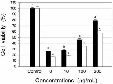
Fig. 1. The effect of A. tegmentosum extracts on HepG2 cell viability treated with As2O3. HepG2 cells were treated with the noted concentrations of A. tegmentosum extracts for 4 hours, and then exposed to As2O3 (■: 10 μg/mL, □: 20 μ g/mL) for 24 hours. Cell viability was determined using MTT method. Each bar represents the mean±SD of triplicate determinations. abcdValues with different letters are significantly different at p<0.05. *p<0.05 when compared with the extract within same group.
(1 mg/mL)을 주입하여 chromatogram의 peak를 얻고, 각 표준 화합물의 회귀방정식을 이용하여 농도를 측정함 으로써, 추출물중 표준 화합물의 함량을 측정하였다.
2.12. 통계 분석
추출물 농도별 항산화 활성은 일원 분산분석을 사용 하여 조사하였고, 농도별 평균값의 차이는 Duncan's multiple range test(Steel and Torrie, 1980)를 사용하 여 p<0.05에서 유의성을 조사하였다. 또한, 추출물과 양 성대조군의 항산화 효능 비교는 대응표본 T-검정을 사용 하여 p<0.05에서 유의성을 조사하였다.
3. 결과 및 고찰 3.1. 세포독성 억제효과
3.1.1. 비소로 유도된 세포독성 억제효과
HepG2 세포는 정상적인 사람의 간세포 기능과 거의 유사하기 때문에 생체외 대사와 독성 연구의 모델로 사 용되고 있다(Knasmuller et al., 1998). 산겨릅나무 추출 물의 비소로 유발된 세포독성에 대한 억제효과는 Fig. 1
에 나타나 있다. 대조군(비소 무첨가군)의 세포생존율을 100%로 기준하였을 때, HepG2 세포에 10 및 20 μ g/mL 농도의 비소 첨가는 각각 26.6 및 16.8 %의 세포 생존율을 나타내어, 비소에 의한 세포독성 유발을 확인 하였다. 본 연구결과와 유사하게, Tchounwon et al.(2002) 은 10 및 20 μg/mL 농도의 arsenic trioxide로 48 시간 처리시 HepG2 세포의 생존율이 각각 52.3 및 24.1%로 감소하였다고 보고한 바 있다. 10 μg/mL 농도의 추출물 로 전처리한 후, 10 및 20 μg/mL 농도의 비소 처리시 세 포생존율은 각각 28.1 및 18.7%로 나타나, 추출물 무첨 가군에 비해 유의적(p>0.05) 차이는 없었다. 그러나, 100 및 200 μg/mL 농도의 추출물로 전처리한 후, 10 μ g/mL 농도의 비소 처리시 세포생존율은 각각 46.4 및 79.3%로 나타났으며, 20 μg/mL 농도의 비소 처리시 각 각 35.0 및 57.5%로 나타나, 산겨릅나무 추출물이 농도 의존적으로 비소 독성에 의한 세포 손상을 유의적으로 (p<0.05) 억제한 것으로 나타났다. Sinha et al.(2007)은 마우스 간세포 실험에서 taurine 또는 ascorbic acid가 sodium arsenite에 의한 세포생존율 감소를 억제시켰고, 산화 활성도 억제시켰다고 보고하였고, Selvaraj et
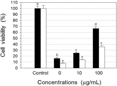
Fig. 2. The effect of A. tegmentosum extracts on HepG2 cell viability treated with t-BHP. HepG2 cells were treated with the noted concentrations of A. tegmentosum extracts for 4 hours, and then exposed to t-BHP (■: 0.2 mM, □: 0.5 mM) for 6 hours. Cell viability was determined using MTT method. Each bar represents the mean±SD of triplicate determinations. abcdValues with different letters are significantly different at p<0.05. *p<0.05 when compared with the extract within same group.
al.(2012)은 항산화제인 selenium 또는 라디칼소거제인 N-acetylcysteine이 arsenic trioxide에 의해 유발된 세 포독성을 억제시켜, 어류 hepatoma의 증식을 증가시켰 다고 보고한 바 있어, 본 연구에서 관찰된 산겨릅나무의 비소로 유발된 세포독성에 대한 억제효과가 탁월함을 알 수 있었다.
3.1.2. t-BHP로 유도된 세포독성 억제효과 t-BHP는 유기태 hydroperoxide로써 산화스트레스로 인한 세포 손상의 기전 연구의 모델로써 사용되고 있다.
산겨릅나무 추출물의 t-BHP로 유발된 세포독성에 대한 억제효과는 Fig. 2에 나타나 있다. HepG2 세포에 0.2 및 0.5 mM 농도의 t-BHP 첨가는 각각 16.4 및 8.2%의 세 포 생존율을 나타내어, t-BHP에 의한 세포독성을 확인 하였다. 10 및 100 μg/mL 농도의 추출물로 전처리한 후, 0.2 mM 농도의 t-BHP 처리시 세포생존율은 각각 25.2 및 66.3%로 나타났고, 0.5 mM 농도의 t-BHP 처리시에 는 각각 14.1 및 35.7%로 나타나, 산겨릅나무 추출물이 농도 의존적으로 t-BHP에 의해 유발된 세포독성을 유의
적으로(p<0.05) 억제시켰다. Vidyashankar et al.(2010) 은 0.2 mM 농도의 t-BHP로 24시간 처리시 HepG2 세 포생존율은 5%이었으나, 항산화제인 ascorbic acid로 30 분 전처리시 세포생존율은 약 25% 이상으로 증가하 였다고 보고한 바 있어, 본 연구에서 관찰된 산겨릅나무 의 t-BHP로 유발된 세포독성에 대한 억제효과가 탁월함 을 알 수 있었다.
3.2. 라디칼 소거활성
3.2.1. ABTS radical 소거활성
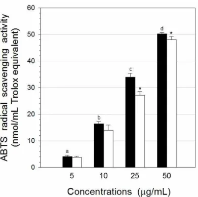
산겨릅나무 추출물의 농도별 ABTS radical 소거활성 은 Fig. 3에 나타나 있다. 5 μg/mL 농도의 추출물의 ABTS radical 소거활성은 8.70 nmol/mL Trolox equivalent이었으며, 추출물 농도가 증가함에 따라 소거 활성도 비례적으로 증가하여, 10, 25 및 50 μg/mL 농도 에서는 각각 16.39, 33.94 및 50.25 nmol/mL Trolox equivalent를 나타내었다. 반면에, 양성대조군으로 사용 한 α-tocopherol의 소거활성은 25 및 50 μg/mL 농도에 서 각각 27.21 및 48.07 nmol/mL Trolox equivalent로
Fig. 3. ABTS radical scavenging activity of A. tegmentosum extracts. Data results were expressed as in terms of nmol/mL Trolox equivalent. Each bar represents the mean±SD of quadraplicate determinations. ■: extract, □: α-tocopherol (positive control). abcdValues with different letters are significantly different at p<0.05. *p<0.05 when compared with the extract within same group.
측정되어, 산겨릅나무 추출물의 ABTS 소거활성이 α -tocopherol에 비해 유의적으로(p<0.05) 높게 나타났다.
3.2.2. Superoxide 소거활성
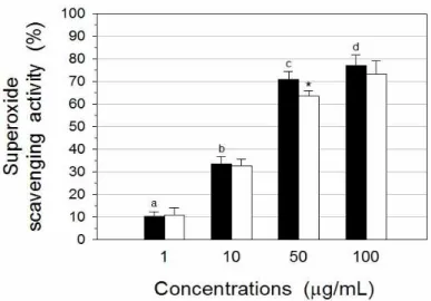
산겨릅나무 추출물의 농도별 superoxide 소거활성 은 Fig. 4에 나타나 있다. 1 μg/mL 농도의 추출물의 superoxide 소거활성은 10.3%이었으며, 추출물 농도가 증가함에 따라 소거활성도 증가하여, 10, 50 및 100 μ g/mL 농도의 추출물의 superoxide 소거활성은 각각 33.5, 70.9 및 77.2%로 나타났다. 반면에, 양성대조군으 로 사용한 catechin의 소거활성은 1, 10, 50 및 100 μ g/mL 농도에서 각각 11.0, 32.6, 63.5 및 73.2%로 측정 되어, 산겨릅나무 추출물의 superoxide 소거활성은 전반 적으로 catechin의 활성과 유사하게 나타났다. 본 연구에 서 관찰된 산겨릅나무의 뛰어난 superoxide 소거활성은 기존에 발표된 연구결과와 일치하고 있다. Kim et al.(2012)은 산겨릅나무 가지 메탄올추출물의 에틸아세
테이트 분획과 부탄올 분획의 superoxide 소거활성이 ascorbic acid에 비해 높게 나타났다고 보고한 바 있다.
3.2.3. ORAC
ORAC assay는 AUC를 측정함으로써, free redical 손상에 대한 억제 시간과 억제율을 모두 반영하는 항산 화능 측정 방법이다. ORAC는 Trolox를 표준물질로 사 용하여 AAPH에 의해 생성된 peroxyl radical에 대한 소 거활성을 나타낸다. 산겨릅나무 추출물의 농도별 ORAC 는 Fig. 5에 나타나 있다. 추출물 2.5 μg/mL 농도의 ORAC는 5.31 nmol/mL Trolox equivalent이었으며, 추출물 농도가 증가함에 따라 ORAC도 증가하여, 5, 10 및 25 μg/mL 농도의 추출물의 ORAC는 각각 9.83, 17.90 및 25.94 nmol/mL Trolox equivalent로 나타났 다. 반면에, 양성대조군으로 사용한 ascorbic acid의 ORAC는 2.5, 5, 10 및 25 μg/mL 농도에서 각각 1.61, 5.97, 11.64 및 20.16 nmol/mL Trolox equivalent로 측
Fig. 4. Superoxide radical scavenging activity of A. tegmentosum extracts. Data results were expressed as % inhibition of the activity. Each bar represents the mean±SD of quadraplicate determinations. ■: extract, □: catechin (positive control). abcdValues with different letters are significantly different at p<0.05. *p<0.05 when compared with the extract within same group.
Fig. 5. Oxygen radical absorbance capacity of A. tegmentosum extracts. Data results were expressed as in terms of nmol/mL Trolox equivalent. Each bar represents the mean±SD of triplicate determinations. ■: extract, □: ascorbic acid (positive control). abcdValues with different letters are significantly different at p<0.05. *p<0.05 when compared with the extract within same group.
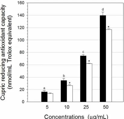
Fig. 6. Cupric reducing antioxidant capacity of A. tegmentosum extracts. Data results were expressed as in terms of nmol/mL Trolox equivalent. Each bar represents the mean±SD of triplicate determinations. ■: extract, □: α -tocopherol (positive control). abcdValues with different letters are significantly different at p<0.05. *p<0.05 when compared with the extract within same group.
정되었다. 모든 농도에서, 산겨릅나무 추출물의 ORAC 가 ascorbic acid에 비해 유의적으로(p<0.05) 높게 나타 났다. Tung et al.(2008)은 산겨릅나무 메탄올추출물의 에틸아세테이트 분획으로부터 분리한 catechin, quercitrin 등 플라보노이드의 peroxyl radical 소거활성이 Trolox 에 비해 높게 나타났다고 보고한 바 있어, 본 연구에서 관 찰된 산겨릅나무의 탁월한 peroxyl radical 소거활성은 기존 연구결과와 일치하고 있다.
3.3. CUPRAC
산겨릅나무 추출물의 농도별 CUPRAC는 Fig. 6에 나타나 있다. 5 μg/mL 농도의 추출물의 CUPRAC는 16.15 nmol/mL Trolox equivalent이었으며, 추출물 농 도가 증가함에 따라 CUPRAC도 비례적으로 증가하여, 10, 25 및 50 μg/mL 농도에서는 각각 34.54, 74.60 및 139.72 nmol/mL Trolox equivalent를 나타내었다. 반
면에, 양성대조군으로 사용한 α-tocopherol의 CUPRAC 는 5, 10, 25 및 50 μg/mL 농도에서 각각 13.50, 26.40, 62.13 및 116.96 nmol/mL Trolox equivalent로 측정되 었다. 10, 25 및 50 μg/mL 농도에서 산겨릅나무 추출물 의 CUPRAC이 α-tocopherol에 비해 유의적으로 (p<0.05) 높게 관찰되어, 산겨릅나무 추출물의 구리이온 환원력이 α-tocopherol에 비해 탁월함을 알 수 있었다.
3.4. 지질과산화 억제효과
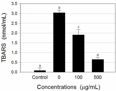
지질과산화 유발을 위해 사용한 t-BHP는 세포내에서 free radical로 대사되어 지질과산화를 개시한다. HepG2 세포를 이용하여, 산겨릅나무 추출물의 지질과산화 억제 효과는 Fig. 7에 나타나 있다. 대조군(t-BHP 무첨가군) 에서 배양액의 TBARS 농도는 0.08 nmol/mL로 나타났 으며, 10 mM 농도의 t-BHP 처리에 의해 TBARS의 농 도는 3.04 nmol/mL로 증가되었다. 100 및 500 μg/mL
Fig. 7. The effect of A. tegmentosum extracts on lipid peroxidation in HepG2 culture. HepG2 cells were treated with the noted concentrations of A. tegmentosum extracts for 4 hours, and then exposed to 10 mM of t-BHP for 2 hours.
Lipid peroxidation was determined by measuring the release of TBARS. Each bar represents the mean±SD of triplicate determinations. abcdValues with different letters are significantly different at p<0.05.
농도의 추출물 전처리시 TBARS 농도는 각각 1.91 및 0.65 nmol/mL로 나타나, 산겨릅나무 추출물은 지질과 산화를 각각 38.2 및 80.7% 억제시켰다. 양성 대조군으 로 사용한 50 μg/mL 농도의 BHT에 의한 TBARS 농도 는 0.78 nmol/mL로 나타났다(data not shown). Hong et al.(2007)은 ferric-thiocyanate법을 사용한 연구에서, 산겨릅나무 줄기 메탄올추출물의 지질과산화 억제효과 가 α-tocopherol과 유사하였고, 에틸아세테이트 분획 중 glucopyranoside의 지질과산화 억제효과는 BHT와 유 사하다고 보고된 바 있다. 또한, Kim et al.(2008)은 랫 트에 산겨릅나무 목부 부탄올추출물 또는 목부에서 분리 한 salidroside의 투여가 D-galactosamine 투여로 증가 된 간 세포질의 MDA 함량을 억제시켜, 산겨릅나무가 지질과산화 형성 저해활성을 나타내었다고 보고한 바 있어, 본 연구에서 관찰된 산겨릅나무의 지질과산화 억 제효과는 기존 발표된 연구결과와 일치하고 있다.
3.5. Supercoiled DNA strand 절단 억제효과
DNA 전기영동을 이용하여, 산겨릅나무 추출물의 농 도별 hydroxyl 및 peroxyl radical에 의해 유도된 DNA
strand 절단에 미치는 효과는 Fig. 8과 Table 1에 나타나 있다.
3.5.1. Hydroxyl radical에 의한 DNA stramd 절단 억제효과
Fenton 시약인 hydrogen peroxide와 ferrous sulfate 처리에 의해 생성되는 hydroxyl radical 존재하에, pBR322 DNA의 supercoiled form은 nicked open circular form 및 linear form으로 전환되었다(lane 1 vs 2, Fig. 8A). Hydroxyl radical 존재하에 plasmid DNA 를 산겨릅나무 추출물과 함께 배양하였을 때, 1 μg/mL 농도의 추출물 첨가에 의한 supercoiled DNA strand의 retention percent는 15.0%로 나타나, hydroxyl radical 에 의해 유도된 supercoiled DNA strand의 절단을 억제 하였다(lane 2 vs 3, Fig. 8A 및 Table 1). 추출물 농도가 증가함에 따라 nicked open circular form은 감소한 반 면 supercoiled form은 증가하여, 산겨릅나무 추출물은 농도 의존적으로 hydroxyl radical에 의해 유도된 single-strand 절단을 억제하였다. 100 μg/mL 농도의 산 겨릅나무 추출물 첨가시 hydroxyl radical에 대한 supercoiled DNA strand의 retention percent는 74.2%
(A)
1 2 3 4 5 6 7
N→
L→
S→
(B)
1 2 3 4 5 6 7
N→
L→
S→
Fig. 8. Electrophoresis of pBR322 DNA treated with hydroxyl radical and peroxyl radical in the presence of A.
tegmentosum extracts. (A) hydroxyl radical was generated by 0.1 mM H2O2 and 0.1 mM FeSO4; (B) peroxyl radical was generated by 5 mM AAPH. Lane 1, DNA alone; Lane 2, DNA+radical; Lane 3, DNA+radical+1 μg/mL extracts; Lane 4, DNA+radical+10 μg/mL extracts; Lane 5, DNA+radical+50 μg/mL extracts; Lane 6, DNA+radical+100 μg/mL extracts; Lane 7, DNA+radical+100 μg/mL sesamol(A) or 10 μg/mL Trolox(B). N, nicked DNA; L, linear DNA; S, supercoiled DNA.
Table 1. Retention percent of supercoiled DNA strand with A. tegmentosum extracts in hydroxyl radical- and peroxyl radical-induced pBR322 plasmid DNA breakage
Retention percent
Hydroxyl radical Peroxyl radical
Extract (μg/mL)
0 8.0±0.3a 8.1±0.2a
1 15.0±0.7b 8.7±0.4a
10 48.0±1.3c 31.6±1.9b
50 64.3±4.2d 80.9±2.3c
100 74.2±5.2e 85.6±5.2c,d
Positive Control1) 85.8±6.1f 87.8±6.0d
Supercoiled pBR322 DNA were treated with 0.1 mM H2O2, 0.1 mM FeSO4 (for hydroxyl radical generation) or 5 mM AAPH (for peroxyl radical generation) in the presence of A. tegmentosum extracts or positive control. The supercoiled and open circular forms of plasmid DNA were separated on a 0.8% agarose gel. Retention percent of supercoiled DNA band was calculated as described in Materials and Methods. The values are means±SD of triplicate determinations.
1)100 μg/mL sesamol for hydroxyl radical- and 10 μg/mL Trolox for peroxyl radical-induced supercoiled DNA strand breakage.
abcdefValues in the same column with different superscripts are significantly different (p<0.05).
(A) (B)
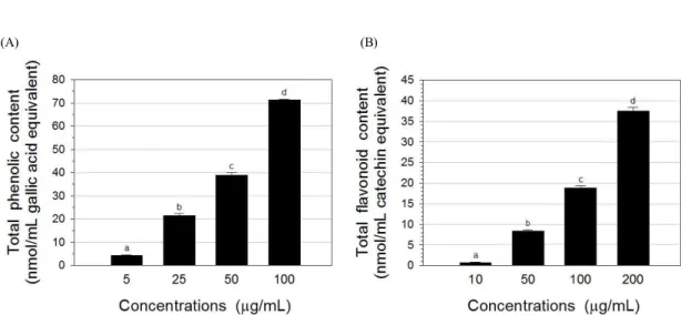
Fig. 9. Total phenolic content(A) and total flavonoid content(B) of A. tegmentosum extracts. Data results were expressed as in terms of nmol/mL gallic acid and catechin equivalents. Each bar represents the mean±SD of quadraplicate determinations. abcdValues with different letters are significantly different at p<0.05.
로 나타나, hydroxyl radical에 의해 유도된 DNA strand 절단을 억제하였으나, 동일한 농도의 양성대조군인 sesamol에 의한 retention percent(85.8%)에 비해 유의 적으로(p<0.05) 낮게 나타났다(lane 6 vs 7, Fig. 8A 및 Table 1).
3.5.2. Peroxyl radical에 의한 DNA strand 절단 억제효과
AAPH 처리에 의해 생성되는 peroxyl radical 존재하 에, pBR322 DNA의 supercoiled form은 nicked open circular form 및 linear form으로 전환되었다(lane 1 vs 2, Fig. 8B). Peroxyl radical 존재하에 plasmid DNA를 산겨릅나무 추출물과 함께 배양하였을 때, 1 μg/mL 농 도의 추출물 첨가는 supercoiled DNA strand의 절단 억 제에 영향을 주지 않았다(lane 2 vs 3, Fig. 8B 및 Table 1). 그러나, 100 μg/mL 농도의 추출물 첨가시 peroxyl radical에 대한 supercoiled DNA strand의 retention percent는 85.6%로 나타나, 10 μg/mL 농도의 양성대조 군인 Trolox의 retention percent(87.8%)와 차이가 없었 고(p>0.05) (lane 6 vs 7, Fig. 8B 및 Table 1), hydroxyl radical에 대한 supercoiled DNA strand의 retention percent(74.2%)에 비해 높게 나타났다(lane 6, Fig. 8A vs lane 6, Fig. 8B 및 Table 1). 따라서 산겨릅나무 추출 물이 hydroxyl radical에 비해 peroxyl radical에 대한
supercoiled DNA strand의 절단 억제효과가 탁월함을 알 수 있었다.
3.6. 총페놀, 총플라보노이드 함량 및 HPLC 분석 산겨릅나무 추출물의 농도별 총페놀 함량은 Fig. 9A 에 나타나 있다. 추출물 5 μg/mL 농도의 총페놀 함량은 4.40 nmol/mL gallic acid equivalent이었으며, 추출물 농도가 증가함에 따라 총페놀 함량도 비례적으로 증가하 여, 25, 50 및 100 μg/mL 농도에서 각각 21.50, 38.98 및 71.30 nmol/mL gallic acid equivalent로 나타났다.
산겨릅나무 추출물의 농도별 총플라보노이드 함량은 Fig. 9B에 나타나 있다. 추출물 10 μg/mL 농도의 총플 라보노이드 함량은 0.64 nmol/mL catechin equivalent 이었으며, 추출물 농도가 증가함에 따라 총플라보노이드 함량도 비례적으로 증가하여, 50, 100 및 200 μg/mL 농 도에서 각각 8.31, 18.83 및 37.42 nmol/mL catechin equivalent로 나타났다.
HPLC 분석에 사용된 4종의 표준 화합물(gallic acid, (-)-epigallocatechin, (+)-catechin 및 ellagic acid)의 표준 검량선 회귀방정식을 이용하여 산겨릅나무 추출물 을 분석한 결과, gallic acid, epigallocatechin, catechin 및 ellagic acid의 함량은 각각 5.4, 47.6, 15.7 및 0.2 μ g/mg으로 측정되어, epigallocatechin의 함량이 가장 높 은 것으로 나타났다.
4. 결 론
본 연구에서는 산겨릅나무 줄기 에탄올추출물의 세포 독성 억제 및 항산화 효과를 조사하였다. HepG2 세포배 양에서 200 μg/mL 농도의 추출물은 10 및 20 μg/mL 비 소로 유도된 세포독성을 각각 79.3 및 57.5% 감소시켰 다. 200 μg/mL 농도의 추출물은 0.2 및 0.5 mM t-BHP 로 유도된 세포독성을 각각 66.3 및 35.7% 감소시켰다.
추출물의 ABTS 라디칼 소거활성은 α-tocopherol에 비 해 높게 나타났고, superoxide 소거활성은 catechin에 비해 높게 나타났으며, peroxyl 라디칼 소거활성은 ascorbic acid에 비해 높게 나타났다. 추출물의 구리이온 환원력은 α-tocopherol에 비해 높게 나타났다. HepG2 세포배양에서 100 및 500 μg/mL 농도의 추출물은 10 mM t-BHP로 유도된 지질과산화를 각각 38.2 및 80.7%
감소시켰다. 또한, 추출물은 hydroxyl 및 peroxyl 라디 칼로 유발된 supercoiled DNA strand의 절단을 억제시 켰다. 100 μg/mL 농도의 추출물의 총페놀 및 총플라보 노이드 함량은 각각 71.3 nmol/mL gallic acid 및 18.8 nmol/mL catechin과 동등한 수준이었다. 따라서 본 in vitro 연구 결과들은 산겨릅나무 추출물의 강력한 세포 독성 억제와 항산화 효과를 나타내며, 이러한 효능은 적 어도 자유라디칼의 산화 억제와 높은 총페놀 함량에 기 인하는 것으로 사료된다. 산겨릅나무 추출물의 생리학적 작용기전을 규명하기 위해서는 in vivo 실험을 포함한 추가적인 연구가 요구된다.
감사의 글
이 논문은 2013년도 상지대학교 교내연구비 지원에 의한 것임.
REFERENCE
Ahn, D. K., 1998, Illustrated Book of Korean Medicinal Herbs, Kyohak Publishing, Seoul, 523-524.
Apak, R., Güçlü, K., Özyürek, M., Karademir, S. E., 2004, Novel total antioxidant capacity index for dietary polyphenols and vitamins C and E, using their cupric ion reducing capability in the presence of neocuproine: CUPRAC method, J. Agric. Food Chem., 52, 7970-7981.
Cha, Y., Park, D., Lee, C. H., Baek, S., Kim, S., Kim, J., Kim, J. H., 2006, Arsenic trioxide induces apoptosis in human colorectal adenocarcinoma HT-29 cells through ROS, Cancer Res. Treat., 38, 54-60.
Erel, O., 2004, A novel automated direct measurement method for total antioxidant capacity using a new generation, more stable ABTS radical cation, Clin.
Biochem., 37, 277-285.
Gao, S., Duan, X., Wang, X., Dong, D., Liu, D., Li, X., Sun, G., Li, B., 2013, Curcumin attenuates arsenic- induced hepatic injuries and oxidative stress in experimental mice through activation of Nrf2 pathway, promotion of arsenic methylation and urinary excretion, Food Chem. Toxicol., 59, 739-747.
Gebel, T. W., 1999, Arsenic and drinking water conta -mination, Science, 283, 1458-1459.
Hiramoto, K., Ojima, N., Sako, K. I., Kikugawa, K., 1996, Effect of plant phenolics on the formation of the spin-adduct of hydroxyl radical and the DNA strand breaking by hydroxyl radical, Biol. Pharm.
Bull., 19, 558-563.
Hong, B. K., Eom, S. H., Lee, C. O., Lee, J. W., Jeong, J. H., Kim, J. K., Cho, D. H., Yu, C. Y., Kwon, Y.
S., Kim, M. J., 2007, Biological activities and bioactive compounds in the extract of Acer tegmen -tosum Maxim. stem, Korean J. Medicinal Crop Sci., 15, 296-303.
Hu, C., Zhang, Y., Kitts, D. D., 2000, Evaluation of antioxidant and prooxidant activities of bamboo Phyllostachys nigra var. Henonis leaf extract in vitro, J. Agric. Food Chem., 48, 3170-3176.
Huang, D., Ou, B., Hampsch-Woodill, M., Flanagan, J.
A., Prior, R. L., 2002, High-throughput assay of oxygen radical absorbance capacity (ORAC) using a multichannel liquid handling system coupled with a microplate fluorescence reader in 96-well format, J.
Agric. Food Chem., 50, 4437-4444.
Kim, S., Hur, S. J., Kim, K. H., Gi, K. S., Whang, W. K., 2012, Antioxidant and anti-inflammatory compounds isolated from Acer tegmentosum, J.
Medicinal Plants Res., 6, 3971-3976.
Kim, S. H., Park, H. J., Choi, J. W., 2008, Hepatopro -tective activity of salidroside isolated from Acer tegmentosum Max on D-galactosamine induced hepatotoxicity in rats, Korean J. Orient. Physiol.
Pathol., 22, 1525-1531.
Knasmuller, S., Parzefall, W., Sanyal, R., Ecker, S., Schwab, C., Williamson, G., Hietsch, G., Langer, T., Darroudi, F., Natarajan, A. T., 1998, Use of metabolically competent human hepatoma cells for the detection of mutagens and anti-mutagens, Mutat.
Res., 402, 185-202.
Kwon, H. N., Park, J. R., Jeon, J. R., 2008, Antioxidative and hepatoprotective effects of Acer tegmentosum M. extracts, J. Korean Soc. Food Sci.
Nutr., 37, 1389-1394.
Liu, F., Ooi, V. E. C., Chang, S. T., 1997, Free radical scavenging activities of mushroom polysaccharide extracts, Life Sci., 60, 763-771.
Liu, M., Li, X. Q., Weber, C., Lee, C. Y., Brown, J., Liu, R. H., 2002, Antioxidant and antiproliferative activities of raspberries, J. Agric. Food Chem., 50, 2926-2930.
Liu, Q., Shin, E., Ahn, M, Hwang, B. Y., Lee, M. K., 2011, Anti-adipogenic activity of Acer tegmentosum and its constituent, catechin in 3T3-L1 cells, Nat.
Product Sci., 17, 212-215.
Mosmann, T., 1983, Rapid colorimetric assay for cellular growth and survival: application to proliferation and cytotoxicity assays, J. Immunol. Methods, 65, 55-63.
Selvaraj, Y., Yeager-Armstead, M., Murray, E., 2012, Protective and antioxidant role of selenium on arsenic trioxide-induced oxidative stress and genotoxicity in the fish hepatoma cell line PLHC-1, Environ. Toxicol. Chem., 31, 2861-2869.
Shim, K., Kim, D, Song, S., Qi, X., Yoon, Y., Kim, H., Lee, J., Oh, H., Kim, S., Lee, K., 2011, Hepatoprotection of different water extracts from Acer tegmentosum M. on CCl4-induced acute hepatotoxicity in mice: comparative efficacies between the extracts of boughs, twigs, and leaves, Mol. Cell. Toxicol., 7, 405-413.
Shin, I., Sa, J., Shim, T., Lee, J., 2006, The physical and chemical properties and cytotoxic effects of Acer tegmentosum Maxim. extracts, J. Korean Soc. Appl.
Biol. Chem., 49, 322-327.
Singleton, V. L., Orthofer, R., Lamuela-Raventós, R.
M., 1999, Analysis of total phenols and other oxidation substrates and antioxidants by means of Folin-Ciocalteu reagent, Methods Enzymol., 299,
152-178.
Sinha, M., Manna, P., Sil, P. C., 2007, Taurine, a conditionally essential amino acid, ameliorates arsenic-induced cytotoxicity in murine hepatocytes, Toxicol. in Vitro, 21, 1419-1428.
Soria, E. A., Eynard, A. R., Bongiovanni, G. A., 2010, Cytoprotective effects of silymarin on epithelial cells against arsenic-induced apoptosis in contrast with quercetin cytotoxicity, Life Sci., 87, 309-315.
Steel, R. G. D., Torrie, J. H., 1980, Principles and Procedures of Statistics, 2nd ed., McGraw-Hill, New York, 186-187.
Tchounwou, P. B., Wilson, B. A., Abdelghani, A. A., Ishaque, A. B., Patlolla, A. K., 2002, Differential cytotoxicity and gene expression in human liver carcinoma (HepG2) cells exposed to arsenic trioxide, and monosodium acid methanearsonate (MSMA), Int. J. Mol. Sci., 3, 1117-1132.
Tirmenstein, M. A., Nicholls-Grzemski, F. A., Zhang, J.
G., Fariss, M. W., 2000, Glutathione depletion and the production of reactive oxygen species in isolated hepatocyte suspensions, Chem. Biol. Inter., 127, 201-217.
Tung, N. H., Ding, Y., Kim, S. K., Bae, K., Kim, Y. H., 2008, Total peroxyl radical-scavenging capacity of the chemical components from the stems of Acer tegmentosum Maxim, J. Agric. Food Chem., 56, 10510-10514.
Valko, M., Leibfritz, D., Moncol, J., Cronin, M. T. D., Mazur, M., Telser, J., 2007, Free radicals and antioxidants in normal physiological functions and human disease, Intl. J. Biochem. Cell. Biol., 39, 44-84.
Vidyashankar, S., Mitra, S. K., Nandakumar, K. S., 2010, Liv.52 protects HepG2 cells from oxidative damage induced by tert-butyl hydroperoxide, Mol.
Cell. Biochem., 333, 41-48.
Wang, C. H., Hsiao, C. K, Chen, C. L., Hsu, L. I., Chiou, H. Y., Chen, S. Y., Hsueh, Y. M., Wu, M.
M., Chen, C. J., 2007, A review of the epidemiologic literature on the role of environmental arsenic exposure and cardiovascular diseases, Toxicol. Appl.
Pharmacol., 222, 315-326.
WHO, 2011, Guidelines for drinking-water quality, 4th ed., World Health Organization, Geneva, 315-318.
Yu, T., Lee, J., Lee, Y. G., Byeon, S. E., Kim, M. H., Sohn, E., Lee, Y. J., Lee, S. G., Cho, J. Y., 2010, In vitro and in vivo anti-inflammatory effects of ethanol
extract from Acer tegmentosum, J. Ethnopharmacol., 128, 139-147.