Effect of Testosterone Replacement Therapy on Bone Mineral Density in Patients with Klinefelter Syndrome
Dae Gi Jo,
1Hyo Serk Lee,
1Young Min Joo,
2and Ju Tae Seo
11Department of Urology, Cheil General Hospital, Kwandong University College of Medicine, Seoul; 2Daewoo General Hospital, Geoje, Korea.
Received: January 17, 2013 Revised: February 18, 2013 Accepted: March 13, 2013
Corresponding author: Dr. Ju Tae Seo, Department of Urology, Cheil General Hospital, Kwandong University College of Medicine, 17 Seoae-ro 1-gil, Jung-gu,
Seoul 100-380, Korea.
Tel: 82-2-2000-7585, Fax: 82-2-2000-7787 E-mail: [email protected]
∙ The authors have no financial conflicts of interest.
© Copyright:
Yonsei University College of Medicine 2013 This is an Open Access article distributed under the terms of the Creative Commons Attribution Non- Commercial License (http://creativecommons.org/
licenses/by-nc/3.0) which permits unrestricted non- commercial use, distribution, and reproduction in any medium, provided the original work is properly cited.
Purpose: Klinefelter syndrome (KS) is related to testicular insufficiency, which causes low testosterone levels in serum. Generally, sex hormone levels and bone mineral density (BMD) are lower in patients with KS than normal. We investigat- ed the effects of testosterone replacement on serum testosterone levels and BMD in KS patients. Materials and Methods: From December 2005 to March 2008, 18 KS patients with a 47, XXY karyotype were treated with initial intramuscular in- jections of long-acting testosterone undecanoate (Nebido®, 1000 mg/4 mL) at baseline and second injections after six weeks. An additional four injections were administered at intervals of 12 weeks after the second injection. BMD was mea- sured at the lumbar spine (L2-4), the left femoral neck and Ward’s triangle, using dual energy X-ray absorptiometry. Medical histories, physical examinations and prostate specific antigen, hematology and serum chemistry were conducted for each patient. In addition, total testosterone and sex hormone-binding globulin lev- els were measured. Results: Following testosterone replacement, mean serum to- tal testosterone increased significantly from baseline (0.90 vs. 4.51 ng/mL, p<0.001), and total testosterone rose to normal levels after replacement in all patients. The mean BMD of the lumbar spine increased significantly (0.91 vs. 0.97 g/cm2, p<0.001). Similar increases of BMD were also observed at the femoral neck, but this increase was not significant. Conclusion: These findings suggest that testos- terone replacement therapy may be effective in treating BMD deficiency in men with testosterone deficiency, especially those with Klinefelter syndrome.
Key Words: Bone mineral density, Klinefelter syndrome, testosterone
INTRODUCTION
Osteoporosis and osteopenia in men are considered to be less frequent than in women. It might be due in part to a man’s greater bone size, greater body mass, greater accrual of bone during growth, absence of a clear decrease in endogenous sex hormones as seen in menopause, and shorter average life span compared with women.1 However, it is frequent in men with secondary osteoporosis due to hypo- gonadism, glucocorticoid excess, alcoholism, hypercalciuria, malabsorption and hyperthyroidism.
Klinefelter syndrome (KS) is a common sex chromosome disorder in which
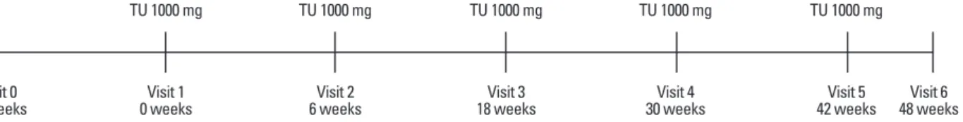
(baseline), week 6 and every 12 weeks thereafter (Fig. 1).
Blood samples were obtained at baseline and at injection time points for prostate specific antigen (PSA), hematology (including hemoglobin and hematocrit) and serum chemis- try. Measurements of height and weight were taken, and body mass index was calculated [body mass index (BMI):
kg/m2].
Hormonal measurements
Baseline sex hormone-binding globulin (SHBG) and serum or total testosterone (TT) levels were measured at 2 weeks before injection, and 30 weeks and 48 weeks after the first injection. Venous blood samples were collected between 08:00 and 10:00 hours. The serum was separated by cen- trifugation and subsequently stored at -20°C until assayed.
Serum TT level was measured by enzyme sandwich immu- noassay using Vitros® ECi Immunodiagnostic System (Or- tho Clinical Diagnostics, Rochester, NY, USA). Free and bioavailable testosterone was calculated by using the “Free
& Bioavailable Testosterone calculator” (http://www.issam.
ch/freetesto.htm). SHBG in serum was measured by using IRMA-Count® (a solid-phase immunoradiometric assay de- signed for the quantitative measurement of SHBG in se- rum) (Diagnostic Products Corporation, Los Angeles, CA, USA).
Bone density determination
BMD (the area density in g/cm2) was assessed using dual-en- ergy X-ray absorptiometry operating in the fan-beam mode (QDR-2000 densitometer, Hologic Inc., Waltham, MA, USA). Quality control scans were taken daily, using an an- thropometric spine phantom supplied by the manufacturer;
the long-term (>1 year) coefficient of variation was 0.40%.
BMD was measured on visit 0 and 48 weeks. Lumbar BMD was assessed at L2-4 on the basis of the postero-anterior view. Fractured vertebrae were excluded from analysis. The femur neck and Ward’s triangle BMD was measured at the upper left femur. The mean precision error of absorptiome- try measurements was <1.5% for the lumbar spine, and
<2% for the femur neck and Ward’s triangle BMD. T scores were calculated using the manufacturer’s references. Indi- males are born with an extra copy of the X chromosome. It
occurs at a frequency of approximately 1 in 500 men and re- sults in impaired spermatogenesis and androgen deficiency.2 KS is a type of primary hypogonadism, and 44-48% of KS patients have osteopenia and 6-14% osteoporosis that is a well known complication of male hypogonadism.2-4 Andro- gen deficiency in men is known to lead to a significant de- crease in bone mineral density (BMD).5 Similarly, osteopo- rosis is related to lower level of serum testosterone in KS.6,7 Although increasing the testosterone levels is presumed to have a beneficial effect on BMD, only few data are avail- able in these patients after testosterone replacement therapy, and data are still controversial.8-12
The aim of this study was to measure BMD, as well as biochemical and hormonal parameters in men with Kline- felter syndrome undergoing testosterone replacement thera- py and determine whether testosterone replacement therapy reversed the detrimental effects of hypogonadism on bone density.
MATERIALS AND METHODS
Patients
From December 2005 to March 2008, 18 Korean KS pa- tients, ranging from 30 to 44 years of age, were studied.
They had a careful medical history and physical examina- tion taken, and the accuracy of data was verified by check- ing all available medical records. The diagnosis of KS was based on clinical features including androgen deficiency symptoms, low testosterone levels, and karyotype showing 47, XXY. Exclusion criteria were treatment with any medi- cation known to affect the skeleton, known prostate cancer or clinically significant benign prostate hyperplasia, andro- gen-dependent carcinoma of the male mammary gland, oth- er significant medical or psychological conditions, sleep ap- nea or snoring requiring treatment, or diabetes mellitus.
Informed consent to the study was obtained from all the sub- jects. Intramuscular long-acting testosterone undecanoate (Nebido®, Bayer Pharma, Berlin, Germany, 1000 mg/4 mL) was administered to the deep gluteus muscle at week 0
Fig. 1. Testosterone injection schedule. TU, testosterone undecanoate.
TU 1000 mg
Visit 1 0 weeks Visit 0
-2 weeks
TU 1000 mg
Visit 2 6 weeks
TU 1000 mg
Visit 3 18 weeks
TU 1000 mg
Visit 4 30 weeks
TU 1000 mg
Visit 5
42 weeks Visit 6 48 weeks
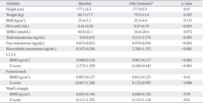
normal limit. SHBG decreased from 44.8 nmol/L to 36.6 nmol/L, but it was not statistically significant (p=0.072). To- tal testosterone rose to normal levels after replacement (0.9 ng/mL to 4.511 ng/mL, p<0.001). Likewise, free and bio- available testosterones increased from 0.015 ng/mL to 0.974 ng/mL, and 0.347 ng/mL to 2.284 ng/mL, respectively, and these were all statistically significant.
The results of BMD (g/cm2) and Z-scores at the lumbar spine, femoral neck, and Ward’s triangle in the treated 18 KS patients are shown in Table 1. At the lumbar spine, BMD and Z-score significantly increased from 0.908 g/cm2 to 0.967 g/cm2 and from -1.272 to -0.244, respectively. Similar increases were also observed at femoral neck, but signifi- cant difference only in Z-score (BMD: 0.807 g/cm2 to 0.812 g/cm2, p=0.42, Z-score: -0.467 to 0.122, p=0.006). At the BMDs and Z-score of the Ward’s triangle, there were no significant changes (BMD: 0.683 g/cm2 to 0.668 g/cm2, p=0.5, Z-score: -0.111 to 0.133, p=0.81).
DISCUSSION
We previously suggested that testosterone replacement may be an effective treatment for increasing BMD in men with KS and a low serum testosterone level.7 In this study, we evaluated whether testosterone replacement therapy re- vidual values of vertebral and femoral BMD in KS patients
were also expressed as Z-score, calculated on the basis of this reference range. T-score compares with young normal mean BMD, and the Z-score compares with age-matched controls and race-matched controls.
Statistical analysis
The characteristics of the patients were summarized by de- scriptive statistics. Student’s t-test (paired) and Wilcoxon rank sum test were used to compare continuous variables.
Data storage and statistical analysis were performed using the SPSS 12.0K statistical program (SPSS Inc., Chicago, IL, USA). A p-value <0.05 was used to define statistical signifi- cance. Results are presented as mean±SD unless otherwise indicated.
RESULTS
Eighteen patients were followed up (mean age 35.9±3.3 years). During follow-up period, there was a slight decrease in weight and a slight increase in height, which resulted in a slight decrease in BMI without significance.
The effects of testosterone replacement on Klinefelter syn- drome are presented in Table 1. A small and significant in- crease was observed in PSA, but PSA levels were lower than
Table 1. Effects of Testosterone Replacement on Bone Mineral Density in Patients with Klinefelter’s Syndrome
Variables Baseline After treatment* p value
Height (cm) 177.1±6.3 177.9±5.9 0.67
Weight (kg) 80.7±17.7 79.9±15.4 0.585
BMI (kg/m2) 25.6±5.1 25.2±4.4 0.133
PSA (mlU/mL) 0.41±0.24 0.87±0.39 <0.001
SHBG (nmol/L) 44.8±22.1 36.6±20.4 0.072
Total testosterone (ng/mL) 0.9±0.633 4.511±2.278 <0.001
Free testosterone (ng/mL) 0.015±0.012 0.974±0.058 <0.001
Bioavailable testosterone (ng/mL) 0.347±0.284 2.284±1.352 <0.001
L2-L4
BMD (g/cm2) 0.908±0.110 0.967±0.117 <0.001
Z-score -1.272±1.209 -0.244±0.843 <0.001
Femoral neck
BMD (g/cm2) 0.807±0.127 0.812±0.125 0.42
Z-score -0.467±1.246 0.122±0.995 0.006
Ward’s triangle
BMD (g/cm2) 0.683±0.146 0.668±0.142 0.50
Z-score -0.111±1.182 0.133±1.110 0.81
BMI, body mass index; PSA, postate-specific antigen; SHBG, sex hormone-binding globulin; BMD, bone mineral density.
Data are given as means±SD.
*After treatment: 48 weeks after the first injection.
We observed in the present study that the testosterone re- placement definitely increased BMD and Z-score of the lumbar spine, whereas it was not effective or minimal in femoral neck and Ward’s triangle. This finding may imply that the loss of bone occurs mainly in trabecular bone rather than cortical bone since trabecular bone is metabolically more active than cortical bone.
The potential weakness of this study is that diet such as calcium, vitamin D and the like were not assessed among the subjects, which could influence BMD. In addition, tes- tosterone replacement was supplemented after puberty.
Therefore, for better results, it would be necessary to give testosterone replacement before patients’ puberty.
In conclusion, male hypogonadism including KS has been recognized as one of the major causes of secondary osteoporosis in men, nevertheless, most cases seem to be left undiagnosed. Bone loss is a distinctive feature of KS.
We suggest that testosterone replacement may be effective for preventing BMD deficiency. Therefore, early diagnosis and testosterone replacement therapy coupled with specific treatment for osteoporosis could be useful in preventing fu- ture severe bone loss and associated skeletal morbidity.
ACKNOWLEDGEMENTS
This study was supported by a grant of the Korea Health- care Technology R&D Project, Ministry for Health, Wel- fare & Family Affairs, Republic of Korea (A084318).
REFERENCES
1. Seeman E. During aging, men lose less bone than women because they gain more periosteal bone, not because they resorb less end- osteal bone. Calcif Tissue Int 2001;69:205-8.
2. Klinefelter HF. Background of the recognition of Klinefelter’s syndrome as a distinct pathologic entity. Am J Obstet Gynecol 1973;116:436-7.
3. Seeman E, Melton LJ 3rd, O’Fallon WM, Riggs BL. Risk factors for spinal osteoporosis in men. Am J Med 1983;75:977-83.
4. van den Bergh JP, Hermus AR, Spruyt AI, Sweep CG, Corstens FH, Smals AG. Bone mineral density and quantitative ultrasound parameters in patients with Klinefelter’s syndrome after long-term testosterone substitution. Osteoporos Int 2001;12:55-62.
5. Orwoll ES, Klein RF. Osteoporosis in men. Endocr Rev 1995;
16:87-116.
6. Horowitz M, Wishart JM, O’Loughlin PD, Morris HA, Need AG, Nordin BE. Osteoporosis and Klinefelter’s syndrome. Clin Endo- crinol (Oxf) 1992;36:113-8.
7. Seo JT, Lee JS, Oh TH, Joo KJ. The clinical significance of bone
versed the detrimental effects of KS on bone density. Sev- eral previous reports indicate that testosterone replacement increased BMD in middle-aged men or androgen deficien- cy.13,14 Isidori, et al.14 reported that testosterone improved BMD among 1083 subjects at lumbar spine by +3.7% (CI:
1.0-6.4%) compared to placebo, but not at the femoral neck after a minimum of 12-36 months of treatment. However, the effects of androgen replacement on bone mineral densi- ty in patients with KS have been studied very little. When studying 29 men with KS, there was no correlation between serum testosterone levels and bone density.15 Wong, et al.12 suggested that sufficient testosterone replacement does not reverse the decrease of bone mass, associated with hypogo- nadism, in patients with KS. They pointed out several pos- sible explanations for these findings as follows: first, short- acting testosterone replacement by parenteral and oral replacement could not dispense physiological testosterone concentrations and normal circadian patterns; and many pa- tients with KS were diagnosed after pubertal development.
After pubertal development, epiphyseal bone closure and skeletal maturation had taken place. Therefore, androgen replacement was instituted late and after puberty. The third possibility is that it may be take a long time for restore the bone density and mass to normal. Contrary to these find- ings, only few available data indicate that increasing the testosterone levels has a beneficial effect on BMD.8-12
Our present findings suggest that testosterone replace- ment is an effective treatment for increasing BMD in men with KS and a low serum testosterone level. In this study, long-acting testosterone undecanoate was administered. The use of a long-acting form of the hormone, which provides a constant level of testosterone, may be effective compared with the use of a shorter acting formulation. The undecano- ate ester of natural testosterone is a long acting, intramuscu- lar, injectable testosterone that extends the current maximum treatment dosing interval approximately 4-fold compared with that of other injectable products, thus eliminateing the need of daily topical application of gels and patches. Tes- tosterone undecanoate (TU: 1000 mg) given at a variable dosing interval is used for treating men with hypogonad- ism. Studies in Europe have demonstrated that 1000 mg TU administered as an intramuscular injection at 10 to 14- week intervals is adequate to sustain normal testosterone levels in men with hypogonadism.16,17 This long acting for- mulation has been shown to be safe and well tolerated. In this study, follow-up over 48 weeks, when all subjects re- ceived TU, showed a good level of safety.
Adequacy of androgen replacement influences bone density re- sponse to testosterone in androgen-deficient men. Eur J Endocri- nol 2005;152:881-6.
14. Isidori AM, Giannetta E, Greco EA, Gianfrilli D, Bonifacio V, Isidori A, et al. Effects of testosterone on body composition, bone metabolism and serum lipid profile in middle-aged men: a meta- analysis. Clin Endocrinol (Oxf) 2005;63:280-93.
15. Smith DA, Walker MS. Changes in plasma steroids and bone den- sity in Klinefelter’s syndrome. Calcif Tissue Res 1977;22 Sup- pl:225-8.
16. Schubert M, Minnemann T, Hübler D, Rouskova D, Christoph A, Oettel M, et al. Intramuscular testosterone undecanoate: pharma- cokinetic aspects of a novel testosterone formulation during long- term treatment of men with hypogonadism. J Clin Endocrinol Metab 2004;89:5429-34.
17. von Eckardstein S, Nieschlag E. Treatment of male hypogonadism with testosterone undecanoate injected at extended intervals of 12 weeks: a phase II study. J Androl 2002;23:419-25.
mineral density and testosterone levels in Korean men with non- mosaic Klinefelter’s syndrome. BJU Int 2007;99:141-6.
8. Choi HR, Lim SK, Lee MS. Site-specific effect of testosterone on bone mineral density in male hypogonadism. J Korean Med Sci 1995;10:431-5.
9. De Rosa M, Paesano L, Nuzzo V, Zarrilli S, Del Puente A, Oriente P, et al. Bone mineral density and bone markers in hypogonado- tropic and hypergonadotropic hypogonadal men after prolonged testosterone treatment. J Endocrinol Invest 2001;24:246-52.
10. Stepan JJ, Burckhardt P, Hána V. The effects of three-month intra- venous ibandronate on bone mineral density and bone remodeling in Klinefelter’s syndrome: the influence of vitamin D deficiency and hormonal status. Bone 2003;33:589-96.
11. Wang C, Swerdloff RS. Androgen replacement therapy. Curr Ther Endocrinol Metab 1997;6:331-7.
12. Wong FH, Pun KK, Wang C. Loss of bone mass in patients with Klinefelter’s syndrome despite sufficient testosterone replacement.
Osteoporos Int 1993;3:3-7.
13. Aminorroaya A, Kelleher S, Conway AJ, Ly LP, Handelsman DJ.