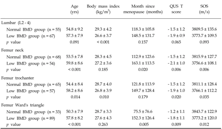
Does Quantitative Tibial Ultrasound Predict Low Bone Mineral Density Defined by Dual Energy X-Ray Absorptiometry?
7
0
0
전체 글
(2)
(3)
(4)
(5)
(6)
(7)
수치

관련 문서
In this paper, we extend approximation method of conic section by Bezier curve to sphere case by polynomial surfaces and we analyze the Hausdorff
Bone mineral content (BMC) and bone mineral density (BMD) of the lumbar spine, total hip, and proximal femur were measured before and after exercise... Results : 1) Body
The purpose of this study was to collect the traffic flow characteristics(volume, spot speed, headway, occupancy, and density, etc.) at the basic segments
The purpose of this study was to collect the traffic flow characteristics (volume, speed, occupancy, and density, etc.) in expressway basic segments, analyze
Keywords: Trabecular bone score, End stage renal disease, Hemodialysis, Chronic kidney disease-mineral and bone disorder, Fracture, Cardiovascular
Background: As the genetic variants of trabecular bone microarchitecture are not well-understood, we performed a genome-wide association study to identify genetic determinants
최대정수함수가 극댓값을 갖는 점과 극솟값을 갖는 점을 모두 구하고, 그 점에서 극값을 구하 시오.. 함수가 미분 가능하면 극값을
그러므로 이러한 표현은 함수의 이름과 미분계수를 구하는 점이 명확하여 혼동할 염려가 없을
