INTRODUCTION
Diffuse large B-cell lymphoma (DLBCL), the most prevalent type of non-Hodgkin lymphoma, constitutes 30–40% of all cases in different geographic regions.1 The two subtypes of DLBCL contained activated B cell-like and germinal center B cell-like, classified by distinct pathogenesis.2 Although the development of therapeutic methods has improved the survival of DLBCL patients, about 30% of patients still develop relapse/refractory disease.3 Therefore, it is important to search for new therapeu- tic markers for DLBCL treatment.
Curcumin, chemically called as diferuloylmethane, is a yel-
Curcumin Inhibits the Proliferation, Migration, Invasion, and Apoptosis of Diffuse Large B-Cell
Lymphoma Cell Line by Regulating MiR-21/VHL Axis
Ling Chen1*, Cheng-Zhi Zhan1*, Tao Wang2, Hua You3, and Rui Yao4
Departments of 1Pathology and 2Gastrointestinal Surgery, Xianning Central Hospital, The First Affiliated Hospital of Hubei University of Science and Technology, Hubei;
3Cancer Center of Guangzhou Medical University, Guangzhou;
4Administrative Department, Xianning Central Hospital, The First Affiliated Hospital of Hubei University of Science and Technology, Hubei, China.
Purpose: Curcumin exerts its anti-cancer effects, partly by targeting special microRNAs, in human cancers. MiR-21 is a key on- comir in carcinogenesis of multiple human cancers. Here, we aimed to further explore the mechanistic insight into the link be- tween curcumin and miR-21 on diffuse large B-cell lymphoma (DLBCL).
Materials and Methods: Quantitative real-time PCR assays were performed to assess the levels of miR-21 and Von Hippel-Lindau (VHL) mRNA. In situ hybridization assay was used for miR-21 expression visualization in lymphoma tissues. Western blot was used for determination of VHL protein, Ki-67, caspase-3, and cleaved caspase-3 levels. Dual-luciferase reporter assay and RNA immu- noprecipitation assay were employed to confirm the direct target of miR-21. MTT assay, flow cytometric analysis, and transwell as- say were used to evaluate cell proliferation, apoptosis, and migration and invasion capacities, respectively.
Results: Curcumin repressed the proliferation, migration, and invasion abilities and promoted apoptosis in SU-DHL-8 cells. Cur- cumin inhibited miR-21 expression and curcumin exerted its anti-proliferation, anti-migration, anti-invasion, and pro-apoptosis effects by miR-21 in SU-DHL-8 cells. VHL was a direct target of miR-21. Moreover, curcumin exerted its regulatory effects on SU- DHL-8 cells by VHL.
Conclusion: Curcumin exerted its anti-proliferation, anti-migration, anti-invasion, and pro-apoptosis functions, at least partly, by repressing miR-21 and regulating VHL expression in DLBCL cell line. Our findings provided a possible molecular mechanism of curcumin-mediated anti-cancer effect.
Key Words: Diffuse large B-cell lymphoma (DLBCL), curcumin, miR-21, Von Hippel-Lindau (VHL)
pISSN: 0513-5796 · eISSN: 1976-2437
Received: December 17, 2018 Revised: March 14, 2019 Accepted: March 18, 2019
Corresponding author: Rui Yao, MD, Administrative Department, Xianning Cen- tral Hospital, The First Affiliated Hospital of Hubei University of Science and Tech- nology, 228 Jingui Road, Xianan District, Xianning, Hubei 437100, China.
Tel: 86-0715-8896139, Fax: 86-0715-8896139, E-mail: [email protected]
*Ling Chen and Cheng-Zhi Zhan contributed equally to this work.
•The authors have no potential conflicts of interest to disclose.
© Copyright: Yonsei University College of Medicine 2020
This is an Open Access article distributed under the terms of the Creative Com- mons Attribution Non-Commercial License (https://creativecommons.org/licenses/
by-nc/4.0) which permits unrestricted non-commercial use, distribution, and repro- duction in any medium, provided the original work is properly cited.
Yonsei Med J 2020 Jan;61(1):20-29 https://doi.org/10.3349/ymj.2020.61.1.20
low-colored polyphenol that has been widely used as food ad- ditive and traditional medicine in Asian countries.4 The bene- ficial functions of curcumin are thought to attribute to its anti- microbial,5 anti-oxidant,6 anti-inflammatory,7 hepatoprotective,8 and hypoglycemia protective9 effects. Growing evidence have suggested that curcumin plays a protective role in preventing and treating numerous human cancers by inhibiting tumori- genesis, progression, metastasis, angiogenesis, chemoresis- tance, and radioresistance.10-12
MicroRNAs (miRNAs), evolutionarily conserved single- stranded RNAs of about 22 nucleotides in length, play a critical role in a series of physiopathology processes.13 Mature miRNAs, loaded into RNA-induced silencing complex (RISC), negatively regulate target gene expression by binding to the 3'-untrans- lated region (3'-UTR) of target mRNAs, leading to translational repression and mRNA degradation.14 Recent studies have elu- cidated that miRNAs are involved in tumor growth, progres- sion, metastasis, angiogenesis, and immune evasion.15 MiR- 21, a commonly overexpressed miRNA, is a key oncomir in carcinogenesis of multiple human cancers, such as gastric can- cer,16 breast cancer,17 and ovarian cancer.18 Moreover, circulat- ing miR-21 was demonstrated as a new disease response bio- marker with differential response stratified by interim-positron emission tomography/CT in DLBCL patients.19 High expres- sion of miR-21 was associated with worse overall survival of DLBCL patients.20 Additionally, miR-21 contributed to DLBCL cells chemoresistance through targeting FOXO1 and regulat- ing PI3K/AKT pathway.21 Downregulation of miR-21 promoted cell apoptosis and tumor-suppressor PTEN expression in DLB- CL, highlighting its role as a potential useful approach for DLB- CL treatment.22
In the present study, our data indicated that curcumin in- hibited the proliferation, migration, and invasion, while pro- moting the apoptosis of DLBCL cell line. Moreover, curcumin suppressed miR-21 expression, and Von Hippel-Lindau (VHL) was a direct target of miR-21. In all, curcumin exerted its anti- proliferation, anti-migration, anti-invasion, and pro-apoptosis functions, at least partly, by repressing miR-21 and regulating VHL expression in DLBCL cell line.
MATERIALS AND METHODS
Specimens and cells
Forty-five cases of lymphoma tissues were obtained from DLB- CL patients who were diagnosed and had undergone surgical resection at Xianning Central Hospital (Hubei, China) be- tween August 2010 to March 2016. Additionally, 23 cases of re- active lymphoid hyperplasia tissues were collected in the same period, and were selected as negative controls. No local or systemic therapy was conducted in these participators before operation. Primary CD19+ cells were obtained from the serum of healthy volunteers through positive selection with CD19+
MicroBeads antibody (Miltenyi Biotec, Auburn, CA, USA).
Written informed consent was obtained from all participants, and the study was approved by Institutional Review Board of Xianning Central Hospital.
Three DLBCL cell lines (SU-DHL-8, OCI-LY1, and SU-DHL-10) were purchased from American Type Culture Collection (ATCC, Manassas, VA, USA), and were cultured in RPMI-1640 medi- um (Thermo Fisher Scientific, Tempe, AZ, USA) plus 10% fetal bovine serum (FBS, Thermo Fisher Scientific), 2 mmol/L L-glu- tamine (Thermo Fisher Scientific) and 100 units/mL penicillin/
streptomycin (Thermo Fisher Scientific), at 37°C in 5% CO2. Cell transfection and treatment
The modified miR-21 mimics and the control (NC mimics);
miR-21 inhibitor (anti-miR-21) and negative control (anti-NC);
and siRNA targeting VHL (si-VHL) and corresponding control (si-NC) were all designed and synthesized by Sangon Biotech (Shanghai, China). 50 nM of the indicated oligonucleotide was transiently transfected into cells by LipofectamineTM 2000 Transfection Reagent (Invitrogen, Breda, the Netherlands) ac- cording to the directions of manufacturers.
For curcumin treatment, SU-DHL-8 cells were treated with different concentrations (0, 5, 10, 20, 40, and 60 μmol/L) of curcumin (Sigma-Aldrich, St. Louis, MO, USA), or transfected cells were treated with 20 μmol/L of curcumin, followed by the detection of cell proliferation, migration, invasion, and apop- tosis capacities.
MTT assay of cell proliferation ability
Cell proliferation ability was assessed by MTT assay. In brief, cells were treated with curcumin or transfected with miR-21 mimics in 96-well microplates. At 0, 24, 48, 72, and 96 h, a total of 50 μL of MTT solution (2 mg/mL, Sigma-Aldrich) was pi- petted into each well, and incubated at 37°C for 3 h to stain the living cells. After MTT solution was removed, 150 μL of dissolving solution (11 g SDS, 50 mL 0.02 M HCl, 50 mL isobu- tanol) was added to dissolve the formazan crystals. Absorbance at 490 nm was measeured with a microplate reader (Bio-Rad, Richmond, CA, USA).
Flow cytometric analysis of cell apoptosis
Cell apoptosis was determined by flow cytometry with Annex- in V-FITC/PI Apoptosis Detection Kit (PharMingen, San Di- ego, CA, USA) according to the instruction of manufacturers.
At 24 h of curcumin treatment or transfection, cells were har- vested and washed twice with cold PBS. Following resuspen- sion in 1× binding buffer at a concentration of 5.0×10⁵ cells/
mL, 5 μL of Annexin V-FITC and 10 μL of PI were added to 100 μL of cells. Cell apoptosis rate was analyzed by FACScan (BD Biosciences, Franklin Lakes, NJ, USA) using CellQuest Pro (in vitro diagnostic) software (BD Biosciences).
Transwell assay of cell migration and invasion
Transwell assays were performed to detect cell migration and invasion capacities by using 8 µm polycarbonate nucleopore filter-containing Boyden chambers (Millipore, Billerica, MA, USA). For migration detection, a total of 2.0×10⁴ cells was plat- ed in the upper chamber with non-coated membrance (Milli- pore) for 6-8 h to attach. For invasion detection, 2.0×10⁴ cells were seeded into the upper chamber with Matrigel-coated membrance (Millipore) to attach. In both assays, serum-free medium was changed in the upper chambers and complete me- dium containing 10% FBS was added to the lower chambers.
Twenty-four hours later, migrated or invaded cells were fixed with 4% paraformaldehyde (Sigma-Aldrich), stained with 0.1% crystal violet (Sigma-Aldrich), and visualised under a mi- croscope (Leica, Wetzlar, Germany) in random fields.
RNA isolation, reverse transcription PCR, and quantitative real-time PCR
Total RNAs were isolated from tissues and cells with Isogen II (Nippon Gene, Toyama, Japan). Concentration of RNA was measured by Nano-drop 2000 spectrophotometer (Thermo Fisher Scientific). To quantify VHL mRNA expression, 100 ng RNA was reversely transcribed into cDNA using iScript cDNA Synthesis Kit (Bio-Rad Laboratories, Hercules, CA, USA). Then, quantitative real-time PCR (qRT-PCR) was performed using SYBRTM Green qPCR Master Mix (Thermo Fisher Scientific) on a 7900HT system (Applied Biosystems, Foster City, CA, USA). Fold changes were calculated by 2-ΔΔCt method. For nor- malization, the transcript levels were compared to that of GAPDH. To quantify miR-21 expression, miScript II RT Kit (Qiagen, Hilden, Germany) was used for reverse transcrip- tion, and qRT-PCR was performed by miScript SYBR Green PCR Kit (Qiagen) and miScript Primer Assay specific for miR- 21 (Qiagen), with U6 as endogenous control.
In situ hybridization of miR-21
The visualization of miR-21 expression in lymphoma tissues was detected using in situ hybridization (ISH) analysis with miRCURY LNATM microRNA ISH Detection Probes & Kit (Exiqon, Toronto, Canada). In brief, a 4-µm section of paraf- fin-embedded lymphoma tissues was dewaxed and hydrated, followed by the treatment with proteinase K (15 µg/mL, New England Biolabs, Ipswich, MA, USA) at 37°C for 15 min. After being washed three times with PBS and dried with ethanol, the section was hybridized using 40 nM DIG-labeled LNA- miR-21-based probe at 55°C for 1 h. After being washed with SCC buffer and PBST (PBS cotaining 0.1% Tween-20), the sec- tion was incubated with anti-DIG-AP (1:300) at 4°C overnight.
Then, the section was stained with NBT/BCIP (Thermo Fisher Scientific) substrates at 30°C for 2 h, and the reaction was stopped with stop-buffer. When the section was dried with ethanol, the expression of miR-21 was determined based on the staining intensity using a microscope (Leica).
Western blot
Cells were lysed with lysis buffer comprised of pH 7.4 Tris- HCl (50 mM), NaCl (150 mM), EDTA (1 mM), Triton X-100 (1%), phenylmethylsulfonyl fluoride (PMSF; 1 mM), SDS (1%), Sodium deoxycholate (1%), and protease inhibitor cocktail (10 μM, Roche Applied Science, Mannheim, Germany). The con- centration of protein extraction was determined by BCA Pro- tein Assay Kit (Thermo Fisher Scientific). An equal amount of protein (50 μg) was separated on 10% SDS-PAGE and trans- ferred onto ployvinylidene difluoride (PVDF, Millipore) mem- branes. Blocked by 5% non-fat milk, the membranes were in- cubated with anti-VHL (1:1000, Cell Signaling Technology, Danvers, MA, USA), anti-Ki-67 (1:5000, Abcam, Cambridge, UK), anti-caspase-3 (1:5000, Abcam), anti-cleaved caspase-3 (1:5000, Abcam), and anti-β-actin (1:1000, Cell Signaling Tech- nology) overnight at 4°C, followed by incubation with HRP-la- beled secondary antibody (1:1000, Cell Signaling Technology).
Protein bands were visualized using Odyssey Imaging System (LI-COR, Lincoln, NE, USA).
Dual-luciferase reporter assay
The VHL wild-type reporter plasmid (VHL-WT) containing the predicted binding sites of miR-21, and its mutant-type (VHL- MUT) were designed and constructed by Sangon Biotech.
Cells were cotransfected with 10 ng of VHL-WT or VHL-MUT constructs and 50 nM of miR-21 mimics or anti-miR-21 using LipofectamineTM 2000 Transfection Reagent. The relative lu- ciferase activities were evaluated using a dual-luciferase re- porter assay system (Promega, Madison, WI, USA), and Renil- la luciferase was used as an internal control.
RNA immunoprecipitation assay
RNA immunoprecipitation (RIP) assay was employed to deter- mine the potentially endogenous interaction between miR-21 and VHL in RISC using Magna RIPTM RNA-Binding Protein Immunoprecipitation Kit (Millipore). In brief, cells were trans- fected with miR-21 mimics or NC mimics. Then, cells lysates were incubated with magnetic beads conjugated with anti- Ago2 (Abcam, Cambridge, UK) or anti-IgG (Abcam). Magnetic bead bound complexes were digested with Dnase (New Eng- land Biolabs) and proteinase K, and the relative enrichment of VHL mRNA was measured by qRT-PCR.
Statistical analysis
All data were analyzed by SPSS 19.0 software (IBM Corp, Ar- monk, NY, USA) and expressed as means±SD at least separate experiments performed in triplicate. Differences between two groups were compared using the two-tailed Student’s t-test.
ANOVA was used for comparison differences among groups.
p-values <0.05 were considered statistically significant.
RESULTS
Curcumin inhibited proliferation and promoted apoptosis of SU-DHL-8 cells
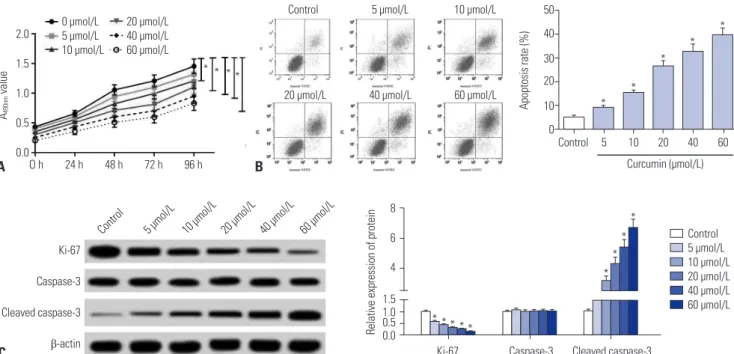
SU-DHL-8 cells were treated with different concentrations (0, 5, 10, 20, 40, and 60 μmol/L) of curcumin, followed by the de- tection of cell proliferation by MTT assay. These data revealed that curcumin suppressed the proliferation of SU-DHL-8 cells in a dose-dependent manner (Fig. 1A). Then, flow cytometric analysis was performed to detect the apoptosis rate of cur- cumin-treated (0, 5, 10, 20, 40, and 60 μmol/L) SU-DHL-8 cells. Results presented that curcumin treatment led to a signifi- cant promotion of cell apoptosis in a dose-dependent manner (Fig. 1B). Moreover, western blot results showed that curcum- in treatment in SU-DHL-8 cells resulted in a decrease of pro- liferation marker Ki-67 expression and an increase of cleaved caspase-3 level (Fig. 1C).
Curcumin inhibited migration and invasion of SU-DHL-8 cells
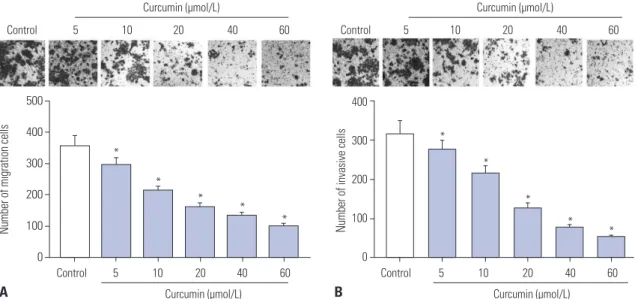
Next, the migration and invasion abilities of curcumin-treated (0, 5, 10, 20, 40, and 60 μmol/L) SU-DHL-8 cells were evaluat- ed by transwell assay. These results indicated that, compared to negative control, curcumin treatment resulted in decreased migration and invasion abilities of SU-DHL-8 cells in a dose- dependent manner (Fig. 2).
Curcumin inhibited miR-21 expression of SU-DHL-8 cells
Previous studies have reported that curcumin downregulates miR-21 expression to repress tumor progression in human cancers.10,23 Therefore, we further evaluated whether curcum- in regulated miR-21 expression in SU-DHL-8 cells. Firstly, the expression levels of miR-21 in lymphoma tissues were evalu- ated by qRT-PCR and ISH assays. Results revealed that com- pared with lymphoid hyperplasia tissues, miR-21 was highly elevated in lymphoma tissues (Fig. 3A and B). Also, miR-21 expression was significantly upregulated in lymphoma cell lines compared to normal control (Fig. 3C). Then, miR-21 ex- pression was examined in SU-DHL-8 cells after different con- centrations (0, 5, 10, 20, 40, and 60 μmol/L) of curcumin treat- ment. As expected, curcumin dose-dependently inhibited miR-21 expression in SU-DHL-8 cells (Fig. 3D).
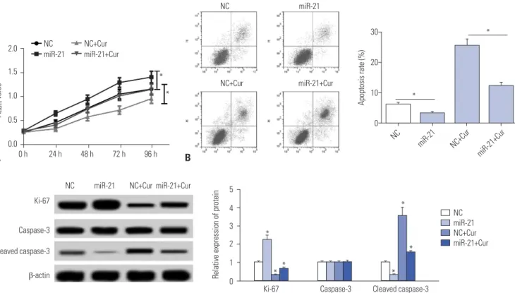
Curcumin exerted its anti-proliferation and pro-apoptosis effects by miR-21 in SU-DHL-8 cells To further investigate the effect of miR-21 on the proliferation and apoptosis of SU-DHL-8 cells, gain-of-function experi- ments were performed by transfecting with miR-21 mimics into SU-DHL-8 cells. Subsequently, MTT assay showed that miR-21 mimics transfection resulted in a marked promotion of cell proliferation compared to negative control (Fig. 4A). More- over, flow cytometric analysis revealed that the apoptosis of SU-DHL-8 cells was strikingly repressed in the presence of miR-21 mimics (Fig. 4B). Moreover, transfection of miR-21 mim-
50 40 30 20 10 0 2.0
1.5 1.0 0.5
0.0 Control 5 10 20 40 60
Control 5 µmol/L 10 µmol/L
20 µmol/L 40 µmol/L 60 µmol/L
Curcumin (µmol/L) O h 24 h 48 h 72 h 96 h
Apoptosis rate (%)
A490nm value
A B
0 µmol/L 20 µmol/L 5 µmol/L 40 µmol/L 10 µmol/L 60 µmol/L
8 6 4 1.5 1.00.5 Relative expression of protein 0.0 C
10 µmol/L 5 µmol/L
Control 20 µmol/L 40 µmol/L 60 µmol/L
Ki-67 Caspase-3 Cleaved caspase-3
β-actin Ki-67 Caspase-3 Cleaved caspase-3
Control 5 µmol/L 10 µmol/L 20 µmol/L 40 µmol/L 60 µmol/L
*
*
*
*
*
*
*
*
*
* * * * *
* * * *
Fig. 1. Curcumin inhibited proliferation and promoted apoptosis of SU-DHL-8 cells. SU-DHL-8 cells were treated with different concentrations (0, 5, 10, 20, 40, and 60 μmol/L) of curcumin. (A) At 0, 24, 48, 72, and 96 h treatment, cell proliferation was detected by MTT assay. (B) After 24 h treatment, cell apoptosis rate was measured by flow cytometric analysis. (C) Western blot was performed to determine the levels of Ki-67, caspase-3, and cleaved caspase-3 in curcumin-treated cells. *p<0.05 vs. control.
ics, but not NC control, significantly increased Ki-67 expres- sion and decreased cleaved caspase-3 level in SU-DHL-8 cells (Fig. 4C).
Next, to provide further mechanistic insight into the link
between curcumin and miR-21 on the proliferation and apop- tosis of SU-DHL-8 cells, SU-DHL-8 cells were transfected with NC mimics or miR-21 mimics, and then treated with curcum- in (20 μmol/L). Results demonstrated that, in comparison to
500 400 300 200 100 0
Control 5 10 20 40 60 Curcumin (µmol/L)
Number of migration cells
*
*
* *
*
400
300
200
100
0
Control 5 10 20 40 60 Curcumin (µmol/L)
Number of invasive cells
B
B
D A
A
C
*
*
*
* *
Control 5 10 20 40 60 Control 5 10 20 40 60
Curcumin (µmol/L) Curcumin (µmol/L)
Fig. 2. Curcumin repressed the migration and invasion of SU-DHL-8 cells. SU-DHL-8 cells were treated with different concentrations (0, 5, 10, 20, 40, and 60 μmol/L) of curcumin for 24 h, followed by the measurement of cell migration (A) and invasion (B) abilities by transwell assay (×100 magnification).
*p<0.05 vs. control.
6 5 4 3 2 1 0
Normal Lymphoma
Relative expression of miR-21
*
Normal Lymphoma
1.5
1.0
0.5
0.0 6
4
2
0
CD19+
SU-DHL-8 OCI-L
Y1
SU-DHL-10
Relative expression of miR-21
Relative expression of miR-21
*
*
*
Control 5 10 20 40 60 Curcumin (µmol/L)
*
* *
* *
Fig. 3. Curcumin inhibited miR-21 expression of SU-DHL-8 cells. (A) MiR-21 expression was detected in 45 cases of DLBCL tissues and 23 cases of re- active lymphoid hyperplasia tissues. (B) Representative ISH of miR-21 in DLBCL tissues and reactive lymphoid hyperplasia tissues (×100 magnification).
(C) MiR-21 expression was assessed in DLBCL cell lines (SU-DHL-8, OCI-LY1 and SU-DHL-10) and normal control CD19+ cells by qRT-PCR. (D) SU-DHL-8 cells were treated with different concentrations (0, 5, 10, 20, 40, and 60 μmol/L) of curcumin for 24 h, followed by the determination of miR-21 expression.
*p<0.05 vs. corresponding control. DLBCL, diffuse large B-cell lymphoma; ISH, in situ hybridization.
NC mimics, the effects of curcumin-treated anti-proliferation and pro-apoptosis on SU-DHL-8 cells were antagonized by miR-21 mimics transfection (Fig. 4).
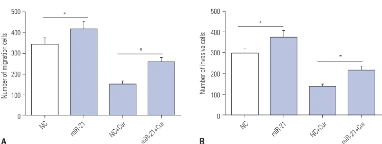
Curcumin-treated anti-migration and anti-invasion effects was mediated by miR-21 in SU-DHL-8 cells Given the data on cell proliferation and apoptosis, we explored whether curcumin exerting its regulatory effect on cell migra- tion and invasion was mediated by miR-21. Hence, SU-DHL-8 cells were transfected with NC mimics or miR-21 mimics, and then treated with or without curcumin (20 μmol/L). Transwell assays revealed that the numbers of migration and invasion cells were more in miR-21 mimics transfection group than those in control group (Fig. 5). Moreover, compared to NC mimics control, miR-21 mimics transfection in SU-DHL-8 cells remarkably abated the effects of curcumin-treated anti- migration and anti-invasion (Fig. 5).
VHL was a direct target of miR-21
Then, online software TargetScan (http://www.targetscan.
org/) was used to predict the targets of miR-21. The data re- vealed that 3'-UTR of VHL mRNA harbored a putative binding site for miR-21 (Fig. 6A). To verify whether VHL was a target of miR-21, dual-luciferase reporter assay was performed by co- transfecting with VHL-WT or VHL-MUT constructs into SU- DHL-8 cells and miR-21 mimics or anti-miR-21. Results pre-
sented that the relative luciferase activity of VHL-WT was highly reduced by miR-21 mimics introduction, while it was strikingly increased when transfected with anti-miR-21 (Fig.
6B). However, the relative luciferase activity of VHL-MUT was almost at the same level, and it failed to respond to miR-21 ex- pression alteration (Fig. 6B). In addition, RIP assay was per- formed to confirm the endogenous interaction between miR- 21 and VHL in SU-DHL-8 cells. The data indicated that miR-21 mimics transfection resulted in a substantial enrichment of VHL mRNA with anti-Ago in SU-DHL-8 cells (Fig. 6C).
In addition, we observed whether miR-21 regulated VHL ex- pression in SU-DHL-8 cells. qRT-PCR results revealed that VHL mRNA expression was significantly decreased by miR-21 mimics introduction, while it was strikingly increased follow- ing anti-miR-21 transfection (Fig. 6D). Consistently, western blot analysis demonstrated that VHL protein expression was inversely correlated with miR-21 level (Fig. 6E). Additionally, we assessed the expression of VHL protein in lymphoma tis- sues and cell lines. These results showed that, in comparison to its counterpart, VHL protein was highly downregulated in lym- phoma tissues and cell lines (Fig. 6F and G).
Curcumin exerted its regulatory effects on
the proliferation, migration, invasion, and apoptosis by VHL in SU-DHL-8 cells
Lastly, we evaluated the expression levels of VHL mRNA in 2.0
1.5
1.0 0.5 0.0
0 h 24 h 48 h 72 h 96 h A490nm value
A
NC miR-21
NC+Cur miR-21+Cur
30
20
10
0
Apoptosis rate (%)
NC miR-21
miR-21+Cur NC+Cur
*
*
NC NC+Cur
miR-21 miR-21+Cur
*
*
5 4 3 2 1 Relative expression of protein 0 C
NC miR-21 NC+Cur miR-21+Cur Ki-67
Caspase-3 Cleaved caspase-3 β-actin
Ki-67 Caspase-3 Cleaved caspase-3
*
*
*
*
* *
NC miR-21 NC+Cur miR-21+Cur B
Fig. 4. The effects of curcumin on proliferation and apoptosis were mediated by miR-21 in SU-DHL-8 cells. SU-DHL-8 cells were transfected with NC mimics or miR-21 mimics, and then were treated with or without curcumin (20 μmol/L). (A) At 0, 24, 48, 72, and 96 h treatment, MTT assay was performed to detect cell proliferation ability. (B) At 24 h treatment, flow cytometric analysis was used to assess cell apoptosis. (C) Western blot was performed to determine the levels of Ki-67, caspase-3, and cleaved caspase-3 in treated cells. *p<0.05 vs. NC mimics or NC mimics+curcumin.
500
400 300
200
100
0 500
400
300
200
100
0
Number of invasive cells
B
Number of migration cells
A
NC
NC miR-21 miR-21
miR-21+Cur
miR-21+Cur NC+Cur
NC+Cur
*
*
*
*
Fig. 5. Curcumin exerted its anti-migration and anti-invasion effects by miR-21 in SU-DHL-8 cells. SU-DHL-8 cells were transfected with NC mimics or miR-21 mimics, and then were treated with or without curcumin (20 μmol/L) for 24 h, followed by the determination of cell migration ability (A) and inva- sion capacity (B) by transwell assay. *p<0.05 vs. NC mimics or NC mimics+curcumin.
4 3 2 1 0
8 6 4 2 0
3
2
1
VHL-WT VHL-MUT 0
Relative luciferase activity Enrichment relative to input (%) Relative expression of VHL mRNA
B A
C D
1.5
1.0
0.5
0.0
1.5
1.0
0.5
0.0 3
2
1
0 Normal Lymphoma
Relative expression of VHL protein
Relative expression of VHL protein Relative expression of VHL protein
G E F
*
* NC
anti-NC miR-21 anti-miR-21
VHL VHL
RIP-Ago2
RIP-IgG NC miR-21
NC miR-21
anti-miR-21 anti-NC
*
*
*
VHL β-actin
VHL β-actin
VHL β-actin
NC miR-21
anti-miR-21
anti-NC CD19+
SU-DHL-8 OCI-L Y1
SU-DHL-10
*
*
*
*
*
*
Fig. 6. VHL was a direct target of miR-21. (A) Schematic illustration of the putative binding sites between miR-21 and VHL, and mutation in binding se- quence in 3’-UTR of VHL. (B) VHL wild-type or mutant-type reporter plasmids (VHL-WT or VHL-MUT) were constructed and transfected into SU-DHL-8 cells together with NC mimics, miR-21 mimics, anti-NC, or anti-miR-21. Relative luciferase activities were detected. (C) SU-DHL-8 cells were transfected with miR-21 mimics, followed by the detection of VHL mRNA enrichment with anti-Ago2 or anti-IgG. SU-DHL-8 cells were transfected with NC mimics, miR-21 mimics, anti-NC, or anti-miR-21, followed by the measurement of VHL mRNA by qRT-PCR assay (D) and VHL protein by western blot (E). The ex- pression of VHL protein was detected by western blot in DLBCL tissues and reactive lymphoid hyperplasia tissues (F), and in DLBCL cell lines (SU- DHL-8, OCI-LY1, and SU-DHL-10) and normal control CD19+ cells (G). *p<0.05 vs. NC mimics or anti-NC. VHL, Von Hippel-Lindau; DLBCL, diffuse large B- cell lymphoma.
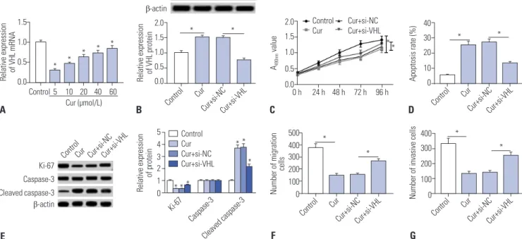
curcumin-treated SU-DHL-8 cells. As shown in Fig. 7A, cur- cumin treatment in SU-DHL-8 cells led to a significant promo- tion of VHL mRNA expression in a dose-dependent manner.
To further investigate the molecular mechanism of curcumin on the progression of SU-DHL-8 cells, siRNA targeting VHL (si- VHL) were transfected into SU-DHL-8 cells prior to curcumin treatment (20 μmol/L). Western blot data presented that, com- pared to negative control, curcumin-treated promotion effect on VHL expression was strikingly reversed by si-VHL transfec- tion (Fig. 7B). Subsequently, functional experiments results demonstrated that curcumin-treated anti-proliferation, anti- migration, anti-invasion, and pro-apoptosis effects on SU- DHL-8 cells were highly antagonized by VHL expression res- toration (Fig. 7C–G).
DISCUSSION
Accumulating evidence have suggested that curcumin pos- sesses anti-cancer properties in a series of human cancers, in- cluding head and neck squamous cell carcinoma,24 non-small cell lung cancer,25 and advanced pancreatic cancer.26 Moreover, curcumin was found to function as a potential chemopreven- tive agents in tumorigenesis.27 Curcumin was also demonstrat- ed to target cancer stem cells and specific miRNAs, and thus served as a promising adjunct to traditional cancer treatments.28 A previous document manifested that the combination of cur-
cumin with epigallocatechin-3 gallate led to a synergistic anti- cancer activity by repressing angiogenesis, metastasis, and entering in complete remission in B cell lymphoma.29 In the present study, we firstly found that curcumin treatment resulted in a inhibition of the proliferation and a suppression of migra- tion and invasion abilities, as well as a promotion of apoptosis in SU-DHL-8 cells. All these results suggested that curcumin might play anti-cancer roles in the development and progres- sion of DLBCL.
Dysregulation of miRNAs has been widely acknowledged to implicate in cancer development and progression.14 More- over, curcumin was verified to exert its anti-cancer effects partly by targeting special miRNAs in human cancers.28 For example, curcumin repressed bisphenol A-induced proliferation of MCF- 7 cells through inhibition of miR-19 expression.30 Curcumin targets miR-21 expression to repress invasion and metastasis of colorectal cancer.10 Conversely, curcumin facilitated tumor- suppressor miR-203 expression, resulting in decreased prolif- eration and increased apoptosis of bladder cancer cells.31 In our study, we validated that curcumin treatment led to a reduction of miR-21 expression in SU-DHL-8 cells, consistent with for- mer works.10,23,32
MiR-21 has been demonstrated as a cancer-associated tran- script of oncogenesis in DLBCL, providing a possibility of miR-21 as biomarkers of diagnose, therapy, and prognosis of DLBCL.20,33,34 In the study, our data supported a significant up- regulation of miR-21 in DLBCL tissues and cell lines, and miR-
Fig. 7. Curcumin exerted its regulatory effects by VHL in SU-DHL-8 cells. (A) SU-DHL-8 cells were treated with different concentrations (0, 5, 10, 20, 40, and 60 μmol/L) of curcumin for 24 h, followed by the measurement of VHL mRNA expression. SU-DHL-8 cells were transfected with si-VHL or si-NC and then were treated with 20 μmol/L of curcumin. (B) After 24 h treatment, VHL protein expression was determined by western blot analysis. (C) At 0, 24, 48, 72, and 96 h treatment, cell proliferation ability was assessed by MTT assay. (D) At 24 h treatment, cell apoptosis was detected by flow cytomet- ric analysis. (E) Western blot was performed to determine the levels of Ki-67, caspase-3, and cleaved caspase-3. At 24 h treatment, cell migration ability was determined by transwell assay (F) and cell invasion capacity was evaluated by transwell assay (G). *p<0.05 vs. control or si-NC+curcumin. VHL, Von Hippel-Lindau.
1.5 1.0 0.5 0.0
2.0 1.5 1.0 0.5 0.0
2.0 1.5 1.0 0.5 0.0
40 30 20 10 0
400 300 200 100 0 500
400 300 200 100 0 5
4 3 2 1 0
0 h 24 h 48 h 72 h 96 h
Relative expression of VHL mRNA Relative expression of VHL protein A490nm value Apoptosis rate (%)Number of invasive cells
Number of migration cells Relative expression of protein
A
E
B C D
G F
Control 5 10 20 40 60 Cur (µmol/L)
VHL β-actin
Control Control
Control Control
Ki-67 Caspase-3
Cleaved caspase-3 Control
Cur Cur
Cur Cur
Cur
Cur+si-NC Cur+si-NC
Cur+si-NC Cur+si-NC
Cur+si-NC
Cur+si-VHL Cur+si-VHL
Cur+si-VHL Cur+si-VHL
Cur+si-VHL
* * * * *
* *
Ki-67 Caspase-3 Cleaved caspase-3 β-actin
Control Cur+si-NC Cur Cur+si-VHL
* * * *
*
* *
* * *
* *
* *
Control Cur Cur+si-NC Cur+si-VHL
21 overexpression enhanced the proliferation, migration, and invasion, and inhibiting the apoptosis of SU-DHL-8 cells, in accordance with previous results.20,22,34 In addition, we validat- ed that curcumin exerted its anti-proliferation, anti-migration, anti-invasion, and pro-apoptosis effects by miR-21 in SU-DHL-8 cells. Similar with our findings, Mudduluru, et al.10 confirmed that curcumin repressed miR-21 expression, and thus inhibit- ed invasion and metastasis of colorectal cancer cells.
Then, online software TargetScan was employed to predict the targets of miR-21. Among these candidates, VHL was se- lected for further study due to its important tumor suppres- sive role in a variety of cancers, including sporadic clear cell renal cancer35 and triple-negative breast cancer.36 Moreover, VHL hypermethylation was reported to be associated with ag- gressive phenotype and worse prognosis in DLBCL.37 In our study, we found a drastic downregulation of VHL protein in DLBCL tissues and cell lines. We also confirmed that VHL was a direct target of miR-21 in SU-DHL-8 cells. Similar with our finding, previous documents demonstrated that miR-21 target- ed VHL in glioblastomas cells38 and head and neck squamous cell carcinomas cells.39 Lastly, our study was the first to verify that curcumin promoted VHL expression and curcumin exert- ed its anti-proliferation, anti-migration, anti-invasion, and pro- apoptosis effects by VHL in SU-DHL-8 cells.
Additionally, earlier document revealed that curcumin (50 µmol/L) inhibited the proliferation of rat thymocytes.40 In the present study, we demonstrated that curcumin repressed the proliferation of SU-DHL-8 cells at a concentration of 40–60 µmol/L. We speculated that higher concentrations of curcum- in might have the capacity for inhibition of DLBCL and normal cells proliferation. Therefore, further studies about the effect of 20 µmol/L of curcumin on normal cells proliferation are need- ed. In conclusion, our study indicated that curcumin exerted its anti-proliferation, anti-migration, anti-invasion, and pro-apop- tosis effects, at least partly, through miR-21/VHL axis. Our find- ings provided a possible molecular mechanism of curcumin- mediated anti-cancer effect.
AUTHOR CONTRIBUTIONS
Conceptualization: Ling Chen, Cheng-Zhi Zhan. Data curation: Ling Chen, Cheng-Zhi Zhan. Formal analysis: Ling Chen, Cheng-Zhi Zhan. Funding acquisition: Ling Chen, Cheng-Zhi Zhan. Investiga- tion: Rui Yao, Tao Wang. Methodology: Ling Chen, Cheng-Zhi Zhan.
Project administration: Tao Wang. Resources: Ling Chen, Tao Wang.
Software: Hua You, Rui Yao. Supervision: Hua You. Validation: Ling Chen, Tao Wang. Visualization: Cheng-Zhi Zhan, Ling Chen. Writ- ing—original draft: Ling Chen, Cheng-Zhi Zhan. Writing—review &
editing: Ling Chen, Cheng-Zhi Zhan.
ORCID iDs
Ling Chen https://orcid.org/0000-0002-5010-3943 Cheng-Zhi Zhan https://orcid.org/0000-0002-3017-4920
Tao Wang https://orcid.org/0000-0002-5449-9447 Hua You https://orcid.org/0000-0003-0865-3295 Rui Yao https://orcid.org/0000-0002-3137-860X
REFERENCES
1. Martelli M, Ferreri AJ, Agostinelli C, Di Rocco A, Pfreundschuh M, Pileri SA. Diffuse large B-cell lymphoma. Crit Rev Oncol Hematol 2013;87:146-71.
2. Shaffer AL 3rd, Young RM, Staudt LM. Pathogenesis of human B cell lymphomas. Annu Rev Immunol 2012;30:565-610.
3. Tilly H, Gomes da Silva M, Vitolo U, Jack A, Meignan M, Lopez- Guillermo A, et al. Diffuse large B-cell lymphoma (DLBCL): ESMO Clinical Practice Guidelines for diagnosis, treatment and follow- up. Ann Oncol 2015;26 Suppl 5:v116-25.
4. Nabavi SF, Thiagarajan R, Rastrelli L, Daglia M, Sobarzo-Sánchez E, Alinezhad H, et al. Curcumin: a natural product for diabetes and its complications. Curr Top Med Chem 2015;15:2445-55.
5. Liu CH, Huang HY. Antimicrobial activity of curcumin-loaded myristic acid microemulsions against Staphylococcus epidermi- dis. Chem Pharm Bull (Tokyo) 2012;60:1118-24.
6. Meng B, Li J, Cao H. Antioxidant and antiinflammatory activities of curcumin on diabetes mellitus and its complications. Curr Pharm Des 2013;19:2101-13.
7. Lüer S, Troller R, Aebi C. Antibacterial and antiinflammatory ki- netics of curcumin as a potential antimucositis agent in cancer patients. Nutr Cancer 2012;64:975-81.
8. Hashish EA, Elgaml SA. Hepatoprotective and nephroprotective effect of curcumin against copper toxicity in rats. Indian J Clin Biochem 2016;31:270-7.
9. Lin J, Chen A. Curcumin diminishes the impacts of hyperglycemia on the activation of hepatic stellate cells by suppressing mem- brane translocation and gene expression of glucose transporter-2.
Mol Cell Endocrinol 2011;333:160-71.
10. Mudduluru G, George-William JN, Muppala S, Asangani IA, Ku- marswamy R, Nelson LD, et al. Curcumin regulates miR-21 ex- pression and inhibits invasion and metastasis in colorectal can- cer. Biosci Rep 2011;31:185-97.
11. Toden S, Okugawa Y, Jascur T, Wodarz D, Komarova NL, Buhrmann C, et al. Curcumin mediates chemosensitization to 5-fluorouracil through miRNA-induced suppression of epithelial-to-mesenchy- mal transition in chemoresistant colorectal cancer. Carcinogenesis 2015;36:355-67.
12. Pandey A, Vishnoi K, Mahata S, Tripathi SC, Misra SP, Misra V, et al. Berberine and curcumin target survivin and STAT3 in gastric cancer cells and synergize actions of standard chemotherapeutic 5-fluorouracil. Nutr Cancer 2015;67:1293-304.
13. Hammond SM. An overview of microRNAs. Adv Drug Deliv Rev 2015;87:3-14.
14. Reddy KB. MicroRNA (miRNA) in cancer. Cancer Cell Int 2015;
15:38.
15. Hayes J, Peruzzi PP, Lawler S. MicroRNAs in cancer: biomarkers, functions and therapy. Trends Mol Med 2014;20:460-9.
16. Simonian M, Mosallayi M, Mirzaei H. Circulating miR-21 as novel biomarker in gastric cancer: diagnostic and prognostic biomark- er. J Cancer Res Ther 2018;14:475.
17. Frankel LB, Christoffersen NR, Jacobsen A, Lindow M, Krogh A, Lund AH. Programmed cell death 4 (PDCD4) is an important functional target of the microRNA miR-21 in breast cancer cells. J Biol Chem 2008;283:1026-33.
18. Chan JK, Blansit K, Kiet T, Sherman A, Wong G, Earle C, et al. The inhibition of miR-21 promotes apoptosis and chemosensitivity in
ovarian cancer. Gynecol Oncol 2014;132:739-44.
19. Cui Q, Vari F, Cristino AS, Salomon C, Rice GE, Sabdia MB, et al.
Circulating cell-free miR-494 and miR-21 are disease response bio- markers associated with interim-positron emission tomography response in patients with diffuse large B-cell lymphoma. Oncotar- get 2018;9:34644-57.
20. Li J, Fu R, Yang L, Tu W. miR-21 expression predicts prognosis in diffuse large B-cell lymphoma. Int J Clin Exp Pathol 2015;8:15019- 24.
21. Go H, Jang JY, Kim PJ, Kim YG, Nam SJ, Paik JH, et al. MicroR- NA-21 plays an oncogenic role by targeting FOXO1 and activating the PI3K/AKT pathway in diffuse large B-cell lymphoma. Onco- target 2015;6:15035-49.
22. Fu R, Li C, Shao Z. Aberrant overexpression and regulatory mech- anism of mir-21 in diffuse large B cell lymphoma. Am Soc Hema- tology 2014;124:5171.
23. Ali S, Ahmad A, Banerjee S, Padhye S, Dominiak K, Schaffert JM, et al. Gemcitabine sensitivity can be induced in pancreatic cancer cells through modulation of miR-200 and miR-21 expression by curcumin or its analogue CDF. Cancer Res 2010;70:3606-17.
24. Wilken R, Veena MS, Wang MB, Srivatsan ES. Curcumin: a review of anti-cancer properties and therapeutic activity in head and neck squamous cell carcinoma. Mol Cancer 2011;10:12.
25. Ye M, Zhang J, Zhang J, Miao Q, Yao L, Zhang J. Curcumin pro- motes apoptosis by activating the p53-miR-192-5p/215-XIAP path- way in non-small cell lung cancer. Cancer Lett 2015;357:196-205.
26. Epelbaum R, Schaffer M, Vizel B, Badmaev V, Bar-Sela G. Curcum- in and gemcitabine in patients with advanced pancreatic cancer.
Nutr Cancer 2010;62:1137-41.
27. Bansal SS, Goel M, Aqil F, Vadhanam MV, Gupta RC. Advanced drug delivery systems of curcumin for cancer chemoprevention.
Cancer Prev Res (Phila) 2011;4:1158-71.
28. Li Y, Zhang T. Targeting cancer stem cells by curcumin and clinical applications. Cancer Lett 2014;346:197-205.
29. Bassiouny AR, Atteya MA, El-Rashidy FAH, Neenaa HM. Curcum- in and EGCG suppress apurinic/apyrimidinic endonuclease 1 and induce complete remission in B-cell non-Hodgkin’s lymphoma patients. Funct Foods Health Dis 2011;1:525-44.
30. Pancione M, Giordano G, Remo A, Febbraro A, Sabatino L, Man- frin E, et al. Immune escape mechanisms in colorectal cancer
pathogenesis and liver metastasis. J Immunol Res 2014;2014:
686879.
31. Saini S, Arora S, Majid S, Shahryari V, Chen Y, Deng G, et al. Cur- cumin modulates microRNA-203-mediated regulation of the Src- Akt axis in bladder cancer. Cancer Prev Res (Phila) 2011;4:1698- 709.
32. Taverna S, Giallombardo M, Pucci M, Flugy A, Manno M, Raccos- ta S, et al. Curcumin inhibits in vitro and in vivo chronic myeloge- nous leukemia cells growth: a possible role for exosomal disposal of miR-21. Oncotarget 2015;6:21918-33.
33. Chen W, Wang H, Chen H, Liu S, Lu H, Kong D, et al. Clinical sig- nificance and detection of microRNA-21 in serum of patients with diffuse large B-cell lymphoma in Chinese population. Eur J Hae- matol 2014;92:407-12.
34. Liu K, Du J, Ruan L. MicroRNA-21 regulates the viability and apop- tosis of diffuse large B-cell lymphoma cells by upregulating B cell lymphoma-2. Exp Ther Med 2017;14:4489-96.
35. Moore LE, Nickerson ML, Brennan P, Toro JR, Jaeger E, Rinsky J, et al. Von Hippel-Lindau (VHL) inactivation in sporadic clear cell renal cancer: associations with germline VHL polymorphisms and etiologic risk factors. PLoS Genet 2011;7:e1002312.
36. Kong W, He L, Richards EJ, Challa S, Xu CX, Permuth-Wey J, et al.
Upregulation of miRNA-155 promotes tumour angiogenesis by tar- geting VHL and is associated with poor prognosis and triple-nega- tive breast cancer. Oncogene 2014;33:679-89.
37. Amara K, Trimeche M, Ziadi S, Laatiri A, Hachana M, Korbi S.
Prognostic significance of aberrant promoter hypermethylation of CpG islands in patients with diffuse large B-cell lymphomas. Ann Oncol 2008;19:1774-86.
38. Zhang KL, Han L, Chen LY, Shi ZD, Yang M, Ren Y, et al. Blockage of a miR-21/EGFR regulatory feedback loop augments anti-EGFR therapy in glioblastomas. Cancer Lett 2014;342:139-49.
39. Wang Y, Wang S, Wu Y, Ren Y, Li Z, Yao X, et al. Suppression of the growth and invasion of human head and neck squamous cell car- cinomas via regulating STAT3 signaling and the miR-21/β-catenin axis with HJC0152. Mol Cancer Ther 2017;16:578-90.
40. Sikora E, Bielak-Zmijewska A, Piwocka K, Skierski J, Radziszewska E. Inhibition of proliferation and apoptosis of human and rat T lymphocytes by curcumin, a curry pigment. Biochem Pharmacol 1997;54:899-907.