The principle of laser refractive surgery is modification of the refractive power of the cornea by means of photoablation of the stromal tissue. The biomechanical strength of the cornea is compromised by surgical tissue subtraction and the loss of Bowman's membrane integrity after laser refractive surgery.
1Forward shift of posterior corneal surface resulting from anterior bulging of the cornea after photorefractive keratectomy (PRK) and laser in situ keratomileusis (LASIK) have bee reported.
2-6Several cases of iatrogenic keratectasia after LASIK and laser-assisted subepithelial keratectomy (LASEK) surgery have been reported.
7-10In addition, forward shift of the cornea can cause myopic regression after laser
refractive surgery.
4-7Especially iatrogenic keratectasia has come to rise as an important complication of refractive surgery, and it is thought to be correlated with preoperative corneal thickness, the amount of laser ablation, preoperative refraction and preoperative intraocular pressure.
4Anterior corneal surface is directly influenced by refractive surgery itself and healing process, so it is difficult to estimate the amount of corneal forward shift by analyzing the elevation map of the anterior surface. So, the posterior surface which is not directly affected by surgical process is usually used to evaluate the anteroposterior movement of the cornea. And the height elevation data relative to best-fit sphere has been measured by scanning-slit corneal topography.
11And changes of elevation should be evaluated at the center of the difference map due to inconsistency of preoperative and postoperative best-fit spheres.
2,3On the other hand, 'standard' laser refractive surgery corrects only spherical and regular astigmatism, and may
Aberrations after Refractive Surgery in Moderate Myopia
Min Joung Lee, MD,
1,2Sang Mok Lee, MD,
1,2Hyun Ju Lee, MD,
1,2Won Ryang Wee, MD,
1,2Jin Hak Lee, MD,
1,2,3Mee Kum Kim, MD,
1,2Department of Ophthalmology, Seoul National University College of Medicine
1, Seoul, Korea Seoul Artificial Eye Center, Seoul National University Hospital Clinical Research Institute
2, Seoul, Korea
Department of Ophthalmology, Seoul National University Bundang Hospital
3, Seongnam, Korea
Purpose: To compare forward shift of posterior corneal surface and higher-order aberration (HOA) changes after LASIK, LASEK, and wavefront-guided LASEK surgery in moderate myopia
Methods: One hundred eighty four eyes undergoing LASIK, LASEK and wavefront-guided LASEK with VISX STAR S4 were included in this study. The posterior corneal elevation was measured with Orbscan before, 2 and 4 months after surgery. Changes of the elevation were assessed using the difference map generated from preoperative and postoperative elevation maps. The values of higher-order aberrations were evaluated preoperatively and 2 months postoperatively with Wavefront aberrometer.
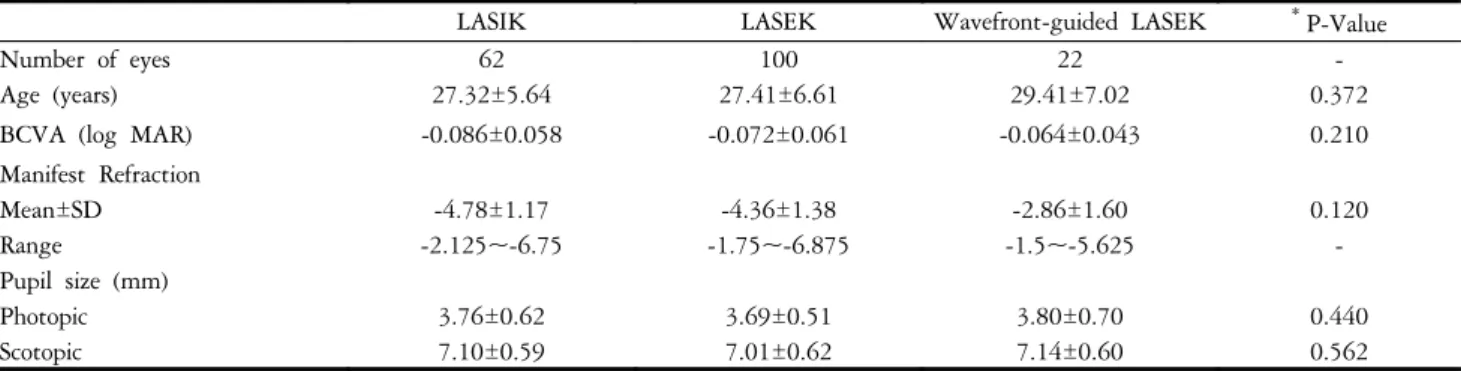
Results: The posterior corneal surface displayed forward shift of 27.2±11.45 µm, 24.3±9.76 µm in LASIK group, 23.4±10.5 µm, 23.6±10.55 µm in LASEK group, 24.0±14.95 µm, 28.4±14.72 µm in wavefront- guided LASEK group at 2 months and 4 months, respectively. There were no statistically significant differences among those three groups, and between 2 and 4 months. The root mean score (RMS) of HOA was increased after LASIK and LASEK (p=0.000, p=0.000, respectively). The mean change of HOA-RMS was significantly smaller in wavefront-guided LASEK than LASIK or LASEK (p=0.000, p=0.000, respectively, Bonferroni-corrected).
Conclusions: The changes of posterior corneal surface forward shift showed no difference among LASIK, LASEK and wavefront-guided LASEK in moderate myopia. HOAs were significantly increased after LASIK and LASEK. The changes of HOAs were significant smaller in wavefront-guided LASEK than LASIK or LASEK.
Korean Journal of Ophthalmology 21(3):131-136, 2007
Key Words: Aberration, LASEK, LASIK, Posterior corneal surface, Wavefront-guided ablation
Received: December 6, 2006 Accepted: June 20, 2007
Reprint requests to Mee Kum Kim, M.D. Department of Ophthal-
mology, Seoul National University College of Medicine, 28
Yongon-dong, Chongno-gu, Seoul 110-744, Korea. Tel: 82-2-2072-
2438, Fax: 82-2-741-3187, E-mail: [email protected]
induce higher-order aberrations that will lead to distorted images.
12-15Wavefront-guided laser refractive surgery considers all the aberration including higher-order aberrations. A correlation between visual symptoms and ocular aberration was strongly suggested, for example in monocular diplopia and glare.
16And the greatest gains in correcting higher-order aberration are noted in improved contrast sensitivity particularly under low light condition.
17In this study, we compared the changes of posterior elevation of the cornea from difference map, the higher-order aberrations (RMS-root mean square, coma, trefoil, spherical aberration) among LASIK, LASEK, wavefront-guided LASEK group.
Materials and Methods
Sixty two eyes (40 patients) undergoing LASIK surgery, 100 eyes LASEK surgery (56 patients), 22 eyes (13 patients) undergoing wavefront-guided LASEK surgery were enrolled.
Mean age was 27.95±5.99 years. The preoperative mean spherical equivalent (SE) refractions were -4.78±1.17 diopter (D) (range -2.125 to -6.75 D) in LASIK group, -4.36
±1.38 D (range -1.75 to -6.875 D) in LASEK group, -2.86±
1.60 D (range -1.5 to -5.625 D) in wavefront-guided LASEK group. The preoperative characteristics of the patients are shown in Table 1. The preoperative refractions were not significantly different among three groups (p=0.120, oneway ANOVA). The mean baseline pupil sizes in photopic and scotopic conditions were not statistically significant among three groups (p=0.440, p=0.562, respectively, oneway ANOVA). Patients were enrolled from those having surgery between October 2002 and July 2004, at the Department of Ophthalmology, Seoul National University Hospital, by one surgeon (Kim MK).
The following examinations were performed preoperatively in all patients: uncorrected visual acuity (UCVA), best- spectacle-corrected visual acuity (BCVA), manifest refraction in a dark room and cycloplegic refraction after instillation of cyclopentolate hydrochloride by operator, objective refractions with the automatic refract-keratometry (KR-7100, Topcon,
Japan), intraocular pressure (NCT CT-60, Topcon, Japan), slitlamp examination of the anterior segment, corneal thickness with ultrasonic pachymetry (Pocket, Quantel Medical, France), corneal topography (Orbscan II
Ⓡ, Bausch
& Lomb, U.S.A).
Wavefront analysis (VISX, Santa Clara, CA, U.S.A) was performed preoperatively in all patients to evaluate HOA. All HOAs were measured in the natural scotopic condition after 10-minute dark adaptation. Patients who had higher RMS of high-order aberration more than 2.00 were recommended to receive the wavefront-guided LASEK. Written informed consent was obtained from all patients.
All surgeries were performed using VISX S4 excimer laser system (VISX Inc., Santa Clara, CA). In LASIK surgery, the automated microkeratome (M2, Moria, France) was used to create a hinged corneal flap, 110 µm thickness. In LASEK surgery, an alcohol solution cone was placed on the corneal surface. Twenty percent of the alcohol solution was instilled inside the cone, left for about 20 seconds, and washed with a balanced salt solution. The cornea stroma was ablased using excimer laser (VISX 4, USA), and then the flap was repositioned in both LASIK and LASEK surgery.
The posterior corneal elevations were measured with the corneal topography before, 2 months, and 4 months after surgery. Changes in the elevation of the posterior corneal surface were evaluated at the center of the difference map generated from preoperative and postoperative elevation maps. High-order aberrations were analyzed using Wavefront aberrometer (VISX Inc. Santa Clara, CA, USA) preoperatively, and 2 months after surgery. All HOA values were obtained in the natural scotopic condition. RMS (Root mean square) value of higher-order aberrations, the 3
rdorder aberration - coma, trefoil, spherical aberration values were compared among LASIK, LASEK, and wavefront-guided LASEK group.
Comparison of continuous variables among three groups was analyzed using the oneway ANOVA test. Continuous variables of two groups were evaluated using independent t-test. Comparison of preoperative and postoperative data was done by dependent t-test. Clinical significance was accepted
Table 1. Preoperative independent variables
LASIK LASEK Wavefront-guided LASEK
*P-Value
Number of eyes 62 100 22 -
Age (years) 27.32±5.64 27.41±6.61 29.41±7.02 0.372
BCVA (log MAR) -0.086±0.058 -0.072±0.061 -0.064±0.043 0.210
Manifest Refraction
Mean±SD -4.78±1.17 -4.36±1.38 -2.86±1.60 0.120
Range -2.125~-6.75 -1.75~-6.875 -1.5~-5.625 -
Pupil size (mm)
Photopic 3.76±0.62 3.69±0.51 3.80±0.70 0.440
Scotopic 7.10±0.59 7.01±0.62 7.14±0.60 0.562
BCVA: best-corrected visual acuity, MAR: minimum angle of resolution.
*
Analyzed by oneway ANOVA test.
for P values of <0.05. All statistical analyses were performed using SPSS for Windows, Version 11.0.0 (SPSS Inc., Chicago, IL).
Results
The mean UCVA and manifest refraction changes at 2 months after surgery are shown in Table 2. There is no statistically significant difference of postoperative visual acuity and manifest refraction among three groups. The mean forward shift of the posterior corneal surface was 27.2±
11.45 µm in LASIK eyes (n=53), 23.4±10.5 µm in LASEK eyes (n=75), 24.0±14.9 µm in wavefront-guided LASEK eyes (n=18) at 2 month postoperatively. On the other hand, it was 24.3±9.8 µm in LASIK (n=34), 23.6±10.6 µm in LASEK (n=72), 28.4±14.7 µm in wavefront-guided LASEK (n=16) at 4 month postoperatively. There were no statistically significant difference of posterior elevation among three groups at 2 and 4 months postoperatively (p=0.231, p=0.624, respectively, oneway ANOVA)(Fig. 1).
Time course of changes was also analyzed. There was no statistically significant change of posterior elevation in all three groups (p=0.176, oneway repeated-measured ANOVA) (Table 3).
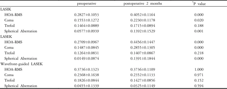
The preoperative and postoperative HOA-RMS, coma, trefoil, and spherical aberration values were shown in Table 4. Preoperatively, HOA-RMS, coma, trefoil were statistically no difference between LASIK and LASEK (p=0.542, p=0.736, p=0.228, respectively, independent t-test), but significantly higher in wavefront-guided LASEK than LASEK (p=0.000, p=0.001, p=0.006, respectively). In case of spherical aberration, there was no statistically significant difference between LASEK and wavefront-guided LASEK (p=0.203).
Postoperatively, there were significant increases of HOA-RMS, coma, spherical aberration in LASIK (p=0.000, p=0.020, p=0.001, respectively, paired t-test) and LASEK group (p=0.000, p=0.000, p=0.000). Trefoil showed no
significant increase in both two groups (p=0.188 in LASIK group, p=0.218 in LASEK group). In wavefront-guided LASEK, coma, spherical aberration and trefoil decreased postoperatively but there were no statistical significances (p=0.971, p=0.152, p=0.594, respectively). The mean changes of HOA-RMS, coma, spherical aberration from preoperative levels to the postoperative 2 month level showed significant difference among three groups (p=0.000, p=0.002, p=0.001, respectively, oneway-ANOVA test). The mean change of HOA-RMS was significantly smaller in wavefront-guided LASEK than LASIK or LASEK (p=0.000, p=0.000, respectively, Bonferroni-corrected). The mean changes of coma and spherical aberration were significantly smaller in wavefront-guided LASEK than LASEK (p=0.002, p=0.001, respectively, Bonferroni-corrected) (Table 5).
Table 3. Time course of change of the posterior elevation
LASIK (n=33) LASEK (n=66) Wavefront-guided LASEK (n=15)
postoperative 2 months (µm) 27.90±12.97 24.40±10.59 25.70±15.39
postoperative 4 months (µm) 24.70±9.52 24.80±11.77 27.70±14.95
*
P value 0.168 0.318 0.713
*
Analyzed by dependent t-test.
Table 2. Uncorrected visual acuity (UCVA) and Manifest refraction (MR) at 2 months after surgery
LASIK LASEK Wavefront-guided LASEK
*P-value
UCVA (log MAR) -0.064±0.112 -0.087±0.095 -0.074±0.085 0.390
MR (Spherical equivalent) 0.059±0.409 0.133±0.333 -0.138±0.363 0.120
MAR: minimum angle of resolution.
MR: manifest refraction.
*
Analyzed by oneway ANOVA test.
Fig. 1. The changes of posterior elevation measured using
difference map showed no statistically significant difference
between LASIK and LASEK, between LASEK and Wavefront-
guided LASEK at 2 and 4 month postoperatively (p>0.05).
Discussion
LASIK & LASEK surgery are all widely accepted useful refractive surgical procedure for the correction of myopia.
18,19LASIK has advantage of reduced postoperative discomfort, improved immediate acuity and less corneal haziness.
20But visual outcomes, contrast sensitivity after LASEK may appear to be superior to those after LASIK in moderate myopia.
21Steepening of the posterior corneal surface and increases in the posterior corneal curvature resulting from anterior bulging of the cornea after photorefractive keratectomy and LASIK have been reported.
4-6Steepening of the posterior surface indicates an increase in the negative power of that surface, so it may reinforce the effect of surgery and be a cause of overcorrection.
4Because the anterior and posterior surfaces face different media - anterior surface contacts air, and posterior surface contacts aqueous humor, the anterior surface exerts far greater refractive changes. So anteroposterior corneal shift results in counteraction of refractive surgery for myopia. Forward shift of the cornea can be a causative factor of myopic regression.
6In the case of iatrogenic keratectasia, the precise causes are not known, but
keratoconus that was not detected preoperatively, insufficient residual corneal thickness, subclinical keratoconus are presumed to be the causes. And it was thought that the residual corneal bed thickness was correlated with bulging of the posterior corneal surface.
9,10We used a difference map generated from the preoperative and postoperative elevation maps to evaluate posterior corneal surface. It was based on the scanning-slit corneal topography. The height data of the scanning-slit corneal topography are expressed as color-coded maps that are compared with best-fit sphere. Forward protrusion of the posterior surface induced steeper best-fit sphere, and there was a correlation between the amount of corneal forward shift and posterior best-fit sphere curvature, so forward protrusion of cornea can be masked if assessed on a single color-coded map of the postoperative elevation map alone.
In our study, all three groups showed some amount of anterior shift of the posterior corneal surface, and iatrogenic keratectasia was not noticed during follow-up period. In LASIK group, the posterior surface shifted anteriorly by 27.9
±12.9 µm at 2 month and 24.7±9.52 µm at 4 month. It is a compatible or favorable result compared with previous studies : Wang Z. et al
2reported 17.2±7.2 µm to 41.0±22.1 Table 4. Preoperative and postoperative HOAs after LASIK (n=40), LASEK (n=78), and Wavefront-guided LASEK surgery (n=22)
preoperative postoperative 2 months
*P value LASIK
HOA-RMS 0.2827±0.1053 0.4052±0.1164 0.000
Coma 0.1553±0.1272 0.2230±0.1178 0.020
Trefoil 0.1464±0.0889 0.1715±0.0894 0.188
Spherical Aberration 0.0577±0.0939 0.1392±0.1529 0.001
LASEK
HOA-RMS 0.2709±0.0967 0.4456±0.1447 0.000
Coma 0.1487±0.0845 0.2855±0.1305 0.000
Trefoil 0.1264±0.0831 0.1407±0.0867 0.218
Spherical Aberration 0.0149±0.0874 0.1391±0.1844 0.000
Wavefront-guided LASEK
HOA-RMS 0.3736±0.1323 0.3736±0.1109 1.000
Coma 0.2368±0.1638 0.2352±0.1133 0.971
Trefoil 0.1826±0.0844 0.1427±0.0856 0.152
Spherical Aberration 0.0455±0.1339 0.0325±0.1149 0.594
HOA-RMS: root mean score of higher-order aberration.
*
Analyzed by dependent t-test.
Table 5. The mean changes of HOAs after LASIK (n=40), LASEK (n=78), and Wavefront-guided LASEK surgery (n=22)
LASIK LASEK Wavefront-guided LASEK
*P-Value
HOA-RMS 0.12±0.16 0.18±0.16 0.00±0.17 0.000
Coma 0.07±0.18 0.14±0.15 -0.01±0.20 0.002
Trefoil 0.03±0.12 0.01±0.10 -0.04±0.13 0.076
Spherical aberration 0.08±0.15 0.13±0.17 -0.01±0.11 0.001
HOA-RMS: root mean score of higher-order aberration.
*