• 검색 결과가 없습니다.

Effects of contamination by either blood or a hemostatic agent on the shear bond strength of orthodontic buttons

N/A
N/A
Protected

Academic year: 2022

Share "Effects of contamination by either blood or a hemostatic agent on the shear bond strength of orthodontic buttons"

Copied!
5
0
0

로드 중.... (전체 텍스트 보기)

전체 글

(1)

Effects of contamination by either blood or

a hemostatic agent on the shear bond strength of orthodontic buttons

Objective: To evaluate the effects of contamination by either blood or a hemostatic agent on the shear bond strength (SBS) of orthodontic buttons.

Methods: We used 45 freshly extracted, non-carious, impacted third molars that were divided into 3 groups of 15. Each tooth was etched with 37% phosphoric acid gel for 30 s. Human blood or the blood stopper agent was applied to the tooth surface in groups I and II, respectively. Group III teeth were untreated (controls). Orthodontic buttons were bonded to the teeth using light-curing composite resin. After bonding, the SBS of the button was determined using a Universal testing machine. Any adhesive remaining after debonding was assessed and scored according to the modified adhesive remnant index (ARI). ANOVA with post-hoc Tukey’s test was used to determine significant differences in SBS and Fisher’s exact test, to determine significant differences in ARI scores among groups. Results: ANOVA indicated a significant difference between groups (p <

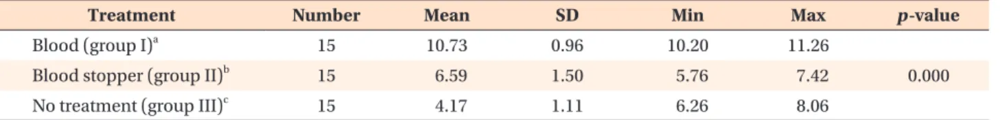
0.001). The highest SBS values were measured in group III (10.73 ± 0.96 MPa).

The SBS values for teeth in groups I and II were significantly lower than that of group III (p < 0.001). The lowest SBS values were observed in group I teeth (4.17

± 1.11 MPa) (p < 0.001). Conclusions: Contamination of tooth surfaces with either blood or hemostatic agent significantly decreased the SBS of orthodontic buttons. When the contamination risk is high, it is recommended to use the blood stopper agent when bonding orthodontic buttons on impacted teeth.

[Korean J Orthod 2013;43(2):96-100]

Key words: Bonding, Soft tissue surgery, Blood stopper agent, Adhesive Ahmet Yalçın Güngör

a

Huseyin Alkis

b

Hakan Turkkahraman

b

a

Department of Orthodontics, Faculty of Dentistry, Akdeniz University, Antalya, Turkey

b

Department of Orthodontics, Faculty of Dentistry, Suleyman Demirel University, Isparta, Turkey

Received March 26, 2012; Revised October 21, 2012; Accepted October 22, 2012.

Corresponding author: Ahmet Yalçın Güngör.

Assistant Professor, Department of Orthodontics, Faculty of Dentistry, Akdeniz University, Antalya 07100, Turkey.

Tel +90-242-227-44-00 e-mail [email protected]

© 2013 The Korean Association of Orthodontists.

The authors report no commercial, proprietary, or financial interest in the products or companies described in this article.

This is an Open Access article distributed under the terms of the Creative Commons Attribution Non-Commercial License (http://creativecommons.org/licenses/by-nc/3.0) which permits unrestricted non-commercial use, distribution, and reproduction in any medium, provided the original work is properly cited.

pISSN 2234-7518 • eISSN 2005-372X

http://dx.doi.org/10.4041/kjod.2013.43.2.96

(2)

INTRODUCTION

Maxillary canines are the most frequently impacted teeth with the exception of the third molars.

1

Since they have great functional and esthetic value, surgical exposure followed by guided eruption with orthodontic treatment is commonly used. In this procedure, the orthodontist bonds an attachment to the surgically exposed area of the impacted tooth. Unfortunately, there is a risk of contamination of the bonding area with blood. Studies have revealed that contamination of the bonding site with blood during the bonding procedure results in a significant and drastic drop in the shear bond strength (SBS) of orthodontic brackets.

2,3

To circumvent this problem, a blood stopper agent can be used to prevent the leakage of blood into the bonding site. Whether blood stopper agents also interfere with the SBS of orthodontic brackets is unclear; to our know- ledge, there is only 1 previous study that addresses this issue.

Ankaferd blood stopper (Ankaferd Drug Inc., Istanbul, Turkey) is a plant extract that has been used in medicine as a hemostatic agent. This product comprises a stan- dardized mixture of the plants Thymus vulgaris, Gly- cyrrhiza glabra, Vitis vinifera, Alpinia officinarum, and Urtica dioica.

4

Trakyali and Oztoprak

4

evaluated the ef f ect of Ankaferd blood stopper on the SBS of ortho- dontic brackets bonded to bovine teeth. They concluded that “Ankaferd blood stopper could be used as a blood stopping agent during application of direct bonding brackets to surgically exposed canines to prevent bond failure due to blood contamination.”

The aim of the present study was to evaluate the ef- fects of blood contamination and a hemostatic agent on the SBS of orthodontic buttons bonded to im- pacted human teeth. The null hypothesis tested was that contamination of the bonding area by blood or a hemostatic agent would not decrease the SBS of ortho- dontic buttons.

MATERIALS AND METHODS

The minimum sample size for 0.75 power in this study was calculated as 15; therefore, 45 freshly-extracted, non-carious, impacted third molars without visible defects were used in this study. After extraction, any residual tissue attached to the root surface was removed mechanically. The teeth were washed under running tap water and stored in distilled water until use. Each tooth was individually embedded in an auto-polymerizing acrylic resin (Meliodent; Heraus Kulzer, Hanau, Ger- many). The facial surfaces of the teeth were cleaned with a mixture of water and pumice. The teeth were rinsed thoroughly with water and dried with compressed

air.

Each tooth was etched with 37% phosphoric acid gel for 30 s, rinsed with a water/spray combination for 30 s, and dried until a characteristic frosty white etched area was observed. All teeth were randomly assigned to 3 groups of 15 each and treated as follows: group I, human blood was applied to the tooth surface and air- dried; group II, blood stopper (patent number 2007- 0-1-114485; Ankaferd Drug Inc.) was applied to the surface and air-dried; and group III, neither blood stopper nor blood was applied (control).

Blood and the blood stopper agent were applied according to the methods previously described by Tra- kyali and Oztoprak.

4

In group I, all tooth surfaces were covered with fresh human blood from a male donor.

The blood was applied to the labial surfaces of the teeth using a brush. In group II, 1 drop of Ankaferd blood stopper solution was applied directly onto the conditioned ena mel surface.

Orthodontic buttons (9.6 mm

2

surface area; G & H Wire Company, Greenwood, IN, USA) were used. Light bond (Reliance Orthodontic Products Inc., Itasca, IL, USA) was used as an orthodontic adhesive. A thin, uniform layer of sealant was applied to the etched ena- mel with a microbrush and cured for 20 s. A thin coat of sealant was also painted onto the metal button base and cured for 10 s before the paste was applied. A syringe tip was used for applying the paste to the button base.

The button was then positioned onto the tooth and pressed lightly into the desired position. Excess adhesive was removed with a sharp scaler, and the adhesive was cured using an LED light curing unit (Ortholux 3M Unitek, Monrovia, CA, USA) for 20 s.

Each specimen was loaded into a Universal testing machine (Instron Universal test machine; Elista, Istan- bul, Turkey), with the long axis of the specimen kept perpendicular to the direction of the applied force.

The standard knife edge was positioned in the oc- clusogingival direction and in contact with the bonded specimen. Bond strength was determined in the shear mode at a crosshead speed of 0.5 mm/min until fracture occurred. The values of failure loads (N) were recorded and converted into megapascals (MPa) by divi ding the failure load (N) by the surface area of the button base (9.6 mm

2

).

After debonding, all teeth and buttons in the test groups were examined under 10× magnification. Any adhesive remaining after debonding was assessed and scored according to the modified adhesive remnant index (ARI).

5

The scoring criteria are as follows: 1, all of the composite, along with an impression of the button base, remained on the tooth; 2, more than 90% of the composite remained on the tooth; 3, more than 10%

but less than 90% of the composite remained on the

(3)

tooth; 4, less than 10% of the composite remained on the tooth; 5, no composite remained on the tooth.

Statistical analysis

The Kolmogorov–Smirnov test was used to check the normality of the SBS distribution. The values indicated that the data were normally distributed (p = 0.772).

Therefore, parametric tests were used. Descriptive stati- stics, including the mean, standard deviation, and mi- nimum and maximum values, were calculated for each of the groups tested. One-way ANOVA and Tukey’s multiple comparison tests were used to compare the SBS

values between the groups. The Fisher’s exact test was used to determine significant differences in ARI scores between groups. Significance for all statistical tests was predetermined at p < 0.05. All statistics were performed using PASW Statistics 18.0 (IBM Co., Armonk, NY, USA).

RESULTS

Descriptive statistics for the SBS (MPa) of all groups are presented as boxplots in Figure 1. ANOVA indicated a significant difference between groups (p < 0.001) (Table 1). The highest SBS values were measured in controls (10.73 ± 0.96 MPa). The SBS in the “blood”

and “blood stopper” groups (4.17 ± 1.11 and 6.59 ± 1.5 MPa, respectively) were significantly lower than that of controls (p < 0.001). The lowest SBS values were measured in the “blood” group (4.17 ± 1.11 MPa; p <

0.001).

The frequency distribution of the ARI scores is pre- sented in Table 2. Chi-square comparison revealed significant differences between the groups. There was a higher frequency of ARI scores of 2, 3, and 4 in the

“blood stopper” and control groups, and ARI scores of 4 and 5 were more frequently observed in the “blood”

group.

DISCUSSION

We observed that the SBS of orthodontic buttons was significantly decreased by the contamination of tooth surfaces with either blood or blood stopper. Several studies

4,6,7

have shown that blood contamination signi- Figure 1. Shear bond strengths (SBS; MPa) of the groups.

Results presented as boxplots. Horizontal line in middle of each boxplot shows median value; horizontal lines in box give 25% and 75% quartiles; lines outside box give 5th and 95th percentiles.

Table 1. Comparison of shear bond strengths between treatment types*

Treatment Number Mean SD Min Max p-value

Blood (group I)

a

15 10.73 0.96 10.20 11.26

Blood stopper (group II)

b

15 6.59 1.50 5.76 7.42 0.000

No treatment (group III)

c

15 4.17 1.11 6.26 8.06

ANOVA and Tukey’s multiple comparison test were used:

a,b,c

significant different groups.

SD, Standard deviation; Min, minimum; Max, maximum

Table 2. Frequency distribution of ARI scores and result of the Fisher’s exact test

Treatment ARI score (%)

n p-value

1 2 3 4 5

Blood (group I) 0 0 2 (16.7) 9 (69.2) 4 (26.7) 15

Blood stopper (group II) 0 6 (40.0) 5 (33.3) 3 (20.0) 1 (6.7) 15 0.000

No treatment (group III) 1 (6.7) 8 (53.3) 5 (33.3) 1 (6.7) 0 15

Values are presented as number or number (%).

ARI, Adhesive remnant index.

(4)

ficantly reduces the SBS of orthodontic brackets. The authors of these studies suggested that the pores of the acid-etched surface could become plugged due to wetting of the surface, resulting in insufficient numbers and lengths of resin tags.

6

They also concluded that contamination by blood adversely affects bond strength by depositing an organic adhesive coating on the tooth.

6

Similar to these results, we found a significant decrease in the SBS of orthodontic buttons due to con tamination by blood.

Extrinsic factors such as bacteria, food, sugar and ab- rasive materials change the enamel surface com position.

Therefore, the enamel may differ between erupted and unerupted teeth. In the present study, freshly extracted unerupted human third molars were used.

Oztürk et al.

8

evaluated the influence of different tooth types on the bond strength of orthodontic adhesives, finding no significant difference between canines and molars. Several authors

9-11

used molars instead of premolars to evaluate the SBS of orthodontic brackets.

Therefore, impacted molars were used in the current study.

Since clinicians commonly use orthodontic buttons rather than brackets on impacted teeth, buttons were used in this study to better simulate the actual clinical con ditions. Orthodontic buttons are usually smaller than brackets and the SBS of the buttons could be lower than that of brackets. Although different attachments were used in this study, our results are comparable to those of other studies. The failure loads were recorded in Newton (N) and converted into MPa by dividing the failure load (N) by the surface area of the bracket base.

Trakyali and Oztoprak

4

evaluated the effect of Ankaferd blood stopper and blood on the SBS of orthodontic brackets bonded to bovine teeth. They found significant differences in the SBS between the three bonding procedures. The authors found that bonding was stron- gest in the control group, followed by the “blood stop- per” group. Brackets bonded to teeth in the “blood”

group had the lowest bonding strength. Similarly, we observed the highest SBS values in the control group, followed by the “blood stopper” and ”blood” groups.

The SBS in the “blood stopper” group may be lower due to the prevention of contact between the tooth surface enamel and the bonding agent and/or the obstruction of resin tags on the etched enamel surface.

Successful orthodontic treatment depends on adequate bonding of the brackets to the enamel.

12

The minimum bond strength required to withstand normal orthodontic forces is believed to be 6 - 8 MPa.

13

In the present study, the SBS was 4.12 ± 1.16 MPa in the “blood”

group and 6.59 ± 1.5 MPa in the “blood stopper”

group. These results indicate that the SBS of the “blood”

group was inadequate, while the SBS of the “blood

stopper” group is adequate for orthodontic treatment.

Although contamination with the blood stopper agent significantly decreased the SBS of the orthodontic buttons, the resulting bond strength was still preferable to that obtained under conditions of contamination by blood. Therefore, our null hypothesis was rejected.

Contamination of either blood or the blood stopper agent significantly decreased the SBS of orthodontic buttons.

CONCLUSION

Contamination of tooth surfaces with either blood or blood stopper significantly decreased the SBS of ortho dontic buttons. Etching and bonding procedures should therefore be delayed until complete removal of the contaminating blood. The blood stopper agent is recommended for bonding orthodontic buttons to impacted teeth where the risk of contamination by blood is high. Remnants of blood and blood stopper agents should be completely removed from the bonding area to enable successful bonding.

REFERENCES

1. Shapira Y, Kuftinec MM. Early diagnosis and inter- ception of potential maxillary canine impaction. J Am Dent Assoc 1998;129:1450-4.

2. Oonsombat C, Bishara SE, Ajlouni R. The effect of blood contamination on the shear bond strength of orthodontic brackets with the use of a new self-etch primer. Am J Orthod Dentofacial Orthop 2003;123:

547-50.

3. van Schalkwyk JH, Botha FS, van der Vyver PJ, de Wet FA, Botha SJ. Effect of biological contamination on dentine bond strength of adhesive resins. SADJ 2003;58:143-7.

4. Trakyali G, Oztoprak MO. Plant extract ankaferd blood stopper effect on bond strength. Angle Orthod 2010;80:570-4.

5. Olsen ME, Bishara SE, Damon P, Jakobsen JR.

Evaluation of Scotchbond Multipurpose and maleic acid as alternative methods of bonding orthodontic brackets. Am J Orthod Dentofacial Orthop 1997;111:

498-501.

6. Sayinsu K, Isik F, Sezen S, Aydemir B. Effect of blood and saliva contamination on bond strength of brackets bonded with a protective liquid polish and a light-cured adhesive. Am J Orthod Dentofacial Orthop 2007;131:391-4.

7. Oztoprak MO, Isik F, Sayinsu K, Arun T, Aydemir B.

Effect of blood and saliva contamination on shear bond strength of brackets bonded with 4 adhesives.

Am J Orthod Dentofacial Orthop 2007;131:238-42.

(5)

8. Oztürk B, Malkoç S, Koyutürk AE, Catalbas B, Ozer F. Influence of different tooth types on the bond strength of two orthodontic adhesive systems. Eur J Orthod 2008;30:407-12.

9. Ostby AW, Bishara SE, Denehy GE, Laffoon JF, Warren JJ. Effect of self-etchant pH on the shear bond strength of orthodontic brackets. Am J Orthod Dentofacial Orthop 2008;134:203-8.

10. Bishara SE, Otsby AW, Ajlouni R, Laffoon J, Warren JJ. A new premixed self-etch adhesive for bonding orthodontic brackets. Angle Orthod 2008;78:1101-4.

11. Izadi MI, Sherriff M, Cobourne MT. A comparative investigation into relative bond strengths of Da- mon3, Damon3MX, and APC II brackets using different primer and adhesive combinations. Eur J Orthod 2012;34:778-82.

12. Adanir N, Türkkahraman H, Yalçin Güngör A. Effects of adhesion promoters on the shear bond strengths of orthodontic brackets to fluorosed enamel. Eur J Orthod 2009;31:276-80.

13. Reynolds IR. A review of direct orthodontic bon ding.

Br J Orthod1975;2:171-8.

수치

Table 2. Frequency distribution of ARI scores and result of the Fisher’s exact test

참조

관련 문서

1 John Owen, Justification by Faith Alone, in The Works of John Owen, ed. John Bolt, trans. Scott Clark, &#34;Do This and Live: Christ's Active Obedience as the

Based on the parametric study on the characteristic of solid NaBH 4 hydrolysis using the NaHCO 3 agent, a prototype of the hydrogen generation system was developed

Effect on the shear bond strength of resin cement to IPS Empress 2 ceramic by plasma surface treatmentD.

The influence of surface conditioning on the shear bond strength of self-adhesive resin cement to zirconia ceramics.. Zirconia as a

The objective of this study is to analyze the shear bond strength of orthodontic buttons according to light tip distance and optic fiber diameter when an extended optic

In this way, it is possible to shorten the time of accident restoration and maintenance by minimizing the expression error of the high-strength facial

The effects of recycling on the tensile bond strength of new and clinically used stainless steel orthodontic brackets: an in vitro study. Material testing of

Within the limitations of this study, it is concluded that pretreatment of desensitizing agent (Seal&amp;Protect) reduced the bond strength of two-step self-etch adhesive