INTRODUCTION
There are two larch species in Korea. : one is Larix kaempferi Carr. brought in Japan in 1904 and another is Larix gmelini var. principisruprechtii which naturally grows in Korea and China. L. kaempferi which grows up to 30 m high and 1 m diameter is one of major softwood tree species in Korea (Kim, 1994; Lee, 1996; Lee, 1980).
Recently there is a great concern on the utilization of softwood needle extractive to study medicinal or func- tional supplementary food using bioactive natural resources.
Many softwood needles contain a large amount of flavo- noids and their glycosides which are useful to formulate some bioactive products.
Some studies on the chemical compositions of L. kaempferi sapwood (Takehara and Sasaya, 1979a, 1979b), inner bark (Miki and Sasaya, 1979) and heartwood (Miki and Sasaya, 1980) have been reported. We have previously been in- vestigated the chemical composition of L. kaempferi green needles (Kim et al., 1997) and fallen needles (Kwon et al., 2006). This study was carried out to investigate the biological activities such as antioxidant, anti-inflammatory
and cytotoxicity of the isolated compounds from L. kaempferi needles.
MATERIALS AND METHODS 1. Plant materials
The green needles (4.6 kg) and fallen needles (4.6 kg) of Larix kaempfperi Carr. were collected from the research forest of Kangwon National University in October and November of 2002, air-dried for 2 weeks at room tem- perature and ground to be extracted.
2. Equipments
Chromatographic column was packed with Sephadex LH-20 to be washed with aqueous methanol and EtOH- hexane mixture as eluting solvents. Eluents were collected using a Gilson FC 204 fraction collector.
TLC was performed on a precoated cellulose plate (25 DC-Plastikfolien Celulose F, Merck) and developed with t-BuOH-HOAc-H2O (3:1:1, v/v/v) or HOAc-H2O (3:47, v/v).
* Corresponding author: (E-mail) [email protected]
Biological Activities of Larix kaempferi Needles
Dong-Joo Kwon and Young-Soo Bae*
Dept. of Wood Science & Engineering, College of Forest and Environmental Sciences, Kangwon National University, Chunchon 200-701, Korea
ABSTRACT : The needles of L. kaempferi was extracted with 95% ethanol and successively partitioned with n-hexane, CH2Cl2
and EtOAc. Repeated column chromatography on the EtOAc and H2O soluble fractions gave three flavan-3-ols, one flavone gly- coside, six flavonol glycosides and one lignan derivative. Their structures were elucidated on the basis of chemical and spec- troscopic evidences. The antioxidant activities of the isolated compounds were evaluated by DPPH (1,1-diphenyl-2-picrylhydrazyl) radical scavenging method. Flavan compounds indicated good antioxidative potentials compared with BHT (butylated hydro- xytoluene) and -tocopherol as controls. In the anti-inflammatory test on most of the isolated compounds, NO (nitric oxide) assayα against the RAW 264.7 (Mouse Macrophage) showed similar inhibitory potentials to NO production of the control. The cytotoxicity was determined by MTT (3-(4,5-dimethylthiazol-2-yl)-2,5-diphenyltetrazolium bromide) assay and most of the isolated compounds indicated no toxicity in various concentration.
Keywords : Larix kaempferi Carr., Needles, Antioxidant activity, Anti-inflammatory activity, Cytotoxicity
Visualization was done by illuminating ultraviolet light (UV, 254 and 365 nm) and by spraying 1% FeCl3 (in EtOH) or vanillin-HCl-EtOH (60:0.15:6, w/v/v) followed by heating.
1H (400 MHz) and 13C-NMR (100 MHz) spectra were obtained using a Bruker Avance DPX 400 NMR spec- trometer in Central Laboratory of Kangwon National Uni- versity. EI-MS and positive FAB-MS were recorded with a Micromass Autospec M363 spectrometer for molecular weight determination.
3. Extraction, Fractionation and Isolation
The ground samples were extracted with 95% EtOH three times at room temperature. Then the extractive were combined together, filtered and evaporated under the reduced pressure. The mixture was sequentially parti- tioned with n-hexane, CH2Cl2, EtOAc and H2O using a separatory funnel. Each fraction was concentrated and freeze dried to give n-hexane soluble (53.3 g), CH2Cl2
soluble (30.1 g), EtOAc soluble (24.9 g), water soluble (189.7 g) from green needles and also to give n-hexane soluble (12.3 g), CH2Cl2 soluble (3.0 g), EtOAc soluble (44.4 g), water soluble (340.1 g) from fallen needles.
A portion of the EtOAc and H2O soluble fractions from both needles were chromatographed on a Sephadex LH-20 using MeOH-H2O (3:1, 1:1, 1:3, 1:4 etc., v/v) and EtOH-hexane (4:1, 4:3, 3:1, 3:2, 2:1 etc., v/v) as eluting solvents. Each fraction was reapplied to a column for further purification and gave 11 compounds from L.
kaempferi needles.
4. Antioxidant activity (DPPH assay)
The antioxidative activity was done on the basis of the scavenging activity of the stable DPPH (1,1-diphenyl-2- picrylhydrazyl) free radical method firstly introduced by M. S. Blois (Blois, 1958) with slight modification. Samples of different concentrations (10~80 g/ml) were added toμ a solution of 1 ml DPPH (0.15 mM) in 4 ml MeOH. After mixing gently and standing at room temperature for 30
min, the optical density was measured at 517 nm with a UV-visible spectrophotometer (Libra S32, Biochrom LTD).
The results were calculated by taking the mean of all triplicated values. IC50 values were obtained through extra- polation from concentration of sample necessary to sca- venge 50% of the DPPH free radicals. BHT (butylated hydroxytoluene) and -tocopherol were used as controls.α 5. Anti-inflammatory activity (NO assay)
NO (nitric oxide) inhibition activity was measured in the culture medium by Griess reaction (Green et al., 1982).
RAW 264.7 (Mouse macrophage) cells were incubated at 37℃ in DMEM (Dulbecco’s modified Eagle’s medium) medium supplemented with 10% FBS (fetal bovine serum), penicillin (100 U/ml) and streptomycin (100 g/ml) in aμ humidified atmosphere of 5% CO2with LPS (Lipoplysac- caride) 0.1 g/ml (Leeμ et al., 2003). Cells were seeded in the growth medium (1 ml) into 96-well plates (1×10⁶cells per each well) and incubated for 24 hr. The test samples were dissolved in DMSO and adjusted to sample concen- trations 50 and 100 g/ml by diluting with the growthμ medium. 50 uM of positive control (NG-monomethyl-L- arginin (NMMA)) was treated with same method. After 24 hr of incubation at 37 , 50 l of cell culture medium℃ μ was mixed with 50 l of Griess reagent (equal volumesμ of 1% sulfanilamide in 5% phosphoric acid and 0.1%
naphtylethylenediamine-HCl) and incubated for 10 min, and then the absorbance at 540 nm was measured in ELISA reader.
6. Cytotoxicity activity (MTT assay)
Cytotoxic activity was measured by a modified Micro- culture Tetrazolium (MTT) assay (Mosmann, 1983). HaCaT (Immortalized Human Keratinocyte) human skin cells were maintained in DMEM medium supplemented with 10%
FBS, penicillin (100 U/mll) and streptomycin (100 g/ml)μ in a humidified 37℃incubator gassed with 5% CO2. Cells were seeded in the growth medium (100 l) into 96-wellμ plates (1×10⁵cells per each well) and incubated for 24 hr.
The test samples were dissolved in DMSO and adjusted to sample concentrations 50, 100 and 200 g/ml by dilutingμ with the growth medium. The culture plates were in- cubated for 24 hr, after which 20 l of MTT solution (5μ mg MTT/ml PBS) was added (final concentration 1 mg/ml), and further incubated at 37℃for 4 hr. 100 l acidμ iso-propanol (0.04N HCl in iso-propanol) added each well, the plates was centrifuged for 30 min at 1500 rpm and then the absorbance at 570 nm was measured in ELISA reader.
RESULTS AND DISCUSSION 1. Isolation of compounds
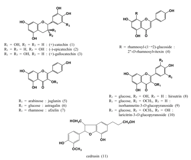
Three flavans: (+)-catechin (1), (-)-epicatechin (2) and
(+)-gallocatechin (3), the flavone glycoside; 2"-O-rhamno- sylvitexsin (4); the six flavonol glycosides: juglanin (kae- mpferol-3-O-α-L-arabinofuranoside) (5), astragalin (kae- mpferol-3-O-β-D-glucopyranoside) (6), afzelin (kaempferol- 3-α-L-rhamnopyranoside) (7), hirsutrin (quercetin-3-O-β- D-glucopyranoside) (8), isorhamnetin-3-O-β-D-glucopyra- noside (9); and laricitrin-3-O-β-D-glucopyranoside (10), and one lignan derivative: cedrusin (11) were isolated from L. kaempferi needles (Fig. 1).
Six compounds (1, 2, 3, 4, 6, 9) were isolated from green needles and eight compounds (1, 2, 4, 5, 7, 8, 10, 11) from fallen needles of L. kaempferi. There was no posi- tive difference in the chemical constituents between green and fallen needles. Compound (1), (2), (4) were isolated from both and supposed to main components of L. kaemp- feri needles.
OH
HO O
OH OH
R1 R2
R3
OH
HO O
OH
O R
R1 = OH, R2 = R3 = H : (+)-catechin (1) R1 = R3 = H, R2 = OH : (-)-epicatechin (2) R1 = R3 = OH, R2 = H : (+)-gallocatechin (3)
R = rhamnosyl-(1 2)-glucoside :→ 2"-O-rhamnosylvitexin (4)
OH
HO O
OH
OR1
O OH
HO O
OH
OR1 O
R2
R3
R1 = arabinose : juglanin (5) R1 = glucose : astragalin (6) R1 = rhamnose : afzelin (7)
R1 = glucose, R2 = OH, R3 = H : hirsutrin (8) R1 = glucose, R2 = OCH3, R3 = H :
isorhamnetin-3-O-glucopyranoside (9) R1 = glucose, R2 = OCH3, R3 = OH :
laricitrin-3-O-glucopyranoside (10)
O
CH2OH
OCH3
HO OH
HOH2C
cedrusin (11)
Fig. 1. Structures of the isolated compounds from L. kaempferi needles.
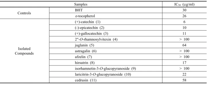
2. Antioxidant activity of the isolated compounds
The DPPH radical scavenging activities (IC50 values) on the isolates were showed in Table 1. Flavan 3-ol com- pounds were indicated the highest activity. Among the flavonol glycosides (5~10), hirsutrin and laricitrin-3-O- glucopyranoside containing the catechol B-ring were more active than juglanin, astragalin afzelin and isorhamnetin- 3-O-glucopyranoside. The catechol B-ring indicated higher antioxidant active than a single OH group in the B-ring (Rice-evans et al., 1996). Substitution with the methoxy group in the 3' position of the B-ring of isorhamnetin-3- O-glucopyranoside showed significantly low activity com- pared with hirsutrin.
3. Anti-inflammatory activity of the isolated compounds
Nitric oxide is a well-known signal in physical and pathological reaction, especially in acute inflammatory response (Surh et al., 2001). It is known that activation of inducible nitric oxide synthase (iNOS) by pro-inflam- matory agent such as LPS can significantly increase nitric oxide (NO) production in macrophages (Kojima et al., 2000). In this study, a LPS-stimulated RAW 264.7 cell assay was employed to evaluate the NO inhibition activity of the compounds isolated from L. kaempferi needles. The results indicated that most of the isolated compounds were effective in NO production inhibition (Fig. 2). (+)-gallo-
Table 1. IC50 values of antioxidant activity of the isolated compounds
Samples IC50 ( g/ml)μ
Controls BHT 30
α-tocopherol 26
Isolated Compounds
(+)-catechin (1) 6
(-)-epicatechin (2) 10
(+)-gallocatechin (3) 11
2"-O-rhamnosylvitexin (4) > 100
juglanin (5) 64
astragalin (6) > 100
afzelin (7) > 100
hirsutrin (8) 17
isorhamnetin-3-O-glucopyranoside (9) > 100
laricitrin-3-O-glucopyranoside (10) 22
cedrusin (11) 58
0 2 0 4 0 6 0 8 0 10 0 12 0
Co n tro l DM S O Co m .1 Co m .2 Co m .3 Co m .4 Co m .5 Co m .6 Co m .7 Co m .8 Co m .9 Co m .10 C ompounds
NO Inhibition (%)
5 0 u g /m l 10 0 u g /m l
Fig. 2. NO inhibition effects of the isolated compounds from L. kaempferi at 50 and 100 g/ml on nitric oxide (NO) productionμ in LPS-stimulated RAW 264.7 cells. Data are the means±S.E. of values from three independent experiments.
catechin (3) and juglanin (5) indicated inhibition activity above 90% of the NO production at a concentration of 100 g/ml.μ
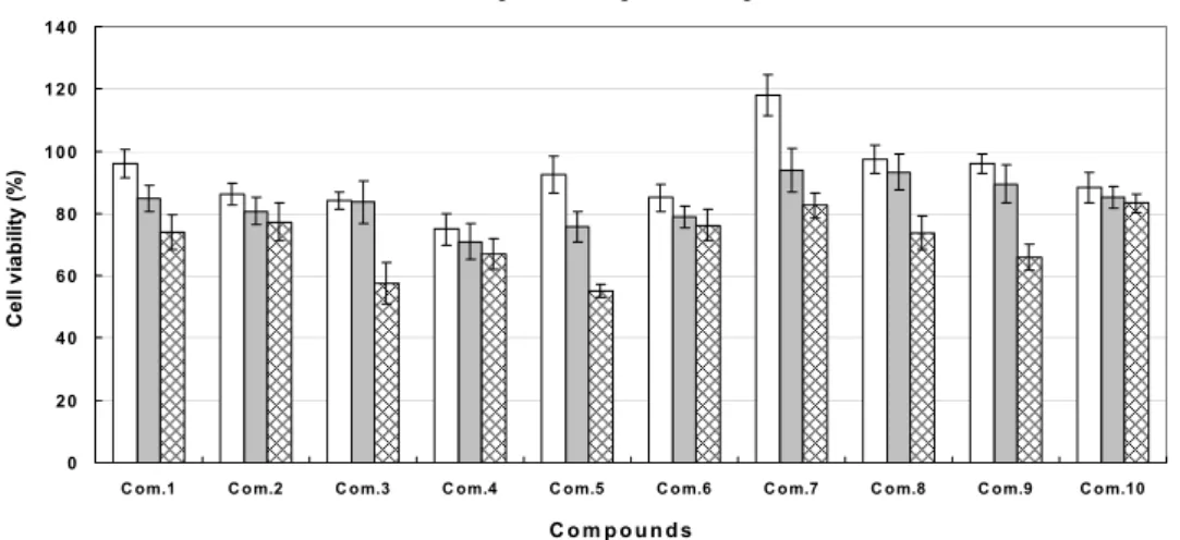
4. Cytotoxic activity of the isolated compounds
The isolated compounds (1~10) were evaluated for their cytotoxic activity against HaCaT normal skin cell lines in vitro by MTT assay method. The results are shown in Fig.
3. Most of the compounds indicated considerable cytoto- xicity against the normal cell lines a dose-dependent man- ner, and showed almost no cytotoxicity, whereas (+)- gallocatechin (3) and juglanin (5) showed only about 50%
cytotoxicity at concentration of 200 g/ml.μ
LITERATED CITED
Blois, M. S. 1958. Antioxidant determinations by the use of a stable free radical. Nature 181(26): 1199-1200.
Green, L. C., D. A. Wagner, J. Glogowsky, P. L. Skipper, J. S. Wishnok and S. R. Tannenbaum. 1982. Analysis of nitrate, nitrite and (15N) nnitrate in biological fluids. Anal. Biochem. 126(1): 131-138.
Kim, J. K., W. G. Park and Y. S. Bae. 1997. Flavonoid glycosides from needles of Larix leptolepis (Pinaceae). Journal of Korean Wood Science and Technology 25(2): 81-87.
Kim, T. W. 1994. The woody plants of Korea in Color. Kyo-Hak Publishing Co., Ltd., Korea.
Kojima, M., T. Morisaki, K. Izuhara, A. Uchiyama, Y. Matsunari, M. Katano and M. Tanaka. 2000. Lipopolysaccharide increases cyclo-oxygenase-2 expression in a colon carcinoma cell line through nuclear factor-KB activation. Oncogene 19: 1225-1231.
Kwon, D. J., J. K. Kim and Y. S. Bae. 2006. Phenolic compounds from fallen needles of Larix kaempferi Carr.. Journal of the Korean Wood Science and Technology 34(6): 72-80.
Lee, K. T., R. K. Kim, S. Y. Ji, K. M. Shin, J. W. Choi, H. J. Jung and H. J. Park. 2003. In vitro antiinflammatory activity of the essential oil extracted from Chrysanthemum sibiricum in murine macrophage RAW 264.7 cells. Natural Product Sciences 9(2):
93-96.
Lee, T. B. 1980. Illustrated Flora of Korea. Hyandg-Mun Publishing Co., Ltd., Korea.
Lee, Y. N. 1996. Flora of Korea. Kyo-Hak Publishing Co., Ltd., Korea.
Miki, K. and T. Sasaya. 1979. Dihydrobenzofuran derivatives in the inner bark of Larix leptolepis Gord. Mokuzai Gakkaishi 25(6):
437-441.
Miki, K. and T. Sasaya. 1980. Lignans from heartwood of Larix leptolepis Gord. Mokuzai Gakkaishi 26(9): 633-636.
Mosmann T. 1983. Rapid colorimetric assay for cellular growth and survival: application to proliferation and cytotoxicity assays. J. of Immunol. Methods 65: 55-63.
Rice-evans, C. A., N. J. Miller and G. Paganga. 1996. Structure- antioxidant activity relationships of flavonoids and phenolic acids.
Free Radical Bio. Med. 20(7): 933-956.
Surh, Y. J., K. S. Chun, H. H. Cha, S. S. Han, Y. S. Keum, K.
K. Park and S. S. Lee. 2001. Molecular mechanisms underlying chemopreventive activities of anti-inflammatory phytochemicals:
down-regulation of COX-2 and iNOS through suppression of NF-KB activation. Mutation Research 480-481: 243-268.
Takehara, T. and T. Sasaya. 1979a. Lignans from sapwood of Larix leptolepis Gord. Mokuzai Gakkaishi 25(7): 516-517.
Takehara, T. and T. Sasaya. 1979b. Dihydrobenzofuran derivatives from Sapwood of Larix leptolepis Gord. Mokuzai Gakkaishi 25 (10): 660-664.
0 20 40 60 80 100 120 140
C om.1 C om.2 C om.3 C om.4 C om.5 C om.6 C om.7 C om.8 C om.9 C om.10
C o m p o u n d s
Cell viability (%)
50 u g /m l 100 u g /m l 200 u g /m l
Fig. 3. Effect of the isolated compounds from L. kaempferi on cell viability of HaCaT cells. Cell viability was determined by MTT assay. Data are the means±S.E. of values from three independent experiments.