The incidence of cancer has been rising expeditiously, and postbiotics have been receiving great attention from researchers for cancer therapy due to the side effects and complications of conventional cancer treatment. Postbiotics encompass a wide range of complex macromolecules such as inactivated microbial cells, cellular fractions or metabolites and provide various physiological health benefits to the host. Postbiotics exert anti- cancer effects by apoptosis, anti-proliferative and anti-inflammatory effects, as well as modulating the composition of the gut microbiota and the immune system. This review provides an overview of the conceptual and safety issues surrounding postbiotics, highlighting their biological role in the prevention and treatment of cancer.
Keywords: apoptosis, cell cycle, gut microbiome, human cancer, immune system, postbiotics
Cancer is considered one of the most important causes of death, because it is a fatal malignancy. Cancer is the second leading cause of mortality in the world, with an estimated 9.6 million deaths in 2018 according to a World Health Organization (WHO) report. The most fatal types of cancer that cause serious deaths are lung, prostate, colorectal and stomach cancer.
Cancer is caused by DNA damage, deficiency in DNA repair,
or genomic instability leading to uncontrolled cell growth and tumor formation (Sung et al., 2021). Current chemotherapy for cancer comes with a variety of undesirable side effects such as diarrhea, bone marrow suppression, peripheral neuropathy and cardiotoxicity (Gegechkori et al., 2017). Therefore, researchers are investigating innovative treatments and preventive measures for cancer.
Postbiotics are metabolic products secreted by probiotics which are live bacteria such as Lactobacillus (L.) and Bifidobacterium (B.), using prebiotics as a nutrient source such as dietary fiber and oligosaccharides (Nataraj et al., 2020).
Probiotics are living microorganisms that metabolize in the gut and provide health benefits (Batista et al., 2020). Recently, research on postbiotics utilizing these advantages has been actively conducted. Postbiotics have been reported to have health-promoting effects such as immunomodulation, anti- carcinogenic, hypolipidemic, and antihypertensive characteristics (Fang et al., 2014; Aguilar-Toalá et al., 2020; Engevik et al., 2021). More recently, postbiotics have emerged as an alter- native to probiotics for high-risk patients or patients with underlying diseases who have difficulty using live probiotic strains. Postbiotics are considered potential anticancer agents due to their properties such as known chemical structure, safe profile, longer shelf life, nontoxicity, resistance to hydrolysis and stability to digestive system conditions compared to probiotics (Nataraj et al., 2020).
Postbiotics for cancer prevention and treatment
SukJin Kim
1, Gun-Hee Kim
1*, and Hyosun Cho
2*
1
Department of Bio-Health Convergence Major, Duksung Women’s University, Seoul 01369, Republic of Korea
2
College of Pharmacy, Duksung Women’s University, Seoul 01369, Republic of Korea
포스트바이오틱스의 암예방 및 치료 효과
김숙진
1・ 김건희
1* ・ 조효선
2*
1
덕성여자대학교 바이오헬스융합 전공,
2덕성여자대학교 약학과
(Received August 18, 2021; Revised September 11, 2021; Accepted September 14, 2021)
*For correspondence. (H. Cho) E-mail: [email protected];
Tel.: +82-2-901-8678; Fax: +82-2-901-8386 / (G.H. Kim) E-mail: [email protected];
Tel.: +82-2-901-8694; Fax: +82-2-901-8661
Anti-cancer effect of postbiotics
∙143
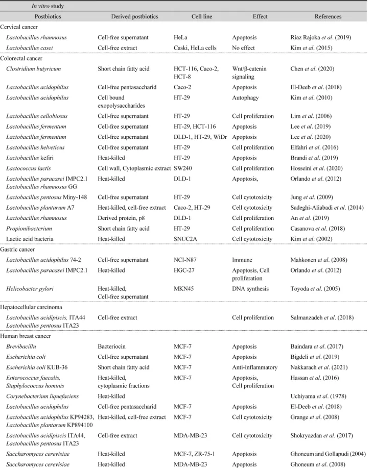
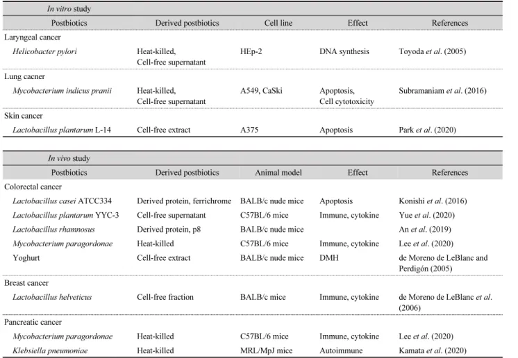
Table 1. In vitro and in vivo studies in the field of cancer and postbiotics
In vitro study
Postbiotics Derived postbiotics Cell line Effect References
Cervical cancer
Lactobacillus rhamnosus Cell-free supernatant HeLa Apoptosis Riaz Rajoka et al. (2019)
Lactobacillus casei Cell-free extract Caski, HeLa cells No effect Kim et al. (2015)
Colorectal cancer
Clostridium butyricum Short chain fatty acid HCT-116, Caco-2,
HCT-8 Wnt/β-catenin
signaling Chen et al. (2020) Lactobacillus acidophilus Cell-free pentasaccharid Caco-2 Apoptosis El-Deeb et al. (2018) Lactobacillus acidophilus Cell bound
exopolysaccharides HT-29 Autophagy Kim et al. (2010)
Lactobacillus cellobiosus Cell-free supernatant HT-29 Cell proliferation Lim et al. (2006) Lactobacillus fermentum Cell-free supernatant HT-29, HCT-116 Apoptosis Lee et al. (2019) Lactobacillus fermentum Cell-free supernatant DLD-1, HT-29, WiDr Apoptosis Lee et al. (2020) Lactobacillus helveticus Cell-free supernatant HT-29 Cell proliferation Elfahri et al. (2016)
Lactobacillus kefiri Heat-killed HT-29 Apoptosis Brandi et al. (2019)
Lactococcus lactis Cell wall, Cytoplasmic extract SW240 Cell proliferation Hosseini et al. (2020) Lactobacillus paracasei IMPC2.1
Lactobacillus rhamnosus GG Heat-killed DLD-1 Apoptosis, Orlando et al. (2012)
Lactobacillus pentosus Miny-148 Cell-free supernatant HT-29 Cell cytotoxicity Jung et al. (2009)
Lactobacillus plantarum A7 Heat-killed, cell-free extract Caco-2, HT-29 Cell cytotoxicity Sadeghi-Aliabadi et al. (2014) Lactobacillus rhamnosus Derived protein, p8 DLD-1 Cell proliferation An et al. (2019)
Propionibacterium Short chain fatty acid HT-29 Cell proliferation Casanova et al. (2018)
Lactic acid bacteria Heat-killed SNUC2A Cell cytotoxicity Kim et al. (2002)
Gastric cancer
Lactobacillus acidophilus 74-2 Cell-free supernatant NCI-N87 Immune Mahkonen et al. (2008)
Lactobacillus paracasei IMPC2.1 Heat-killed HGC-27 Apoptosis, Cell
proliferation Orlando et al. (2012) Helicobacter pylori Heat-killed,
Cell-free supernatant MKN45 DNA synthesis Toyoda et al. (2005) Hepatocellular carcinoma
Lactobacillus acidipiscis, ITA44
Lactobacillus pentosus ITA23 Cell-free extract Cell proliferation Salmanzadeh et al. (2018) Human breast cancer
Brevibacillu Bacteriocin MCF-7 Apoptosis Baindara et al. (2017)
Escherichia coli Cell-free supernatant MCF-7 Apoptosis Bigdeli et al. (2019)
Escherichia coli KUB-36 Short chain fatty acid MCF-7 Anti-inflammatory Nakkarach et al. (2021) Enterococcus faecalis,
Staphylococcus hominis Heat-killed,
cytoplasmic fractions MCF-7 Apoptosis,
Cell proliferation Hassan et al. (2016)
Corynebacterium liquefaciens Heat-killed Uchiyama et al. (1978)
Lactobacillus acidophilus Cell-free pentasaccharid MCF-7 Apoptosis El-Deeb et al. (2018) Lactobacillus acidophilus KP94283,
Lactobacillus plantarum KP894100 Heat-killed, cell-free extract MCF-7 Cell cytotoxicity Grange et al. (2008) Lactobacillus acidipiscis ITA44,
Lactobacillus pentosus ITA23 Cell-free extract MDA-MB-23 Cell cytotoxicity Shokryazdan et al. (2017)
Saccharomyces cerevisiae Heat-killed MCF-7, ZR-75-1 Apoptosis Ghoneum and Gollapudi (2004)
Saccharomyces cerevisiae Heat-killed MDA-MB-23 Apoptosis Ghoneum et al. (2008)
Currently, anti-cancer reviews mainly focus on probiotics and colorectal cancer. However, the present review fully addresses the importance of postbiotics in various cancer types.
Several reports have suggested that postbiotics exhibit anti- cancer activity through anti-proliferative, apoptotic, and anti- inflammatory effects. Consequently, it provides a basic under- standing of the alternative cancer treatment and mechanisms of postbiotics.
Definition, Classification and Safety of Postbiotics
Definition of postbiotics
Probiotics are living microorganisms that are beneficial to health by using prebiotics as nutrients, a type of dietary fiber, to secrete postbiotics (Batista et al., 2020). Postbiotics are bioactive
soluble factors secreted from probiotics or released during the fermentation and lysis of bacteria. They provide physiological benefits to the host as metabiotics, biogenics, or simply meta- bolites. Before the term postbiotics appeared in 2018, researchers referred to them as “biogenic,” “cell-free supernatant,” “abiotic,”
“metabiotic,” “paraprobiotic,” “ghost probiotics,” “pseudo- probiotic,” “postbiotic,” supernatant,” etc. (Aguilar-Toalá et al., 2018). Postbiotics consist of bacteriocins, enzymes, vitamins, amino acids, neurotransmitters, short-chain fatty acids (SCFAs), nitric oxide (NO), organic acids, etc., which have been reported to have immune, antioxidant, and anticancer effects and regulate lipid/cholesterol metabolism (Bönisch et al., 2018;
Hati et al., 2019).
Classification and characteristics of postbiotics The composition and activity of the gut microbiota in the host depends on the nutrients provided by the host (Asnicar et
Table 1. Continued
In vitro study
Postbiotics Derived postbiotics Cell line Effect References
Laryngeal cancer
Helicobacter pylori Heat-killed,
Cell-free supernatant HEp-2 DNA synthesis Toyoda et al. (2005) Lung cacner
Mycobacterium indicus pranii Heat-killed,
Cell-free supernatant A549, CaSki Apoptosis,
Cell cytotoxicity Subramaniam et al. (2016) Skin cancer
Lactobacillus plantarum L-14 Cell-free extract A375 Apoptosis Park et al. (2020)
In vivo study
Postbiotics Derived postbiotics Animal model Effect References
Colorectal cancer
Lactobacillus casei ATCC334 Derived protein, ferrichrome BALB/c nude mice Apoptosis Konishi et al. (2016) Lactobacillus plantarum YYC-3 Cell-free supernatant C57BL/6 mice Immune, cytokine Yue et al. (2020)
Lactobacillus rhamnosus Derived protein, p8 BALB/c nude mice An et al. (2019)
Mycobacterium paragordonae Heat-killed C57BL/6 mice Immune, cytokine Lee et al. (2020)
Yoghurt Cell-free extract BALB/c nude mice DMH de Moreno de LeBlanc and
Perdigón (2005) Breast cancer
Lactobacillus helveticus Cell-free fraction BALB/c mice Immune, cytokine de Moreno de LeBlanc et al.
(2006) Pancreatic cancer
Mycobacterium paragordonae Heat-killed C57BL/6 mice Immune, cytokine Lee et al. (2020)
Klebsiella pneumoniae Heat-killed MRL/MpJ mice Autoimmune Kamata et al. (2020)
Anti-cancer effect of postbiotics
∙145
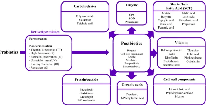
al., 2021). Furthermore, these microbes secrete a myriad of metabolites that contribute to the growth of beneficial organisms, interaction of cells, and promoting production. These inter- actions by metabolites are vital for shaping host-microbial symbiosis and the establishment of stable communities via modulation of cellular metabolism. Bacterial lysis occurs during the production of postbiotics, releasing other intracellular metabolites and various compounds as follows (Fig. 1); SCFs (acetate, butyrate, propionate, lactate), proteins/peptides (bac- teriocin, glutathione, lactocepin), vitamins (biotin, riboflavin, pantothenate, ascorbic acid, thiamine, folic acid, phylloquinone, cobalamin), enzymes (GPx, SOD, peroxidase), cell wall com- ponents (lipoteichoic acid, teichoic acid peptidoglycan-derived, S-layer), and carbohydrates (polysaccharide, galactose) (Hati et al., 2019). Butyrate, a host energy source, is mainly used by intestinal epithelial cells, and acetate is used systemically (Venegas et al., 2019). Acetate and propionate move to the liver and peripheral organs and become substrates for gluco- neogenesis and lipogenesis.
Postbiotics are produced naturally through the fermentation of probiotics, and the various biological properties of post- biotics depend on different production environment conditions (Homayouni Rad et al., 2021). As shown in Fig. 1, in addition to natural methods, pure postbiotics with high functionality are
produced by physical methods including mechanical disruption, heat treatment, UV irradiation, formalin inactivation, high hydrostatic pressure, freeze-drying, sonication, filtration, ohmic heating, supercritical CO
2, pulsed electric field, pH changes and drying (Barros et al., 2021). In general, heat treatment is the most useful method, and a variety of methods are used to improve the nutritional value, shelf life and health functionality for consumers.
Safety of postbiotics
Before discussing the therapeutic benefits of postbiotics, the issue of stability of postbiotics must be addressed. Probiotics treatment uses living microorganisms, which can cause problems due to the generation of antibiotic-resistant genes and virulence factors in vivo. This is especially important for young children with an immature immune system and weak barriers, and postbiotics can bypass this problem.
Seven randomized controlled trials (RCT) of 1,740 children reported that supplementation with heat-killed L. acidophilus LB reduced the duration of diarrhea and prevented pharyngitis, laryngitis, and diarrhea (Malagón-Rojas et al., 2020). Among the RTCs, only one showed dehydration associated with heat- killed L. acidophilus LB administration. It was confirmed that heat-inactivated L. acidophilus LB can activate immune-
Fig. 1. Composition of postbiotics.
inflammatory mechanisms in children related to intestinal cell adhesion and pro-inflammatory chemokines. To evaluate the effect of L. paracasei CNCM I-1572 on intestinal microflora, a clinical study was conducted on children aged 6–14. As a result, it was reported that the function of the gut microbiota was modulated without adverse side effects (Cremon et al., 2018).
Furthermore, Andresen et al. (2020) reported that heat-killed B. bifidum HI-MIMBb75 alleviated irritable bowel syndrome.
The most common adverse event in this clinical trial was suspected abdominal pain occurring in two patients (< 1%) in the B. bifidum HI-MIMBb75 group and one patient (< 1%) in the placebo group. Further studies are necessary to determine the effects and side effects of various postbiotics.
Compared to probiotics, postbiotics have many advantages for industrial production, including ease of use and storage, extended shelf life, stability over a wide range of pH and temperature, and no bioamine production. Nevertheless, further studies on manufacturing, delivery systems and safety para- meters of pharmaceuticals and functional foods are needed to utilize postbiotics as probiotic substitutes (Salminen et al., 2021).
Potential Mechanisms for Postbiotics Action:
Anticarcinogenic Activity
In preclinical and clinical studies, carcinogens of nutritional origin, such as polycyclic aromatic hydrocarbons (PAH), N- nitroso compound (NOCs), heterocyclic amine (HCA), acryl- amide, and mycotoxins, were found to cause breast cancer, colorectal cancer, liver cancer and prostate cancer (Wang et al., 2012). These substances promote uncontrolled cell growth and DNA damage in the mammary gland, colon and prostate (Hebels et al., 2010). In general, PAH components are absorbed by charcoal-grilled, smoked, and processed meats. In this regard, N-nitroso compounds, even in small amounts, can be harmful to the host and are directly related to digestive system cancer (Xu et al., 2015).
The gut microbiota directly or indirectly affects the host's tumorigenesis. Digestive system cancer, especially colorectal cancer, is caused by abnormalities of the intestinal bacteria and the proliferation of certain bacterial pathogens, such as Helico- bacter (H.) pylori, Streptococcus (S.) bovis, Enterococcus
faecalis, Clostridium septicum, Escherichia coli, Fusobacterium spp., Bacteroides fragilis, and Streptococcus gallolyticus (Li et al., 2019). Bacterial pathogens influence host intestinal commensal bacteria and immune system efficiency to induce tumor growth and development. Bacterial pathogens directly contribute to host tumorigenesis by releasing large numbers of toxins and regulating several cellular proliferation and promotion pathways (Mager, 2006). Typically, dysbiosis in the gut environment predominantly affects pathogenic bacteria, and destroys the host’s DNA through the production of toxins, leading to genomic instability, tumor initiation and progression (Sobhani et al., 2011). According to Wei et al. (2010), H. pylori as a class A carcinogen negatively regulates p53 by increasing ubiquiti- nation and proteasomal degradation by activation of serine/
threonine kinase AKT, which phosphorylates and activates the ubiquitin ligase HDM2.
Recent in vitro and in vivo studies have reported that postbiotics have important anticancer effects (Table 1). In this review, the molecular mechanisms of postbiotics in cancer prevention and treatment are involved in pathways such as modulation of immune response, inhibition of mutagenesis and carcinogens, activation of pro-apoptotic pathways, decreased bacterial translocation, and increased apoptosis and autophagy.
Induction of apoptosis in cancer cells
Apoptosis of cancer cells determines the rate of cancer cell development. Postbiotics are of great interest to scientists because they cause the apoptosis of cancer cells without damaging adjacent cells. Postbiotics include modulating apoptosis signaling through mitochondrial-dependent (intrinsic) and death receptor-dependent (extrinsic) pathways (Fig. 2). Riaz et al. (2019) showed that the induction of intrinsic and extrinsic apoptosis of L. rhamnosus was achieved by the up-regulation of Bad, Bax, caspase3, caspase8, and caspase9, and down- regulation of Bcl-2 genes in human cervical cancer HeLa cells.
Phagocytosis of S. cerevisiae in human breast cancer MCF7 cell is associated with the disruption of mitochondrial mem- brane potential and activation of initiator and effector caspases 8, 9, and 3 (Ghoneum and Gollapudi, 2004). It was demon- strated that postbiotics from L. paracasei IMPC2.1 and L.
rhamnosus GG inhibit the growth and progression of human
colon cancer DLD-1 cell and human gastric cancer HGC-27
Anti-cancer effect of postbiotics
∙147
cell lines through the activation of the pro-apoptotic pathway (Orlando et al., 2012). In addition, ferrichrome derived from L.
casei has been reported to have an apoptotic effect on tumor cells through an increase in cleaved PARP and cleaved caspase 3 (Konishi et al., 2016).
Bacteriocin Laterosporulin10 extracted from Brevibacillus spp. has been reported to induce apoptosis of human cancer cells lines MCF-7, HEK293T, HT1080, HeLa and H1299 by in vitro flow cytometry analysis (Baindara et al., 2017). Jan et al.
(2002) have suggested that propionibacteria inhibit human colorectal cancer cell lines HT-29 and caco-2 in vitro by their ability to produce SCFA inducing apoptosis. These studies show that bacterial culture supernatants and pure SCFA exhibited typical apoptosis, including loss of mitochondrial transmembrane potential, generation of reactive oxygen species, caspase-3 treatment, oncoprotein Bcl-2 regulation, and nuclear chromatin condensation. Based on this evidence, it is believed that certain postbiotics upregulate cancer cell proliferation inhibition and apoptosis pathways.
Inhibition of cell cycle progression in cancer cells The cell cycle arrest of cancer cells suppresses cancer cell proliferation by controlling cell division. Cells continue to cycle in the sequence of G1, S, G2, and M phases, among which DNA replication occurs in S phase and cell division occurs in M phase (Fig. 2). The G1 and G2 phases are the stages to ensure that division and replication have completely occurred. Specific regulation between cyclin-dependent kinases (CDKs) and cyclins is important to the progression of the cell cycle. Cyclin D/CDK4 in G1 phase, cyclin A/CDK2 in S phase, and the cyclin B/CDC2 complex in G2/M phase are involved in cell cycle regulation, respectively.
Cell wall components, peptidoglycans, cytoplasmic extracts, and cell supernatants of various postbiotics exhibit anti- proliferative effects against cancer cell lines. SCFAs produced in P. freudenreichii caused the accumulation of sub-G1 phase, and the reduction of S and G2/M phases in human colorectal cancer RKO cells, as measured by FACS (Casanova et al., 2018). It has been reported that cell-free pentasaccharide of L.
Fig. 2. Induction of anti-proliferative and apoptotic pathways.
spp. promotes apoptosis and inhibits S-phase cell cycle progression in human colorectal cancer HT-29 cell and primary colon cells T4056 (Elfahri et al., 2016). Hosseini et al. (2020) showed that nisin, the cytoplasmic extract of L. lactis ssp.
lactis, and the cell walls of this bacterium have an antipro- liferative effect that is associated with the decreased expression of cyclin D1 in human colorectal SW480 cancer cells in vitro.
An et al. (2019) found that the antiproliferation activity of probiotic-derived p8 protein was mediated by inhibition of the p53-p21-Cyclin B1/Cdk1 signal pathway, resulting in growth arrest at the G2 phase of the cell cycle in vitro in human colorectal DLD-1 cells. Thus, it can be suggested that post- biotics inhibit the proliferation of cancer cells.
Induction of autophagic in cancer cells
Autophagy is a cellular process that maintains intracellular homeostasis by delivering misfolded proteins and damaged organelles to lysosomes. Numerous studies have shown that increased autophagy in many cancer cells can inhibit tumori- genesis. A functional blockade of the proteasome induces high levels of GRP78, promoting autophagosome formation through activation of the unfolded protein response (UPR) in the endoplasmic reticulum stress pathway. Beclin-1 induces dis- sociation of the Beclin-1/Bcl-2 complex, thereby promoting autophagy by reducing the Beclin1/PI3K-III complex (Fig. 2).
Tang et al. (2011) have demonstrated that propionate treatment in human colon carcinoma cell lines HCT-116 and SW480 exhibit extensive characteristics of autophagic pro- teolysis by increased LC3-I to LC3-II conversion, acidic vesicular organelle development, and reduced p62/SQSTM1 expression. Kim et al. (2010) reported that cell-bound exopoly- saccharides isolated from L. acidophilus 606 inhibited the proliferation of HT-29 colon cancer cells by directly affecting cell morphology. This anticancer activity is due to the activation of autophagic cell death, which is promoted not only indirectly through the induction of Bcl-2 and Bak, but also directly by the induction of Beclin-1 and GRP78. Therefore, autophagy of cancer cells is a useful approach in postbiotic cancer therapy.
Modulation of immune response
A delicate balance between immune responses depends on
the various mediators released by cancer cells, cancer-associated cells and host inflammatory cells in the tumor microenvironment.
Thus, modulation of immune response determines the overall outcome of the carcinogenesis process. Activated macrophages and neutrophils eliminate tumor cells (Fig. 3). Moreover, anti- inflammatory and pro-apoptotic cytokines such as TRAIL, interleukin (IL)-10 and TGF-β inhibit carcinogenesis depending on the particular tumor microenvironment. Postbiotics upregulate the functioning of the gut immune system and the treatment and prevention of chronic inflammatory diseases (Żółkiexicz et al., 2020).
Kim et al. (2013) reported that SCFA activates GPR41 and GPR43 in intestinal epithelial cells, leading to mitogen-activated protein kinase signaling and the rapid production of che- mokines and cytokines. These pathways mediate immunity and inflammation response in mice. Heat-killed L. gasseri TMC0356 reportedly has immunomodulatory effects through stimulation of IL-12 production in macrophages (Kawase et al., 2012).
SCFA protects the mucosal layer from damage by reducing the levels of immunomodulators, such as prostaglandin, produced by cyclooxygenase 2 (COX-2), which induce tumor inflam- mation and development in human breast cancer MCF7 cells (Nakkarach et al., 2021). Therefore, postbiotics have the same effective immunomodulatory activity as live probiotics, and the optimal level of the inactivation method (mainly the method of preparation by heat treatment) may not adversely affect the immunomodulatory and anti-inflammatory properties.
Heat-killed L. pentosus b240 promoted the production of immunoglobulin A (IgA), IL-6, IL-10, interferon (IFN)-γ, and tumor necrosis factor, but not IL-4, IL-5, B-cell activating factors, IFN-α, IFN-β, and transforming growth factor-β1 (Kotani et al., 2014). L. plantarum strain YYC-3 strongly inhibited human colorectal cancer HT-29 and Caco2 cell lines.
This anticancer effect involved a mechanism that modulated
the immune system and downregulated the expression of
inflammatory cytokines interleukin IL-6, IL-17, and IL-22,
along with reduced infiltration of inflammatory cells (Yue et
al., 2020). Based on these results, studies using cell lines
demonstrated that postbiotics had anticancer effects through
immunomodulation, suggesting that postbiotics-based therapies
can be used for anticancer treatment.
Anti-cancer effect of postbiotics
∙149
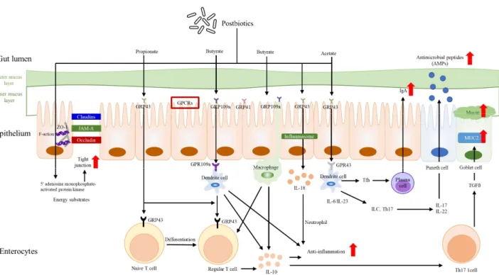
Improved intestinal barrier and cellular junction proteins Under normal conditions, enterocytes in the intestinal wall are a key defense mechanism for maintaining the entire intestine. The gastric mucosal layer protects against destruction of the intestinal epithelium by unsafe compounds. An important component of the intestinal barrier is the mucus layer, which contains various mucin glycoproteins that maintain homeo- stasis and regulate the inflammatory response (Fig. 3). The gut environment contains many different types of bacteria that coexist in balance with the host. Various in vitro studies have demonstrated that SCFA has a protective effect on DNA transcription and has the potential to protect the intestinal barrier by inhibiting histone deacetylase (HDAC), which regulates gene expression and increases the expression of MUC2 in goblet cells (Hatayama et al., 2007). Several studies have reported that SCFA enhances epithelial cell tight junctions by stimulating 5-adenosinmonophosphate (5’-AMP) to activate protein kinase, a fundamental role in regulating energy metabolism in colon cells. According to a report by Zheng et al. (2017), microbial-derived butyrate enhanced IEC barrier formation, induced IL10RA mRNA, IL-10RA protein, and transactivation through activated Stat3 and HDAC
inhibition. It has been demonstrated that P40, a soluble protein derived from L. rhamnosus GG, promotes the intestinal barrier by activating EGFR and synthetic mucin (Wang et al., 2014).
Consequently, postbiotics are effective in the prevention and treatment of colorectal cancer by preserving the intestinal barrier.
Inhibition of growth of bacterial pathogens
Many pathogens have shown to cause human cancer. Studies have reported associations between H. pylori and stomach cancer, bovis streptococci and colorectal cancer, Chlamydia pneumonia and lung cancer, Salmonella and gallbladder cancer, respectively (Mager, 2006). The gut microbiota greatly affects the health of the host. The intestinal environment is pH 5.5–6.5; the upper large intestine is a weakly acidic pH environment, and in the lower large intestine, the pH is more neutral. The growth of bacterial pathogens is reduced when the intestinal pH is below 6.0 and the panel cells release anti- microbial peptides (Kok et al., 2020). A diet supplemented with heat-killed Limosilactobacillus fermentum and L. delbrueckii promotes the growth of beneficial B. spp. and associated metabolic changes in the human fermented fecal community
Fig. 3. Diminution of bacterial translocation and preservation of the intestinal barrier.