J Korean Soc Coloproctol Vol. 20, No. 4, 2004
191
Sulindac-induced Apoptosis without Oli- gonucleosomal DNA Fragmentation in HT-29 Cells - Its Special References to Mitochon- drial Pathway -
Ki-Jae Park, M.D., Yuk Kwon, M.D., Sung-Heun Kim, M.D., Min-Chan Kim, M.D., Hong-Jo Choi, M.D., F.A.C.S., Young-Hoon Kim, M.D., Se-Heon Cho, M.D., Ghap- Joong Jung, M.D., Sung-Hyun Kim, M.D.1, Hyuk-Chan Kwon, M.D.1
Departments of Surgery and 1Internal Medicine, Dong-A University College of Medicine, Busan, Korea
Purpose: This study was undertaken to reveal the molecular mechanism underlying sulindac-induced apoptosis in the human colon cancer cell line HT-29 (mutant p53).
Methods: Apoptosis was determined by using Hoechst 33342 staining, and translocation of proteins was established by using immunofluorescence, immunoelectron microscopy, and Western blotting after ultra- fractionation. Results:
This type of apoptosis was associated with decreased mitochondrial membrane potential, a translocation of the apoptosis-inducing factor (AIF) to the nucleus, and morpho- logical evidence of nuclear condensation. However, DNA electrophoresis did not elucidate the ladder pattern of DNA fragments. Instead, a pulse-field gel electrophoresis showed that sulindac led to disintegration of nuclear DNA into-high- molecular-weight DNA fragments of about 100∼300 kbp.
Conclusions: Our findings indicate that sulindac induces large-scale DNA fragmentation, suggesting a predominantly AIF-mediated cell-death process, through translocation of the AIF to the nucleus in HT-29 cells. J Korean Soc Coloproctol 2004;20:191-198
Key W ords : Apoptosis, Colon cancer cell line HT-29, AIF, HMW DNA fragmentation
세포사멸, 대장암세포주 HT-29, 세포사멸 유 발인자, 고분자 DNA조각화
ꠏꠏꠏꠏꠏꠏꠏꠏꠏꠏꠏꠏꠏꠏꠏꠏꠏꠏꠏꠏꠏꠏꠏꠏꠏꠏꠏꠏꠏꠏꠏꠏꠏꠏꠏꠏꠏꠏꠏꠏꠏꠏꠏꠏꠏꠏꠏꠏꠏ 서 론
많은 연구들을 통해 비스테로이드성 항염증약물(이 하 NSAIDs)은 대장암의 발생 빈도와 대장암에 의한 사망률을 감소시키는 것으로 알려져 있다.1,2 NSAIDs 에 의한 이러한 항종양효과의 기전으로는 COX-2 억제 가 관련이 있는 것으로 알려져 있다.3 그리고 NSAIDs 의 또 다른 항종양효과로는 세포사멸 유발 기전도 많 이 거론되고 있는데, NSAIDs에 의한 세포사멸은 인간 의 대장암 세포를 포함한 여러 세포들에서뿐만 아니 라,4,5 생체(in vivo)에서도 확인되고 있다.6
세포사멸이 발생한 세포에서는 특징적으로 세포 수 축(shrinkage), 세포질과 염색질의 응축(condensation), 그리고 올리고뉴클레오좀 분절화(oligonucleosomal fragmentation)와 같은 소견들이 관찰된다.7 세포사멸의 궁극적인 기전은 caspase에 의한 단백질분해로 여겨지 고, 여기에는 미토콘드리아가 중요한 조절 역할을 하 는 것으로 보인다.8 미토콘드리아의 막간공간(inter- membrane space)에는 여러 가지 세포사멸인자들, 즉 cytochrome c, procaspases (-2, -3, 및 -9), Smac/ DIABLO, 그리고 세포사멸 유발인자(apoptosis-inducing factor: 이 하 AIF) 등이 존재하고, 이들은 세포사멸이 진행될 때 방출되는 것으로 알려져 있다.9,10 결과적으로 caspase
Sulindac에 의한 대장암 세포주 HT-29 세포의 세포사멸 기작 연구-미토콘드리아 통로를 중심으로-
동아대학교 의과대학 외과학교실, 1내과학교실
박기재․권 육․김성흔․김민찬․최홍조․김영훈․조세헌․정갑중․김성현1․권혁찬1
접수: 2004년 5월 20일, 승인: 2004년 8월 13일
책임저자: 최홍조, 602-715, 부산시 서구 동대신동 3가 1번지 동아대학교의료원 외과
Tel: 051-240-5146, Fax: 051-247-9316 E-mail: [email protected]
본 연구는 2002년도 대한대장항문학회 아벤티스-제일제당 학술 연구비로 시행되었음.
Received May 20, 2004, Accepted August 13, 2004
Correspondence to: Hong-Jo Choi, Departments of Surgery, Dong-A University College of Medicine, 3-1 Dongdaesin-dong, Seo-gu, Busan 602-714, South Korea.
Tel: +82-51-240-5146, Fax: +82-51-247-9316 E-mail: [email protected]
가 활성화되면 PARP (poly-ADP-ribose- polymerase)의 절단(cleavage)과 lamins의 분해(degradation) 그리고 엔 도뉴클레아제(endonuclease)의 활성이 초래된다.11 Sulindac은 대장암의 예방에 있어 가장 많이 연구되 고 있는 NSAIDs의 하나다. 그러므로 대장암의 약물학 적 치료의 관점에서 볼 때 sulindac의 세포사멸 유발 기전을 규명하는 것은 매우 중요하다고 할 수 있다. 약 물학적으로나 구조적으로 sulindac과 유사한 indome- thacin에서는 HT-29세포주에서 세포사멸을 유발할 때 흐름세포측정(flow cytometry)에서 subdiploid peak를 보이고, DNA 전기영동에서 세포사멸의 전형적인 소 견으로서 올리고뉴클레오좀 분절화를 시사하는 사다 리 모양(ladder pattern)이 관찰된다.12 그러나 저자들의 이전 연구를 통해 sulindac은 대장암 세포주 HT-29에 서 세포사멸을 유도한다는 것은 확인할 수 있었으나, sulindac의 처리 시간(최대 96시간)이나 용량에 관계없 이 전기영동에서 DNA 분해를 시사하는 사다리 모양 은 관찰되지 않았다.13 이러한 소견과 유사하게 아스피 린도 대장암 세포주 HT-29에서 세포사멸은 유발하지 만 전형적인 세포사멸의 특징적인 소견인 DNA ladder 와 함께 흐름세포측정(flow cytometry)상 subdiploid peak를 보이지 않았다고 보고되었다.12,14 이런 소견들 로 미루어 아스피린은 HT-29 세포주에서 비전형적인 세포사멸을 유발하는 것으로 추정된다.14
이에 저자들은 인간의 대장암 세포주 HT-29에서 sulindac에 의한 유도되는 세포사멸의 기전, 특히 미트 콘드리아와 핵에서 관찰되는 분자생물학적인 변화들 을 관찰하기 위해 연구를 시행하였다.
방 법
1) 시약
(1) 항체: cytochrome c, AIF, β-actin, 그리고 sec- ondary FITC-conjugated antibodies (Santa Cruz Biotech- nology, Santa Cruz, CA, USA)를 사용하였다.
(2) 배지 및 혈청: RPMI medium 1640 및 fetal bovine serum (FBS) (Gibco, Gaithersburg, MD, USA)을 사용하 였다.
(3) 기타: Sulindac, sulindac sulfide, Hoechst 33342, protease inhibitor cocktail, 및 propidium iodide (이상 Sigma Aldrich, St. Louis, MO, USA), 5,5’, 6,6’-tetra- chloro-1,1’, 3,3’-tetraethylbenzimidazol carbocyanine iodide (JC-1) (Molecular Probes, Eugene, OR, USA), 그리고 ECL Western blotting kits (Amersham Pharmacia Biotech
(Piscataway, NJ, USA)를 사용하였다.
2) 세포배양
대장암 세포주인 HT-29 (KCLB 30038)를 100 U/ml penicillin, 100μg/ml streptomycin, 그리고 10% heat- inactivated FBS가 첨가된 DMEM (Dulbecco's modified Eagle's medium) 배지로 5% CO2가 포함된 37oC 배양기 에서 배양하였다. HT-29 세포를 24시간 배양하여 70∼
80%의 융합(confluency)이 된 후 sulindac처리를 하였다.
Sulindac 보존용액은 DMSO에 약제를 용해시켜 -20oC 로 사용 전까지 보관하였다. 그리고 다양한 농도의 sulindac으로 처리하여 trypan blue로 염색을 하고 hemacytometer를 이용하여 그 수를 측정하였다.
3) 세포사멸의 형태학적 변화 관찰
광학현미경 검사를 위하여 세포부유물을 세포원심 분리기로 분리한 후 4μg/ml Hoechst 33342로 염색하여 형광현미경을 이용하여 형태의 변화를 관찰하였다. 그 리고 전자현미경(Hitachi H600-3 electron microscope, Tokyo, Japan) 소견을 관찰하기 위해 24시간 동안 1 mM sulindac을 처리한 세포를 200 g에서 원심 분리하여 1시간 동안 PBS (pH 7.4) 에서 4% paraformaldehyde로 고정하고, 다시 1% osmic acid에 고정한 후 LR white (EM Polysciences, Warrington, PA, USA)에 포매시킨 후 초박 절편(ultrathin section)을 만들어 uranyl acetate와 lead citrate로 염색 후 관찰하였다.
4) 세포사멸(DNA strand breaks)의 관찰을 위한 TdT-mediated dUTP nick end labeling (TUNEL) 분석
원심 분리된 세포들을 1% PBS/BSA로 씻어낸 뒤 4%
paraformaldehyde로 15분 동안 고정하였다. 그리고 다 시 PBS/BSA로 씻어내고서 5분 동안 얼음 위 0.1%
Triton-X 100에서 투과시켰다. 그 후 Apoptosis Detec- tion System kit (Roche Molecular Biochemicals, Mannheim, Germany)를 사용하여 FITC-conjugated dUTP를 처리하 였다.
5) DNA 분절화 분석
세포를 30분간 용해 완충액[10 mM Tris (pH 7.5), 2 mM EDTA, and 0.5% Triton X-100]으로 처리하고, 45 분간 4oC에서 14,000 rpm으로 원심 분리한 뒤 superna- tant fractions를 모은 뒤, 20oC에서 300 mM NaCl과 isopropanol에 DNA를 밤새 침전시켰다. 침전된 DNA
를 30분간 4oC에서 14,000 rpm으로 원심 분리 후 70%
ethanol로 두 번 씻어내고, 10 mM Tris (pH 7.5) 와 100 μg/ml RNase 를 함유한 1 mM EDTA 완충액에서 재용 해하고, 1시간 동안 37oC에서 배양하였다. DNA 절편 들은 1.8% 아가로오즈 겔 전기영동(agarose gel elect- rophoresis)으로 분리 후 자외선 하에서 관찰하였다.
6) Pulse Field Gel Electrophoresis (PFGE) 세포(2×10⁶)를 50μl의 PBS에 부유시키고, 1%의 저 온도 용해 아가로오즈(low melting temperature agarose) 를 함유한 50μl의 PBS를 혼합하였다. 부유세포를 5×
2×10 mm 크기의 형판(template)에 붓고 마개를 덮은 뒤 냉각시켰다. 굳은 아가로오즈 겔 블록(agarose gel block)을 48시간 동안 50oC에서 proteinase K (1 mg/ml), N-lauroyl sarcosine sodium (1% w/v), 그리고 0.5 M EDTA (pH 9.2) 혼합액 250μl를 처리 후 배양하였다.
소화된(digested) 아가로오즈 겔 블록의 반을 0.5×TBE buffer (89 mM Tris-boric acid, 2 mM EDTA, pH 8.0)에 서 1% (w/v) 아가로오즈 겔 (Sigma type II, 150×150×
4.4 mm)에 넣었다. PFGE 장치는 Pulsaphor system (Pharmacia)을 사용하여 28시간(constant, 150 V; the first pulse, 20시간 동안 90초; the second pulse, 8시간 동안 120초) 동안 순환냉각수로 14oC를 유지하면서 0.5× TBE에서 시행하였다. 겔에 있는 DNA는 ethidium bromide로 염색하였고, 302 nm의 자외선 하에서 Polaroid 667 film을 이용하여 촬영하였다. S. cerevisiae (Bio-Rad Lab, Richmond, CA, USA)의 염색체 λ DNA 와 HindIII-digested λ DNA (Sigma Chemical Co., St.
Louis, MO, USA), DNA 혼합물과 그 연쇄동일서열 (concatemers)을 DNA 크기 표지로 사용하였다.15 7) 면역전자현미경(Immunoelectron Microscopy) 4% paraformaldehyde로 고정된 세포는 초박 절편(80 nm)을 1시간 동안 AIF (dilution 1:20)에 대한 토끼의 다클론 항체(rabbit polyclonal antibody)로 처리하여 상 온에서 배양한 뒤, 10 nm gold-conjugated goat antirabbit IgG (1:40)로 1시간 동안 다시 배양하였다. 그리고 나 서 LR White (EM Polysciences, Warrington, PA, USA)에 함입시키고, 전자현미경을 이용하여 검경하였다.
8) 핵추출물의 처리
Trypsin-EDTA로 추출된 세포를 PBS로 세척하고 protease inhibitor cocktail이 첨가된 저장성(hypotonic) lysis buffer (0.2 ml, 10 mM HEPES, pH 7.9, 10 mM KCl,
1.5 mM MgCl2)에서 재부유시켜, 얼음에서 15분간 배 양하였다. 그리고 25μl의 2.5% NP-40을 추가하여 세 포용해 10분 뒤에 4oC에서 500 g으로 원심 분리하여 핵을 채취하였다. 핵 단백은 protease inhibitor cocktail 이 첨가된 적출 완충액(10 mM HEPES, pH 7.9, 100 mM NaCl, 1.5 mM MgCl2, 0.1 mM EDTA, 0.1 mM DTT)에서 재부유시켰다. 그리고 얼음에서 20분간 배 양 후 10분간 16,000 g에서 원심 분리 후 Bradford (Bio- Rad protein assay) 방법으로 단백질 농도를 정량화하였 다.16
9) Western Blot 분석
핵 추출물의 동일 양을 7.5% 혹은 12% SDS/PAGE에 첨가하였다. 겔을 electroblotting apparatus (Bio-Rad, Richmond, CA, USA)를 이용하여 NC (nitrocellulose) 막 에 옮긴 후 각 항체와 반응하게 하였다. 각 항체의 면 역염색은 ECL (enhanced chemiluminescence) Western blotting 시약을 이용하여 LAS-1000PLUS (Fujifilm, Japan) 으로 관찰하였다.
10) 통계학적 분석
독립적 실험을 세 차례에 걸쳐 시행하였고, 통계치 는 각 실험에서 얻은 평균치를 기준으로 평균 ± 표준 편차로 나타내었다.
결 과
1) HT-29 세포에서 Sulindac에 의한 세포사멸 HT-29 세포를 sulindac 및 sulindac sulfide가 첨가된 배지와 대조군을 6, 12, 24, 48, 72시간 동안 각각 배양 관찰하였다. 이때 sulindac 처리 72시간 후의 1/2 최대 억제 (half-maximal inhibition) 용량(1 mM )을 세포사멸 의 표준 실험농도로 하였다(Fig. 1A). Sulindac은 세포 사멸에 부합하는 TUNEL양성, 세포사멸체(apoptotic body) 형성과 같은 특징을 나타내었다. TUNEL 분석법 에서 보이는 TUNEL양성 세포는 DNA가닥이 조각날 때 free 3'-OH 그룹을 감지하는 것으로 세포의 사멸을 시사하는데, 예상했던 대로 72시간 동안 sulindac 처리 를 한 세포의 50%에서 TUNEL양성을 보였다(Fig. 1B).
그리고 Hoechst 염색법으로 세포의 형태학적 변화를 관찰해 본 결과 정상 세포(Fig. 1C, upper left panel)에 서의 전형적인 둥근 핵에 반하여 세포 사멸을 보이는 HT-29세포에서는 핵의 변연부 응축 혹은 비전형의 핵 을 보였다(Fig. 1C, lower panels).
2) Sulindac에 의한 미토콘드리아에서의 cyto- chrome c의 방출
Fig. 2는 sulindac으로 처리한 HT-29 세포에서 시간 에 비례하여 미토콘드리아에서 세포질 내로 cyto- chrome c의 방출이 증가하는 것을 보여주는 Western
% TUNEL-positive cells
0 20 40 60
Control 0.25 1 0.05 0.2
Cell viability % of control)
Sulindac 0
20
(mM)
Sulindac sulfide 40
60 80 100
0 6 12 24 48 72 (hr)
A B
0 24
48 72
C
Fig. 2. Sulindac induces cytochrome c release from mitochondria in HT-29 cells. Cells were treated with 1 mM sulindac for 6, 12, 24, and 48 hr. Cytochrome c protein levels were determined by Western blot analysis using corresponding antibody. μ-actin was used as an internal control to monitor equal protein sample loading.
cyt c
β-actin
0 6 12 24 48 (hr)
Fig. 3. Sulindac induces loss of mitochondrial membrane poten- tial (∆ψm) in HT-29 cells. ∆ψwas quantitated by measurement of J aggregate (JC-1) fluorescence in cells treated with 1 mM sulindac. The percent of control is calculated ∆ψm of treated cells/∆ψm of untreated control cells. Four independent assays were performed and data shown are the mean ± SD of the means obtained from triplicates each experiment.
0 6 12 24 48 72
ψ△-loss % of control
0 20 40 60 80 100 120
(hr) Fig. 1. Sulindac shows several apoptotic indicators of HT-29 cells. HT-29 cells were treated with 1 mM sulindac and 200μM sulindac sulfide for 6, 12, 24, 48, and 72 hr, collected at each time point. (A) Measurement of cell viability. Cells were treated with different concentrations of sulindac and its metabolite sulindac sulfide for 72 hr, and then the percentage of cell survival was determined using MTT assay. (B) TUNEL assay. Each value is a mean ± SD calculated from three independent experiments. Values from each treatment were expressed as a percent relative to the control (100%). (C) Hoechst staining pictures demonstrating nuclear condensation or fragmentation.
blot 소견이다. 이때 Jaggregate (JC-1) fluorescence를 이 용한 미토콘드리아 막전위(mitochondrial membrane potential: ∆ψm)를 측정해 본 결과, sulindac처리를 하고 6시간까지는 미토콘드리아 막전위 하강이 현저하지 않았지만, 12시간이 경과하면서 미토콘드리아 막전위 가 감소하는 소견이 관찰되어 cytochrome c의 방출은 미토콘드리아 막전위 하강에 선행하여 일어나는 것을 관찰할 수 있었다(Fig. 3).
3) Sulindac의 비전형적 세포사멸 유발
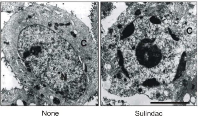
전자현미경으로 세포들을 관찰해 본 결과 sulindac 은 세포막의 주름(fold)을 소실하게 하고, 핵의 주변부 응축(peripheral condensation)을 유발함을 관찰하였다 (Fig. 4). 그리고, 고농도의 sulindac에 의해 세포사멸이 유발된 세포를 1% 아가로오즈겔 하에서 DNA 전기영 동분석을 시행한 결과 사다리 모양(ladder pattern)의 DNA 분절화를 관찰할 수 없었는데, 이는 sulindac으로 96시간 이상을 배양했을 때도 마찬가지였다(Fig. 5A).
4) Pulse Field Gel Electrophoresis (PFGE) 분석 Sulindac 처리를 한 HT-29세포는 PFGE 분석에서 고 분자량의 DNA 분절화가 유발됨을 관찰할 수 있었다.
즉, sulindac 처리를 한 12시간부터 고분자량 DNA조각 (HMW DNA fragments)이 나타났고, 48∼96시간째에 100-300 kbp의 고분자 DNA조각이 생성됨을 확인하였 다(Fig. 5B). 그러나 배양의 후반부(48시간 이상)에서 도 200∼2,000 bp 크기의 분절화는 발생하지 않음을 확인할 수 있었다.
5) Sulindac의 비전형적 세포사멸 기전에 있어 AIF의 역할
Sulindac 처리를 한 세포를 Western blot을 통해 분석 Fig. 4. Transmission electron microphotographs of sulindac
treated cells. Left panel, a control cell with a normal nucleus (N). Right panel, 72 hr after treatment with 1 mM sulindac. An apoptotic cell shows peripheral condensation of the nucleus and disapperance of surface fold. Bar = 5μm.
None Sulindac
Fig. 5. Sulindac induces high molecular weight (HMW) DNA fragmentation associated with apoptosis but no ladder-like DNA fragmentation in HT-29 cells. (A) Cells were exposed in the absence or the presence of sulindac (1 mM), etoposide (20μg/ml), or bile acid HS-1200 (20μM) for the indicated periods. Fragmented DNA was extracted and analyzed on 1.8% agarose gel, and visualized under UV light. (B) Cells were incubated in the absence or the presence of 1 mM sulindac for 0∼96 hr. At different time points, cells were collected and analyzed for HMW DNA cleavage. PFGE analysis was repeated three times, and similar results were obtained in the experiments. Lane M indicates size marker DNAs.
0 24 48 72 96 24 24 (hr)
Sulindac
Etoposide M 0 6 12 24 48 72 96 (hr) Kbp
1,600
750 565
225
A B
Bile acid
한 결과 AIF가 핵에서 점차 증가함을 확인할 수 있었 고(Fig. 6A), 또한 AIF가 미토콘드리아에서 핵으로 이 동하는 것은 면역형광현미경으로 관찰할 수 있었다 (Fig. 6B).
고 찰
세포사멸(apoptosis)이란 병태생리학적으로 불필요 하거나 손상받은 세포를 제거하기 위한 생체의 정상 적이고 능동적 세포 사멸 과정을 말한다.17 미토콘드리 아는 여러 가지의 세포사멸 유발인자(cytochrome c, Smac/DIABLO, Htr2/Omi, AIF, Endo G)를 함유하고 있 으며, 이 인자들의 분비 조절은 Bcl-2 단백 계열이 관 여하는 것으로 알려져 있다.18,19 즉 세포사멸을 유발하 는 Bcl-2계열(pro-apoptotic Bcl-2 members)은 cytochrome c, Smac/DIABLO 그리고 Htr2/Omi 같은 세포사멸 유발 인자들을 선택적으로 미토콘드리아에서 세포질로 방 출하게 하여 caspase의 활성을 유도하게 된다.10,20 이런 활성 caspase에 의해 Endo G 혹은 AIF 통로의 개방을 유발하게 되면서 세포사멸이 진행된다고 알려져 있 다.10,21
ATP생성을 포함한 다양한 기능에 있어 필수적인 미 토콘드리아에서 cytochrome c는 막전위 생성에 결정적 인 역할을 한다. Cytochrome c가 미토콘드리아에서 방 출되는 기전으로는 세포사멸을 유발하는 Bcl-2계열의
일부 혹은 전적인 작용으로 미토콘드리아의 외막에 있는 통로를 통한다고 여겨진다.22 투과이동공(permea- bility transition pore)의 개방으로 미토콘드리아 기질의 종창(swelling)에 기인한 외막의 파괴를 통해 발생하든 지,23 미토콘드리아 외막에 존재하는 전압의존성 음이 온 통로(voltage-dependent anion channels)의 폐쇄 결과 로 cytochrome c가 방출된다고 생각되고 있다.24 세포 사멸의 많은 연쇄단계가 세포질에서 존재하는 세포사 멸 유발 Bcl-2계열인 Bak 혹은 Bax가 세포질에서 미토 콘드리아로 이동함으로서 유도되는데, 이들 단백질들 이 미토콘드리아의 외막으로 들어가게 되면 미토콘드 리아의 막전위의 소실과 cytochrome c를 포함한 세포 사멸 유발 인자들의 방출이 일어나게 된다.25 본 연구 에서도 sulindac 처리 후 시간이 경과함에 따라 미토콘 드리아로부터 cytochrome c의 방출이 증가함을 관찰하 였고, 또한 sulindac에 의한 cytochrome c의 방출은 미 토콘드리아 막전위 하강에 선행함을 확인할 수 있었 다.
플라빈단백질(flavoprotein)인 AIF는 정상적으로 미 토콘드리아의 외막과 내막간의 공간에 존재하며 세포 사멸이 유발되면 핵으로 이동한다. 그리고 재조합 AIF 는 핵의 염색체 응축과 DNA의 고분자 분절화를 유발 하는 것이 실험적으로 설명되고 있는데, 정재된 핵에 재조합 AIF를 첨가하게 되면 caspase와 무관하게 고분 자(∼50 kb)로 DNA 분절화가 유발되면서 핵의 변연부 Fig. 6. AIF localizes from the mitochondria to the nucleus during apoptosis induced by sulindac. (A) Western blot analysis of AIF localization in nucleus induced by sulindac. Cells were treated with 1 mM sulindac for each time point, and samples of nucleus and total cell were subjected to 10% SDS-PAGE and transferred to a nitrocellulose filter. Equivalent protein loading was confirmed by Ponceau S staining. (B) Immunoelectron microscopic analysis of AIF localization to nucleus (N) in HT-29 cells induced by sulindac.
Cells were treated in the absence or the presence of sulindac (1 mM) for 72 hr, fixed in 4% paraformaldehyde, and assessed for immunoelectron microscopy. No background is observed in cytosol (C), mitochondria (M) and nucleus. Right panels are a higher magnification of the left. Bars = 250 nm.
AIF
0 6 12 24 48 72 (hr)
Nucleus
Total cell
None
Sulindac
A B
에 염색질 응축이 발생된 소견들이 관찰되며, 이는 세 포사멸이 진행되는 정상 세포의 첫 단계(stage I)와 유 사하다고 보고되고 있다.10 다양한 세포들에서 뉴클레 오좀 크기로의 DNA 분절화를 보이는 핵의 DNA 붕괴 는 세포사멸 과정의 가장 특징적인 소견으로 보인다.
그러나 고분자량 DNA 분절화는 세포사멸 현상을 재 현하는 데 있어 더욱 용이한 것으로 여겨지고 있으 며,26 염색체의 DNA가 세포사멸의 자극을 받고서 사 멸하는 과정에서 초기 단계에 발생하는 보편적이고 필수적 역할을 할 것이라고 강조하는 보고들이 있
다.27,28 동일 세포에서 세포사멸 유발 인자에 따라 올리
고뉴클레오좀 분절화가 동반되지 않더라도 세포사멸 이 발생함을 본 연구에서도 확인할 수 있었다. 특히 sulindac은 HT-29세포주에서 올리고뉴클레오좀 분절 화가 없이 고분자량의 DNA 분절화만을 유발하는 것 이 확인되어, sulindac은 stage I 세포사멸을 유도한다고 여겨진다. 세포사멸에서 발생하는 고분자 크기의 DNA 분절화는 핵의 기질의 접합부에 있는 DNA의 고 리 영역(loop domain)의 절제에 의한 것으로 보이고, 이는 고염(high salt)으로 추출된 핵에서 보이는 일련의 고분자량 DNA 분절화 과정과 거의 동일한 것으로 생 각된다. HT-29세포에서 다양한 자극에 의해 유발된 세 포사멸에서 서로 다른 양상의 DNA분절화가 관찰되는 현상으로 미루어 세포사멸에 다른 조절 기전이 관여할 가능성이 있을 수 있을 것으로 판단되며 이에 대해서는 더 심층적인 연구가 뒤따라야 할 것으로 생각한다.
결 론
Sulindac은 HT-29세포에서 세포사멸을 유도하고, PFGE에서 100∼300 kbp의 고분자량 DNA 분절화로만 핵의 DNA가 분해됨을 확인하였으나, 세포사멸의 전 형적인 소견인 사다리 모양(ladder pattern)의 DNA 전 기영동 양상은 관찰되지 않았다. 그러므로 sulindac은 HT-29 대장암 세포주에서 비전형적(atypical) 세포사멸 을 유발하는 것으로 생각되며, 여기에는 세포사멸 과 정의 또 다른 조절 기전이 관여할 수 있다는 단서를 제공한다고 여겨진다. 미토콘드리아에서 핵으로 이동 하는 AIF가 이런 비전형적 사멸형태를 보이는 요인의 하나로 생각되지만, AIF 외에 또 다른 어떤 인자가 sulindac에 의한 세포사멸 기전에 관계하는지 규명하 기 위해서는 더 많은 연구가 필요할 것으로 보인다.
REFERENCES
1. Rosenberg L, Palmer JR, Zauber AG, Warshauer ME, Stolley PD, Shapiro S. A hypothesis: nonsteroidal anti- inflam- matory drugs reduce the incidence of large-bowel cancer. J Natl Cancer Inst 1991;83:355-8.
2. Rigas B, Shiff SJ. Nonsteroidal anti-inflammatory drugs and the induction of apoptosis in colon cells: Evidence for PHS- dependent and PHS-independent mechanisms. Appoptosis 1999;4:373-81.
3. Reddy BS, Rao CV, Seibert K. Evaluation of cyclooxyge- nase-2 inhibitor for potential chemopreventive properties in colon carcinogenesis. Cancer Res 1996;56:4566-9.
4. Shiff SJ, Qiao L, Tsai LL, Rigas B. Sulindac sulfide, an aspirin-like compound, inhibits proliferation, causes cell cycle quiescence, and induces apoptosis in HT-29 colon adenocarcinoma cells. J Clin Invest 1995;96:491-503.
5. Piazza GA, Rahm AK, Finn TS, Fryer BH, Li H, Stoumen AL, et al. Apoptosis primarily accounts for the growth- inhib- itory properties of sulindac metabolites and involves a mech- anism that is independent of cyclooxygenase inhibition, cell cycle arrest, and p53 induction. Cancer Res 1997;57: 2452-9.
6. Pasricha PJ, Bedi A, O'Connor K, Rashid A, Akhtar AJ, Zahurak ML, et al. The effects of sulindac on colorectal proliferation and apoptosis in familial adenomatous poly- posis. Gastroenterology 1995;109:994-8.
7. Williams GT. Programmed cell death: apoptosis and onco- genesis. Cell 1991;65:1097-8.
8. Yuan J. Evolutionary conservation of a genetic pathway of programmed cell death. J Cell Biochem 1996;60:4-11.
9. Green DR, Reed JC. Mitochondria and apoptosis. Science 1998;281:1309-12.
10. Susin SA, Lorenzo HK, Zamzami N, Marzo I, Snow BE, Brothers GM, et al. Molecular characterization of mitoc- hondrial apoptosis-inducing factor. Nature 1999;397:441-6.
11. Thornberry NA, Rosen A, Nicholson DW. Control of apop- tosis by proteases. Adv Pharmacol 1997;41:155-77.
12. Shiff SJ, Koutsos MI, Qiao L, Rigas B. Nonsteroidal anti- inflammatory drugs inhibit the proliferation of colon adeno- carcinoma cells: effects on cell cycle and apoptosis. Exp Cell Res 1996;222:179-88.
13. Choi HJ, Kim HH, Lee HS, Huh GY, Seo SY, Jeong JH, et al. Lactacystin augments the sulindac-induced apoptosis in HT-29 cells. Apoptosis 2003;8:301-5.
14. Qiao L, Hanif R, Sphicas E, Shiff SJ, Rigas B. Effect of aspirin on induction of apoptosis in HT-29 human colon adenocarcinoma cells. Biochem Pharmacol 1998;55:53-64.
15. Higuchi Y, Matsukawa S. Appearance of 1-2 Mbp giant DNA fragments as an early common response leading to cell death induced by various substances that cause oxi-
dative stress. Free Radic Biol Med 1997;23:90-9.
16. Wang W, Passaniti A. Extracellular matrix inhibits apoptosis and enhances endothelial cell differentiation by a NfkappaB- dependent mechanism. J Cell Biochem 1999;73:321-31.
17. Kroemer G, Petit P, Zamzami N, Vayssiere JL, Mignotte B.
The biochemistry of programmed cell death. FASEB J 1995;9:1277-87.
18. Wang X. The expanding role of mitochondria in apoptosis.
Genes Dev 2001;15:2922-33.
19. van Loo G, Saelens X, van Gurp M, MacFarlane M, Martin SJ, Vandenabeele P. The role of mitochondrial factors in apoptosis: a Russian roulette with more than one bullet. Cell Death Differ 2002;9:1031-42.
20. Gross A, McDonnell JM, Korsmeyer SJ. BCL-2 family members and the mitochondria in apoptosis. Genes Dev 1999;13:1899-911.
21. Li LY, Luo X, Wang X. Endonuclease G is an apoptotic DNase when released from mitochondria. Nature 2001;412:
95-9.
22. Shimizu S, Ide T, Yanagida T, Tsujimoto Y. Elec- trophysiological study of a novel large pore formed by Bax and the voltage-dependent anion channel that is permeable to cytochrome c. J Biol Chem 2000;275:12321-5.
23. Brenner C, Cadiou H, Vieira HL, Zamzami N, Marzo I, Xie Z, et al. Bcl-2 and Bax regulate the channel activity of the
mitochondrial adenine nucleotide translocator. Oncogene 2000;19:329-36.
24. Vander Heiden MG, Chandel NS, Li XX, Schumacker PT, Colombini M, Thompson CB. Outer mitochondrial membr- ane permeability can regulate coupled respiration and cell survival. Proc Natl Acad Sci U S A 2000;97:4666-71.
25. Heerdt BG, Houston MA, Wilson AJ, Augenlicht LH. The intrinsic mitochondrial membrane potential (∆ψm) is associated with steady-state mitochondrial activity and the extent to which colonic epithelial cells undergo butyrate- mediated growth arrest and apoptosis. Cancer Res 2003;63:
6311-9.
26. Sleiman RJ, Catchpoole DR, Stewart BW. Drug-induced death of leukaemic cells after G2/M arrest: higher order DNA fragmentation as an indicator of mechanism. Br J Cancer 1998;77:40-50.
27. Walker PR, Weaver VM, Lach B, LeBlanc J, Sikorska M.
Endonuclease activities associated with high molecular weight and internucleosomal DNA fragmentation in apop- tosis. Exp Cell Res 1994;213:100-6.
28. Oberhammer F, Wilson JW, Dive C, Morris ID, Hickman JA, Wakeling AE, et al. Apoptotic death in epithelial cells:
cleavage of DNA to 300 and/or 50 kb fragments prior to or in the absence of internucleosomal fragmentation. EMBO J 1993;12:3679-84.