Korean J Gastroenterol Vol. 75 No. 5, 292-295 https://doi.org/10.4166/kjg.2020.75.5.292 pISSN 1598-9992 eISSN 2233-6869
CASE REPORT
Korean J Gastroenterol, Vol. 75 No. 5, May 2020 www.kjg.or.kr
위 내시경 조직 검사로 진단된 위 궤양 천공의 간 침범
이선욱, 지삼룡, 이홍섭, 이상헌
인제대학교 의과대학 부산백병원 내과
Gastric Ulcer Perforation to the Liver Diagnosed by Endoscopic Biopsy
Seon Uk Lee, Sam Ryong Jee, Hong Sub Lee and Sang Heon Lee
Department of Internal Medicine, Busan Paik Hospital, Inje University College of Medicine, Busan, Korea
Peptic ulcer disease is common and can be diagnosed easily if the patient has an ulcer history or characteristic abdominal symptoms. On the other hand, it may take a long time for the patient to visit the hospital due to severe complications if the patient is old or insensitive to symptoms caused by peptic ulcers. In the present case, a 72-year-old female visited the hospital due to general weakness and inadequate oral intake, which started two weeks ago. Endoscopy and abdominal CT revealed huge gastric ulcer findings. Through a tissue examination by endoscopy, hepatic cells were identified, and the patient was diagnosed with pep- tic ulcer perforation to the liver and later received surgical treatment. (Korean J Gastroenterol 2020;75:292-295)
Key Words: Stomach ulcer; Peptic ulcer perforation; Perforation to the liver; Endoscopic biopsy
Received February 18, 2020. Revised March 26, 2020. Accepted March 28, 2020.
CC This is an open access article distributed under the terms of the Creative Commons Attribution Non-Commercial License (http://creativecommons.org/licenses/
by-nc/4.0) which permits unrestricted non-commercial use, distribution, and reproduction in any medium, provided the original work is properly cited.
Copyright © 2020. Korean Society of Gastroenterology.
교신저자: 지삼룡, 47392, 부산시 부산진구 복지로 75, 인제대학교 의과대학 부산백병원 내과
Correspondence to: Sam Ryong Jee, Department of Internal Medicine, Busan Paik Hospital, Inje University College of Medicine, 75 Bokji-ro, Busanjin-gu, Busan 47392, Korea. Tel: +82-51-890-6536, Fax: +82-51-892-0273, E-mail: tokimom@nate.com, ORCID: https://orcid.org/0000-0002-7928-1153
Financial support: None. Conflict of interest: None.
INTRODUCTION
Peptic ulcer disease can be complicated by inflammation, ulceration, bleeding, or perforation. The condition can be diag- nosed easily if there is a history of ulcer or acute characteristic pain in the abdomen. On the other hand, it is difficult to diagnose patients without the characteristic symptoms of peptic ulcer or elderly patients who are insensitive to pain. Penetration into the liver is a rare peptic ulcer disease complication that can lead to severe complications, such as upper gastrointestinal bleeding or abscess formation.1 This paper reports a 72-year-old female who developed a silent gastric ulcer perfo- ration to the liver. The diagnosis was made by a histological examination of endoscopic biopsies, and surgical treatment was performed.
CASE REPORT
A 72-year-old female visited the hospital because of a two-week history of weakness, dizziness, poor oral intake, and vomiting. She appeared pale and had anemic conjunctiva.
Her abdomen was soft without organomegaly or mass, and she did not suffer from abdominal pain or distension. On the other hand, she complained of tenderness on the pressure of the epigastric area. The bowel sounds were normo-active.
The rectal examination did not reveal any palpable mass. Five months before her visit to the hospital, she purchased pain- killers at the pharmacy and used them because of sustained pain on the left shoulder. She had anemia in the blood test at a local hospital 3 months ago and was taking hypertension medication.
Lee SU, et al. Gastric Ulcer Perforation to the Liver 293
Vol. 75 No. 5, May 2020
A B
Fig. 2. (A, B) Abdominal computed tomography shows diffuse wall thickening in the stomach with a suspected ulcerative lesion (arrows) in the lesser curvature side of the gastric antrum and perigastric infiltration.
Fig. 1. Chest X-ray showing no free air.
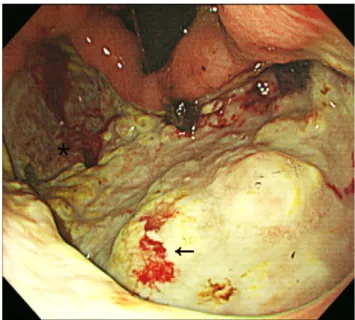
Fig. 3. Endoscopic image shows a large round deep ulcer (A1 stage) at the lesser curvature side of the body. Biopsies were done (arrow for Fig. 4A and asterisk for Fig. 4B).
Upon arrival to the emergency room, her initial vital signs were a regular heart rate at 102 beats/min, a blood pressure of 100/60 mmHg, and 36.5ºC of body temperature. The fol- lowing are the results of her complete blood and biochemistry tests at the time of her visit: complete blood count, white blood cells count 18.75×10⁹ L (95% neutrophils, 4% lympho- cytes); hemoglobin 2.5 g/dL; hematocrit 9.1%, platelet 882 k/mm3; liver function tests; PT 11.7 sec; total bilirubin 0.3 mg/dL; AST 17 U/L; ALT 6 U/L; ALP 199 U/L; BUN 53 mg/dL; creatinine 1.15 mg/dL; glucose 174 mg/dL; amy-
lase 26 U/L; lipase 14 U/L; and CRP 8.11 mg/dL.
There was no free air in the chest X-ray (Fig. 1). CT revealed diffuse wall thickening in the stomach with a suspected ulcer- ative lesion in the lesser curvature side of the gastric antrum, possible sealed-off perforation, and perigastric infiltration (Fig. 2). Upper gastrointestinal endoscopy revealed a large ul- cer (A1 stage), which was identified by CT, approximately 10 cm in size at the lesser curvature side of the body (Fig. 3).
The pylorus had a deformity because of an ulcer scar and had stenosis that impeded the endoscope. The duodenal bulb
294 이선욱 등. 위 내시경 조직 검사로 진단된 위 궤양 천공의 간 침범
The Korean Journal of Gastroenterology
A B
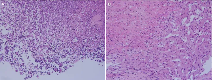
Fig. 4. (A) An endoscopic biopsy demonstrated neutrophils aggregates and necrotic debris, indicative of the findings of an ulcer bed (H&E,
×200). (B) A small piece of biopsied tissue showed fibrotic granulation tissue (upper left) that adhered tightly to hepatic parenchyma (lower right) (H&E, ×200).
and second portion were normal. Biopsies from the center and margin of the gastric ulcer were taken. The rapid urease test for Helicobacter pylori was positive. A histology examina- tion of the biopsies revealed ulcer debris and hepatic paren- chyma (Fig. 4). Considering the size of the ulcer and the risk of developing a liver abscess, it was decided that surgery should be performed. Hemigastrectomy was performed, and the pathology of the surgical specimen was a chronic ulcer with perforation and fibrous adhesion to the liver. The patient started sips of water and diet on the 5th day of surgery and was discharged on the 13th day of surgery without post- operative complications.
DISCUSSION
In a systematic review, the reported annual incidence of peptic ulcer perforation ranged from 3.8 to 14 per 100,000 individuals.1 Thirty-day and 90-day mortality rates of up to 20% and 30%, respectively, have been reported.2 In develop- ing countries, patients tend to be young male smokers. In developed countries, however, patients tend to be elderly with multiple co-morbidities and the associated use of NSAIDs or steroids. NSAIDs, Helicobacter pylori, physiological stress, smoking, corticosteroids, and previous history of peptic ulcer disease are risk factors for perforated peptic ulcer.2 The clin- ical features of peptic ulcer perforation include the sudden onset of abdominal pain, tachycardia, and abdominal rigidity
in the early stages, followed by abdominal distension, pyrexia, hypotension, fever, and vomiting.3 The prognosis for a diag- nosis within the first 6 hours is good, but adverse effects increase markedly when the delay exceeds 12 hours.
Therefore, a rapid diagnosis is essential.4 Abdominal plain film, CT, and ultrasonography may have a diagnostic role in confirming peptic ulcer perforation.5
Organ penetration to the pancreas, gastro-hepatic omen- tum, biliary system, and liver was reported in 8% of 417 pa- tients who had undergone surgical treatment due to peptic ulcer disease.6,7 Liver penetration is a rare but severe compli- cation of peptic ulcer disease. Generally, the diagnosis is made by surgery or endoscopic biopsy.7-12 Non-surgical treat- ment using antibiotics and intravenous proton pump inhibitors may be possible if the patient has no leakage, is clinically stable, and shows signs of improvement. On the other hand, urgent surgery is essential if the patient’s condition deterio- rates, regardless of a leak.
In 2004, Kayacetin and Kayacetin8 described 14 cases of gastric ulcer penetrating to the liver. All the patients showed severe gastrointestinal bleeding, as in the present case.
Abdominal pain was described in only four patients. Therefore, in the present case, abdominal pain did not appear to be a common finding. Most cases were located at the anterior wall or lesser curvature of the corpus or antrum. Nine of the 14 cases were treated with surgery operation.
Most cases had normal liver function tests. This may reflect
Lee SU, et al. Gastric Ulcer Perforation to the Liver 295
Vol. 75 No. 5, May 2020
that local hepatic injury does not cause abnormalities in the liver functions. The diagnostic value of liver function tests in cases of ulcer penetration into the liver is limited.
In this case, the patient showed severe anemia at the time of hospitalization and complained of dizziness, vomiting, in- adequate oral intake, and epigastric tenderness. A physical examination did not show any signs indicating a perforation, such as rebound tenderness and peritoneal irritation sign, and there was no free air in the simple abdominal image.
In the present case, the history of painkiller medication, ad- vanced age, and Helicobacter pylori could be the cause of the gastric ulcer perforation. A small gastric ulcer perforated to the liver. As the liver tissue around the perforation became inflamed and fibrous, it caused adhesion, which concealed the patient’s perforation symptoms. As the size of the gastric ulcer gradually grew, it is believed that ulcer bleeding caused severe anemia.
In conclusion, a high index of suspicion to make a diagnosis is necessary when patients have the risk factors of a perfo- rated peptic ulcer because they may have a silent perforated peptic ulcer that is not accompanied by sudden physical symptoms.
REFERENCES
1. Lau JY, Sung J, Hill C, Henderson C, Howden CW, Metz DC.
Systematic review of the epidemiology of complicated peptic ul- cer disease: incidence, recurrence, risk factors and mortality.
Digestion 2011;84:102-103.
2. Chung KT, Shelat VG. Perforated peptic ulcer - an update. World J Gastrointest Surg 2017;9:1-12.
3. Silen W. Method of diagnosis: the examination of the patient and the grouping of symptoms and signs. In: Silen W, ed. Cope's early diagnosis of the acute abdomen. 22nd ed. New York (NY): Oxford University Press, 2010:28-53.
4. Svanes C, Lie RT, Svanes K, Lie SA, Søreide O. Adverse effects of delayed treatment for perforated peptic ulcer. Ann Surg 1994;
220:168-175.
5. Grassi R, Romano S, Pinto A, Romano L. Gastro-duodenal perfo- rations: conventional plain film, US and CT findings in 166 con- secutive patients. Eur J Radiol 2004;50:30-36.
6. Armbruster C, Dittrich K, Kriwanek S. The place of selective prox- imal vagotomy in complicated duodenal ulcers. Wien Klin Wochenschr 1989;101:615-617.
7. Novacek G, Geppert A, Kramer L, et al. Liver penetration by a duo- denal ulcer in a young woman. J Clin Gastroenterol 2001;33:
56-60.
8. Kayacetin E, Kayacetin S. Gastric ulcer penetrating to liver diag- nosed by endoscopic biopsy. World J Gastroenterol 2004;10:
1838-1840.
9. Brullet E, Campo R, Calvet X, Gimenez A. Gastric ulcer penetrat- ing to the liver: endoscopic diagnosis. Am J Gastroenterol 1993;88:794-795.
10. Oka A, Amano Y, Uchida Y, et al. Hepatic penetration by stomal ulcer: rare complication of a peptic ulcer. Endoscopy 2012;44 Suppl 2 UCTN:e347-e348.
11. Lesquereux-Martínez L, Álvarez AM, Parada-González P, Bustamante-Montalvo M. Gastric ulcer penetrating the liver. Cir Esp 2017;95:46.
12. Hayashi H, Kitagawa H, Shoji M, et al. Duodenal ulcer penetration into the liver at the previous left hemihepatectomy site. Int J Surg Case Rep 2013;4:1110-1112.