물여뀌 에탄올 추출물의 미백 효과
황병수, 이승영, 강창희, 한웅, 오영택, 유상미, 김민진, 김철환, 엄정혜, 정상철, 이욱재, 안영희, 정용태*
국립낙동강생물자원관
Received: October 18, 2018 / Revised: November 2, 2018 / Accepted: November 12, 2018
서 론
물여뀌(Polygonum amphibium L.)는마디풀속여뀌절에 속하는식물로써육상및수중환경모두에서식할수있는 분류군이다. 현재북미, 동아시아, 유럽등북반구의온대에 서아한대지방에주로자라며, 남미, 멕시코및남아프리카 등지에도일부유입되어자라고있는것으로알려져있다[1].
이러한물여뀌에대한생리활성연구는많이이루어져있지 않으며, 림프구활성화, 항암및항산화효과에대한보고가 있을뿐이다[2−4].
미백에대한관심은외모에대한관심이증가된현대인에 게크게증가된양상이다. 이러한미백효과는멜라닌의합
성제어와밀접한관계가있다. 멜라닌은 ultraviolet (UV)와 같은고에너지광원으로부터피부를보호하기위해멜라닌 을합성하지만과생성시에는기미주근깨, 주름과같은피부
질환을야기한다[5, 6]. 이러한멜라닌의과생성을억제하고
피부에침착되는멜라닌을제어하는물질을찾는연구가최 근활발하며현재화장품과같은산업적으로이용되는멜라 닌저해제로대표적인물질은 arbutin과 kojic acid 등이있
다[7]. 그러나과다사용시피부자극및알레르기와같은부
작용이발생할위험성을내포하고있어제한적으로사용되
어지고있다[8]. 따라서안전적이며효과적인멜라닌저해제
를찾기위한노력이계속되고있으며특히천연물로부터 의미백활성을가진물질을찾기위한많은연구들이수행 되고있는실정이다.
따라서본연구를통해서미백에대한연구가전무한물
여뀌에탄올추출물의미백활성을 B16F10 흑색종세포와
제브라피쉬 embryo 모델을이용하여살펴보고자한다. Whitening Activities of Ethanol Extract from Polygonum amphibium L.
Buyng Su Hwang, Seung Young Lee, Chang Hee Kang, Woog Han, Young Taek Oh, Sang Mi Yu, Min Jin Kim, Chul Hwan Kim, Jung Hye Eom, Sang Chul Jeong, Wook Jae Lee, Young Hee Ahn, and Yong Tae Jeong*
Freshwater Bioresources Utilization Bureau, Nakdonggang National Institute of Biological Resources, Sangju 37242, Republic of Korea The purpose of this study was to investigate the melanogenesis inhibiting activity of the ethanol extract from Polygonum amphibium L. Firstly, the n-hexane (Hx), chloroform (CHCl3), ethyl acetate (EA), n-buta- nol (BuOH), and water (Water) fractions were isolated from the P. amphibium L. ethanol extract. The effi- cacy of melanogenesis was found to significantly decrease via the EA and BuOH fractions when compared to the control in B16F10 cells. EA particularly showed the lowest melanin content in B16F10 cells when compared to all the other extracts. Concentration-dependent inhibition of melanin synthesis was also observed in the EA fraction at concentrations below 50 µg/ml, which did not exhibit cytotoxicity in B16F10 cells. Notably, the expression of three key proteins (tyrosinase, tyrosinase-related protein-1 (TRP-1), and TRP-2), which are involved in melanogenesis, were significantly decreased via the EA fraction. EA also inhibited body pigmentation in vivo in a zebrafish model. Overall, we demonstrated melanogenesis sup- pression using the EA fraction from P. amphibium L., which could be a potential candidate for an anti- melanogenesis agent.
Keywords: Polygonum amphibium L., melanogenesis, B16F10, zebrafish, ethanol extract
*Corresponding author
Tel: +82-54-530-0936, Fax: +82-54-530-0949 E-mail: [email protected]
© 2019, The Korean Society for Microbiology and Biotechnology
재료 및 방법
기기 및 시약
본 실험에서 사용된 시약은 L-tyrosine, NaOH, 12-ο- tetradecanoylphorbol-13-acetate, 1-phenyl-2-thiourea (PTU) 는 Sigma Chemical Co. (USA)에서구입하였다. Fetal bovine serum (FBS), penicillin/streptomycin 및 Dulbecco’s modified eagle’s medium (DMEM)은 Gibco Co. (USA)에서구입하여 세포를 배양하였으며 cell counting kit-8 (CCK-8)은 Dojindo Lab. (Japan)에서구입하였다.
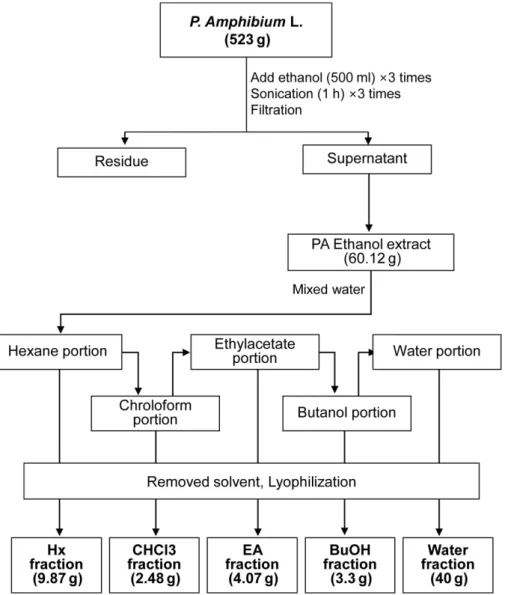
시료추출
물여뀌를 ethanol로먼저 추출한후 water로현탁한다 음, 동량의헥산으로진탕추출하여헥산분획(Hx)을회수
하였다. 증류수층을다시동량의클로로포름으로진탕추출 하여클로로포름분획(CHCl3)을회수하고증류수층을다시 동량의에틸아세테이트로진탕추출하여에틸아세테이트 분획(EA)을회수하며증류수층을위방법과동일하게부탄
올로진탕추출하여부탄올분획(BuOH)을얻었으며, 나머
지증류수층으로네종류의분획을 Fig. 1과같이 분리하
였다. 세포배양
미백 활성 실험에 널리 이용되는 B16F10 mouse
melanoma 세포배양은 37℃, 5% CO2의조건인 CO2 배양기 (Thermofisher, USA)에서배양되었다. 사용된배지는 10%
FBS, 1% penicillin/streptomycin이포함된 DMEM 배지를 사용하였다. 세포독성은 CCK-8을사용하여측정하였다.
Fig. 1. The schematic diagram for the recovery of purified extract produced from the P. amphibium L.
멜라닌 양 측정
멜라닌양은 Hosoi 등의방법을변형하여사용하였다[9].
B16F10 세포 1 × 10⁵개를 24-well plate의각 well에분주한
후 200 nM의α-MSH를첨가하여하룻밤배양하여실험에
사용하였다. 각화합물을농도별로배지에처리하여 4일동 안 배양했다. 이후 phosphate-buffered saline (PBS; pH 7.4)으로세척하고 1 N NaOH를 200 μl씩각 well에처리하 여 용해시켰다. 용해된 세포를 96 well plate로 옮겨 microplate reader (Biotek cytation3)를 사용하여 optical density를 405 nm에서측정하고멜라닌함량은대조군과대 비해 %로계산한다.
Western blot 분석
Western blot 방법을이용하여 melanin 합성에작용하는 단백질(tyrosinase, tyrosinase related protein-1 (TRP-1), TRP-2 등)의발현을분석하였다. 상기의 B16F10 세포를배 양하여 25, 50 μg/ml 농도의 EA 분획을 72 h 처리한후, 상 기 세포를 단백질분해효소 저해제(protease inhibitor cocktail, Sigma, USA)를 첨가한 RIPA 완충용액(10 mM Tris-HCl (pH 7.5), 1% NP-40, 0.1% sodium deoxycholate, 0.1% SDS, 150 mM NaCl, 1 mM EDTA)으로용해시켰다. 이후, 얻어진세포용해물을 4℃에서 5 min 간 12,000 ×g로 원심분리한후, BCA 단백질키트를이용하여단백질의농 도를측정하였다. 세포에서추출한단백질에전기영동용완 충용액을첨가하고 100℃에서 5 min간가열한후 sodium dodecyl sulfatepolyacrylamide gel electrophoresis (SDS-
PAGE)를수행하였다. 전기영동후, 분자량별로분리된단백
질은젤에서 nitrocellulose (NC) membrane에전이시켰다. 이 때, 비특이적반응을최소화하기위해완충용액(phosphate buffered saline; PBS (pH 7.4), 0.1% Tween 20, 5%
skimmed milk)을 1 h 동안상온에서반응시킨후, 1차항체 를다음과같이희석하여사용하였다. 항체희석은 5% Skim
milk 용액을이용하여상온에서 1 h 동안반응시켜희석하
였다. 반응 후, 세척 완충액(0.1% Tween-20, PBS)으로 5 min 간 3−4회세척한후 1:5000으로희석시킨 2차항체 (HRP-conjugated anti-goat IgG antibody) 용액으로 1 h 동 안상온에서반응시켰다. 2차항체반응후, ECL (enhanced
chemiluminescence) 시약을사용하여분석대상단백질밴
드를발색시켜정량하였다. 제브라피쉬 사육
성숙제브라피쉬는 14:10 h의낮과밤의주기로 28.5℃의 수온을유지, 폐쇄순환여과시스템을갖춘수조에서사육 하였다[10, 11]. 성숙제브라피쉬는 20 L 수조에 3−5마리넣 고살아있는 brine shrimp (San Francisco BayBrand, Inc.,
USA)를하루에 3회식이하였다. 적정생육온도인 28.5℃의 수온을유지한어항에서사육하였다.
제브라피쉬 멜라닌 저해 실험
제브라피쉬의멜라닌저해실험은 Choi 등의방법을변형 하여사용하였다[12]. 성숙제브라피쉬암수를알채취전날 알채취용수조에넣고다음날광주기시기 1−2 h 이후에알 을채취하였다. 채취된 알은 zebrafish embryo medium에 넣고 24 h 발생시킨후, 각샘플을농도별로처리하였다. 샘
플처리시코리온(chorion)의샘플투과정도를알수없기
에각알의코리온에구멍을내어처리하였고, 샘플처리후
24 h 이후샘플의독성이나타난알은제거하고, 나머지알
의코리온을완전제거후 2번째샘플을처리하였다. 샘플
처리 48 h 이후에대조군대비샘플처리구의색소발생정
도를실체현미경으로관찰하였다. 제브라피쉬멜라닌생성 량은코리온에구멍낸 100마리의제브라피쉬알에샘플처 리을 48 h 처리하였다. 멜라닌 추출은 동량의 Pro-prep protein extraction solution (Intron) 처리 후 sonication하 고원심분리하여상득액을제거하고침전물에 1 N NaOH (70℃)를 처리하여용해시켰다. 용해된 침전물을 96 well plate로옮겨 microplate reader (Bioteck cytation3)를사용 하여 optical density를 405 nm에서측정하고멜라닌함량 은대조군과대비해 %로계산한다.
통계처리
본연구의모든결과는 3회반복실험에대한평균(mean)
±표준오차(standard deviation, SD)로나타내었으며, 통계 분석은 Student’s t-test를실시하여관찰하였다.
결과 및 고찰
물여뀌 에탄올 추출물의 B16F10 세포의 멜라닌 합성에 미 치는 영향
물여뀌전초 523 g을에탄올로추출하여 60.12 g의에탄 올추출물의회수하였다. 회수한물여뀌에탄올추출물을
B16F10 세포를이용하여멜라닌합성저해능을실험한결
과, 50 μg/ml의농도에서 16.4% 저해하였다(Fig. 2A). 이렇 게멜라닌합성저해능을확인한물여뀌에탄올추출물은다 시유기용매로분획처리하여헥산분획(9.87 g), 클로로포름 분획(2.48 g), 에틸 아세테이트 분획(4.07 g), 부탄올분획 (3.3 g), 그리고 Water 분획(40 g)으로분리하였다. 각분획
중가장높은멜라닌합성저해능은 Fig. 2B에서와같이에
틸아세테이트분획이대조군대비통계적으로유의미하게 감소하여 32.6%로가장높은효과를보였다.
물여뀌 에틸아세테이트(EA) 분획의 B16F10 세포의 멜라 닌 합성에 미치는 영향
EA 분획에의한 B16F10 세포에서의세포독성을 CCK-8 assay kit를이용하여분석하였다(Fig. 3A). 그결과, 100 μg/
ml의농도에서부터세포독성을나타내어 50 μg/ml 이하의
농도에서 B16F10 세포의멜라닌합성저해효과를실험하
였다. Fig. 3B에서와같이농도의존적으로 B16F10 세포내 의멜라닌함량이감소되는경향을보였으며, 이전실험과 동일하게 50 μg/ml의농도에서 대조군대비 32.6% 감소되 는것을확인하였다.
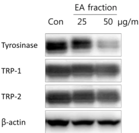
또한 EA 분획의멜라닌합성저해기전을알아보기위하 여멜라닌합성에관련한주요 효소인 tyrosinase, TRP-1, TRP-2에대한단백질발현정도를 western blot을이용하여 확인하였다(Fig. 4). 그결과, tyrosinase 단백질이확연히농 도 의존적으로 감소되고 TRP-1, -2 단백질 발현은
tyrosinase 단백질에비해약하게감소됨을확인하였다. 따
라서 tyrosinase 단백질이주요기전으로작용하여 EA 분획 에의해저해됨으로써멜라닌합성저해효과를나타내는것 으로사료된다.
생체내에서의멜라닌합성과정은 tyrosinase을기질로하 여 tyrosinase에 의해 hydroxylation 반응을 거쳐 3,4- dihydroxyindolephenylamine (L-DOPA)로되고이것은다 시 L-DOPA quinone으로산화된다[13]. 이후아미노산혹은 단백질과중합반응에의해멜라닌이합성되는것으로알려 져있다[14]. 또한 멜라닌의합성에 있어서 key protein은 tyrosinase, tyrosinase related protein-1 (TRP-1), TRP-2 가있으며이들단백질에의해 3,4-dihydroxyphenylalanine (DOPA)를 DOPA quinone으로전환된후일련의과정을거
쳐멜라닌이합성된다[15]. 물여뀌에대한미백효과는보고
되지않았지만여뀌(P. hydropiper L.)에서추출한에틸아 세테이트분획과 aglycon 분획에서 tyrosinase 활성저해효 Fig. 2. Effects of P. amphibium L. ethanol extract on melano-
genesis in B16F10 cells. (A) PA ethanol extract. (B) PA fractions.
Inhibition of melanin synthesis was measured with triplicate experiment. The cells were cultured with 50 µg/ml of PA ethanol extract and PA fractions for 3 days. Each value is expressed a mean ± SD of triplicate determinations. *p < 0.05, ***p < 0.001 versus of control group.
Fig. 3. Effects of EA fraction isolated from P. amphibium L. on cell cytotoxicity and melanogenesis in B16F10 cells. (A) Effect of cell cytotoxicity was measured with triplicate experiment. The cells were cultured with 0-200 µg/ml of EA fraction for 3 days, and cell cytotoxicity was determined by CCK-8 cell counting kit.
(B) Inhibition of melanin synthesis was measured with triplicate experiment. The cells were cultured with 0-50 µg/ml of EA frac- tion for 3 days. Each value is expressed a mean ± SD of triplicate determinations. ***p < 0.001 versus of control group.
과와 elastase의활성저해효과를확인함으로써항미백과항 주름 효과를 보고하였다[16]. 또한 붉은털여뀌(Persicaria orientalis) 추출물이 B16F1 세포에 대한 약한 저해효과 (0.1% 저해율)가특허로보고되었다.
제브라피쉬의 멜라닌 합성 저해 활성
제브라피쉬는한번에다수(100−200개)의알을낳으며발 생이빨라실험결과를빠르게도출할수있고, 세포실험에
서알수없는여러가지결과를알수있는장점이있다. 또 한동물실험을통한동물윤리문제로인해하등동물또는세 포실험으로대체하려하나생리활성의복잡한메커니즘으
로인해연구에한계가있다[17]. 이와같은이유로제브라
피쉬는새로운동물모델로각광을받고있다[18]. 특히새로 운미백제탐색하는데있어여러가지제약을회피할수있 는장점이있으며몸색깔이투명하여색소관찰이용이해 많이이용되고있다[12, 19, 20]. 이러한장점을가지고있는 제브라피쉬모델을이용하여 EA 분획을제브라피쉬 embryo
에 48 h 처리한후멜라닌합성에미치는영향을살펴본결
과 Fig. 5A와같다. 양성대조군으로이미알려진미백인 PTU (phenylthiourea )를사용하였으며, PTU (50 μg/ml을)를처
리하였을때대조군대비 70.4% 저해됨을확인하였다. EA
분획 50 μg/ml을처리했을때요크(yolk) 부분의색소침착 이저해될뿐만아니라 EA 분획처리농도가증가됨으로써 멜라닌합성이저해됨을확인하였고 EA 분획 25 μg/ml 처 리군에서는대조군대비유의미한저해를보이지않았지만 EA 분획 50 μg/ml 처리군에서는통계적으로유의미하게대 조군에비해저해됨(13.1%)을확인하였다(Fig. 5B).
물여뀌에탄올추출물의멜라닌합성저해효과를살펴보 기위해 B16F10 세포와제브라피쉬 embryo를이용하였다.
그결과, B16F10 세포의멜라닌합성이저해되며하위분획
중에틸아세테이트분획이가장높은미백효과를나타내 었다. 또한제브라피쉬 embryo의멜라닌합성이높게저해 됨을확인하였다. 이러한결과를바탕으로에틸아세테이트 분획의활성단일물질규명과기전연구가이루어져야될것 이며이를바탕으로이제껏연구되지않았던담수수변식물 Fig. 4. Effects of EA fraction isolated from P. amphibium L. on
melanogenesis and expression of melanogenesis-related protein in B16F10 cells. The cells were cultured with 0-50 µg/
ml of MSE for 72 h. Whole-cell lysate were then subjected to western blot analysis using antibodies against tyrosinase, TRP-1 and TRP-2. Equal protein loading was confirmed using β-actin antibody.
Fig. 5. Effects of EA fraction isolated from P. amphibium L. on melanogenesis in zebrafish. Synchronized embryos were treated with melanogenic inhibitors at the indicated concentrations. EA fraction were dissolved in 0.1% DMSO then added to the embryo medium. The effects on the pigmentation of zebrafish were observed under the stereomicroscope. Each value is expressed a means
± SD of triplicate determinations. ***p < 0.001 versus of control group
인물여뀌추출물이미백화장품천연소재로서의활용가 능성이높을것으로생각된다.
Acknowledgments
This work was supported by a grant from the Nakdonggang National Institute of Biological Resources (NNIBR), funded by the Minis- try of Environment(MOE) of the Republic of Korea (NNIBR201902105).
Conflict of Interest
The authors have no financial conflicts of interest to declare.
References
1. Yaqian G, Bhandari GS, Park JH, Park CW. 2013. A systematic study of the Polygonum amphibium L. complex (Polygonaceae) based on chloroplast DNA sequences. Korean J. Pl. Taxon. 43:
34-45.
2. Smolarz HD, Surdacka A, Roliński J. 2003. Influence of querce- tin-3-methyl ether from Polygonum amphibium on activation lymphocytes from peripheral blood of healthy donor in vitro.
Phytother. Res. 17: 744-747.
3. Smolarz HD, Budzianowski J, Bogucka-Kocka A, Kocki J, Men- dyk E. 2008. Flavonoid glucuronides with anti-leukaemic activ- ity from Polygonum amphibium L. Phytochem. Anal. 19: 506- 513.
4. Kwon SH, Na HL, Jeong JD, Baek NI, Park SG, Choi HK. 2012. A comparison of radical scavenging activity and cyanobacteria growth inhibition of aquatic vascular plants. Korean J. Limnol.
45: 11-20.
5. Tsatmali M, Ancans J, Thody AJ. 2002. Melanocyte function and its control by melanocortin peptides. J. Histochem. Cytochem. 50:
125-133.
6. Briganti S, Camera E, Picardo M. 2003. Chemical and instru- mental approaches to treat hyperpigmentation. Pigment Cell Res. 16: 101-110.
7. Fujimoto N, Watanabe H, Nakatani T, Roy G, Ito A. 1998. Induc- tion of thyroid tumours in (C57BL/6N x C3H/N)F1 mice by oral administration of kojic acid. Food Chem. Toxicol. 36: 697-703.
8. Hermanns JF, Pierard-Franchimont C, Pierard GE. 2000. Skin colour assessment in safety testing of cosmetics. An overview.
Int. J. Cosmet. Sci. 22: 67-71.
9. Hosoi J, Abe E, Suda T, Kuroki T. 1985. Rgulation of melanin syn- thesis of B16 mouse melanoma cells by 1 a-25-dihydroxyvita- min D3 and retinoic acid. Cancer Res. 45: 1474-1478.
10. Kimmel CB, Ballard WW, Kimmel SR, Ullmann B, Schilling TF.
1995. Stages of embryonic development of the zebrafish. Dev.
Dyn. 203: 253-310.
11. Westerfield M. 1993. The Zebrafish Book: A Guide for the Labora- tory Use of Zebrafish (Brachydanio rerio), pp.385. 4th Ed. M.
Westerfield, Eugene, OR.
12. Choi TY, Kim JH, Ko DH, Kim CH, Hwang JS, Ahn S, et al. 2007.
Zebrafish as a new model for phenotype-based screening of melanogenic regulatory compounds. Pigment Cell Res. 20: 120- 127.
13. Jimbow K, Quevedo WC, Fitzpatric TB, Szabo G. 1993. Biology of Melanocyte. Dermatology in General Medicine, pp. 261-289.
4th Ed. McGraw-Hill Book, New York.
14. Yoon HS, Lee SR, Ko HC, Choi SY, Park JG, Kim JK, et al. 2007.
Involvement of extracellular signal-regulated kinase in nobile- tin-induced melanogenesis in murine B16/F10 melanoma cells. Biosci. Biotechnol. Biochem. 71: 1781-1784.
15. Lee SY, Jun HI, Lee IC, Lee JY. 2013. Down-regulation of tyrosi- nase, MITF, TRP-1, and TRP-2 expressions by Juniperus rigida sieb. in murine B16F10 melanoma. J. Life Sci. 23: 1445-1453.
16. Kim EH, Kim JE, Park SN. 2009. Antioxidative and antiaging effects of Persicaria hydropiper L. extracts. J. Soc. Cosmet. Sci.
Korea 359: 293-300.
17. Baek SH, Park JH, Kim JH. 2013. Screening of medicinal herbs against melanin biosynthesis inhibition in vivo zebrafish model. Korean J. Aesthetics Cosmetol. 11: 505-511.
18. Stanley KA, Curtis LR, Simonich SL, Tanguay RL. 2009. Endosul- fan I and endosulfan sulfate disrupts zebrafish embryonic development. Aquat. Toxicol. 95: 355-361.
19. O’Reilly-Pol T, Johnson SL. 2008. Neocuproine ablates melano- cytes in adult zebrafish. Zebrafish 5: 257-264.
20. Lajis AF. 2018. A zebrafish embryo as an animal model for the treatment of hyperpigmentation in cosmetic dermatology medicine. Medicina 54: 3.