ARTICLE
Vol. 4, No. 2, November 2011논문접수일: 2011년 9월 20일 / 심사(수정)일: 2011년 11월 8일 / 심사완료일: 2011년 11월 8일 교신저자: 곽진영, 서울시 서대문구 신촌로 50, 120-752, 연세대학교 의과대학 영상의학과 방사선의학연구소
Tel: 02-2228-7400, Fax: 02-393-3035, E-mail: [email protected] 본 논문은 2011년도 대한갑상선학회-(유유) 연구비의 지원에 의하여 이루어졌음.
정량적 갑상선 조직 탄성도 분석을 통한 전단파 탄성초음파의 갑상선암 진단 유용성에 관한 연구
연세대학교 의과대학 영상의학과 방사선의학연구소
곽진영, 김은경
Diagnostic Performance of Quantitative Shear Wave Ultrasound Elastography for Thyroid Cancer
Jin Young Kwak, MD and Eun-Kyung Kim, MD
Department of Radiology, Research Institute of Radiological Science, Yonsei University, College of Medicine, Seoul, Korea
Background and Objectives: Thyroid ultrasonography (US) is a useful diagnostic tool for the evaluation of thyroid cancer. And US elastography is a newly developed dynamic technique that can provide an estimation of thyroid nodule stiffness. The aim of this study was to evaluate the role of conventional US and shear wave elastography in the diagnosis of thyroid cancer. Materials and Methods: Eighty one patients who underwent both conventional US and shear wave elastography were included in this study. Patients were classified into benign and malignant groups according to the cytopathologic results. We evaluated and compared the diagnostic performances of conventional US, shear wave elastography, and combination of conventional US with shear wave elastography. Results: Of 81 nodules, 58 were benign and 23 were malignant. All diagnostic performances of conventional US were higher than those of shear wave elastography. The area under the receiver operating characteristic curve (Az) of conventional US was superior (Az=0.792) to that of shear wave elastography (Az=0.609) (p=0.049) and that of combination of conventional US with shear wave elastography (Az=0.72) (p=0.118). Conclusion: Conventional US was good diagnostic modality for diagnosing thyroid cancer.
Shear wave elastography itself or a combination of conventional US with shear wave elastography was not as useful as the use of conventional US alone for predicting thyroid malignancy.
Key Words: Thyroid, Ultrasound (US), Elastography
서 론
최근 고해상도 초음파 기기의 발전과 일반 국민들의 건강에 대한 관심의 증가로 갑상선 결절의 발견 빈도 가 증가하고 있다. 갑상선 결절은 부검에서 50%의 빈 도이고,1) 여성에서 흔하고 나이가 들수록 증가한다.2) 초음파 기기를 이용한 성인의 갑상선 검사에서는 67%
의 환자에서 결절이 발견되었다.3) 발견된 갑상선 결절 중 갑상선암의 빈도는 9∼12%이다.4) 여러 갑상선 관 련 학회에서는 갑상선 결절이 발견되는 경우에서의 초
음파 소견에서 악성이 의심스러운 경우 세침흡인생검 을 시행하도록 권고하고 있다.5-8)
고해상도 실시간 초음파는 양성과 악성 결절의 감별 에 중요한 역할을 한다.5-7,9,10) 악성 결절을 시사하는 초 음파 소견들은 미세석회화,5,11) 미세소엽 또는 불규칙
가장자리,5,7,12,13) 저에코,5,6,8,12) 그리고 가로보다 세로가
긴 모양5,6,14) 등이 있다. 이런 소견들을 기초로 여러 연
구들에서는 세포검사를 하는 기준을 제시하고 있다.5,14) 그러나 초음파 기준들은 악성의 민감도가 올라가면 양 성예측도가 떨어지는 한계가 있다. 지금까지 여러 연 구자들의 다양한 초음파 기준에 따른 악성 음성예측도
는 95.9∼98.1%로 보고되고 있다.5,6,11,15)
앞서 언급한 갑상선 결절의 높은 유병률로 인한 문 제점과 지금까지 검사법의 한계를 고려시 갑상선암의 조기 진단뿐 아니라 불필요한 양성 결절의 침습적인 검사도 피할 수 있는 검사법이 필요하다. 초음파 탄성 영상법은 기능적이고 비침습적인 영상검사로서 조직 탄성의 재조합을 이용한 검사법으로 외부의 힘이 가해 질 때 조직의 변형이나 왜곡을 반영하는 기계적인 성 질을 이용하는 방법이다. 임상적으로 촉진상 결절이 매우 딱딱한 경우 악성을 시사하는 소견이다.16) 그렇지 만 크기가 크거나 피부 가까이 위치한 경우는 촉진이 비교적 수월하지만 피부에서 깊이 위치한 혹은 촉진이 거의 불가능하다. 초음파 탄성영상법은 갑상선 검사시 의사의 손으로 시행하는 촉진과 같은 원리로 압박에 의해 야기되는 조직 변형이나 긴장을 이용하여 압박 전과 후의 초음파 신호를 측정하는 방법이다.17) 지금까 지 몇몇 연구들이18,19) 갑상선 결절의 감별에 초음파 탄 성영상법의 가치에 대해 보고한 바 있었으나, 이들은 고정 탄성초음파를 이용한 방법이었다. 최근 전단파 탄 성초음파(shear wave elastography)를 이용하여 갑상선 결 절의 분석에 이용한 연구가 있었으나20) 실제적으로 초 음파의 여러 소견을 조합으로 하여 전단파 탄성초음파 의 추가적 역할에 대한 규명을 한 연구는 없다. 따라서 이 연구는 갑상선 결절의 진단에 실제적으로 사용되는 여러 초음파소견의 조합에 전단파 탄성초음파의 추가 적 역할이 도움이 되는지를 알아보고자 하였다.
대상 및 방법
본 연구는 2010년 10월부터 11월까지 세브란스병원 에서 갑상선 결절로 인하여 세침흡인검사를 시행하러 온 환자 중 고식적 초음파검사와 전단파 탄성초음파를 모두 시행한 109명의 환자를 대상으로 하였다. 이 중 수술로 확진이 되거나(n=20) 세포검사에서(n=61) 양 성(베데스다 범주 2) 또는 악성(베데스다 범주 6)으로 진단된 환자는 81명이었고 이 사람들을 대상으로 하였다.
이 연구는 74명의 여자환자와 7명의 남자환자로 구 성되었고 환자당 한 개의 결절만 포함되었다. 환자의 평균나이는 50.8세(나이 범위, 26∼79세)였다. 여자환 자의 평균나이는 50.5세(나이 범위, 26∼79세)였고 남 자환자의 평균나이는 54세(나이 범위, 39∼76세)였다.
모든 환자의 갑상선기능은 정상이었다.
고식적 초음파와 전단파 탄성초음파 모두 SuperSonic Imagine (Les Jardins de la Duranne, Aix en Provence,
France)기종을 이용하였다. 이 기종은 버튼 하나만 누 름으로써 검사자가 관심을 가지는 부위에 관심영역을 표시하여 탄성계수를 킬로-파스칼(kilo-Pascal, kPa)로 표시된다. 고식적인 초음파소견은 결절 내부의 미세석 회화 소견, 미세소엽 또는 불규칙 가장자리, 주변 띠 근육보다 떨어지는 현저한 저에코(marked hypoecho- genicity), 그리고 가로보다 세로가 긴 방향성을 가진 경 우를 의심스러운 소견으로 간주하였고 이 소견들 중 의심스러운 소견이 하나라도 있는 경우를 초음파상 악 성이 의심되는 소견으로 간주하였다.5)
환자의 모든 세포학적, 조직학적 결과를 후향적으로 의무기록을 분석하였다. 최종진단은 수술한 결절은 조 직학적 진단을 기준으로 하였고 수술하지 않은 결절은 세포검사 결과를 기준으로 하였다.
자료는 평균±표준 편차 또는 비율(%)로 표현하였 다. 변수들간의 상관관계는 카이제곱 검정 또는 Fisher 의 정확검정법을 이용하여 분석하였다. 연령이나 나이 와 같은 연속변수는 Student t-test를 이용하여 비교하 였다. 갑상선암에 대한 고식적 초음파와 전단파 탄성 초음파의 진단정확도는 receiver operating characteristic (ROC) curve 분석을 이용하였고, under the ROC curve (Az 값)를 이용하여 비교하였다. 전단파 탄성초음파의 탄성계수의 값의 cut-off 값을 구하기 위해 Youden’s index를 이용하였다. 고식적 초음파와 전단파 탄성초음 파의 민감도, 특이도, 양성예측도, 악성예측도를 각각 구하였다. 통계적인 유의성은 p 값으로 표현하였으며 양측으로 0.05 미만인 경우에 유의한 차이를 보이는 것 으로 간주하였다. 통계 분석은 SAS version 9.1 (SAS Inc., Cary, NC, USA)을 이용하였다.
결 과
갑상선암은 23개, 갑상선 양성 결절은 58개였다. 갑 상선암으로 진단받은 환자의 평균 나이는 48.1±13세 로 갑상선 양성 혹으로 진단받은 환자의 평균 나이인 51.8±11.2세 보다 작았다(p=0.199). 갑상선 양성 혹의 크기는 15.1±11.6 cm이고 갑상선암의 크기는 9.5±5.2 cm으로 통계학적으로 유의하게 갑상선암의 크기가 작 았다(p=0.004). 환자의 성별은 양군간에 차이는 없었 다.
전단파 탄성초음파에서 갑상선 양성 결절과 악성 결 절의 탄성계수는 각각 평균 33.7 kPa (범위, 4.8∼122.2), 45.1 kPa (범위, 10.5∼122.2)였다. 전단파 탄성초음파가 기존의 고식적 초음파와 비교하여 갑상선 결절의 구분
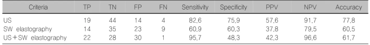
Table 1. Diagnostic performances in the differentiated malignant from benign thyroid nodules according to the diagnostic modalities
Criteria TP TN FP FN Sensitivity Specificity PPV NPV Accuracy
US
SW elastography US+SW elastography
19 14 22
44 35 28
14 23 30
4 9 1
82.6 60.9 95.7
75.9 60.3 48.3
57.6 37.8 42.3
91.7 79.5 96.6
77.8 60.5 61.7 FN: false negative, FP: false positive, NPV: negative predictive value, PPV: positive predictive value, SW elastography: shear wave elastography, TN: true negative, TP: true positive, US+SW elastography: combination US with shear wave elastography
Fig. 1. Receiver operator characteristics for diagnosing thyroid cancers of conventional US, shear wave elastography, and combination of conventional US with shear wave elastography in this study. The cutoff value of shear wave elastography for diagnosing thyroid cancer was 33.34 kPa. The area under the curve is 0.792 (95% confidence interval 0.696∼0.889), 0.609 (95% confidence interval 0.463∼0.755), and 0.72 (95% con- fidence interval [0.642∼0.797]) at conventional US, shear wave elastography, and combination of conventional US with shear wave elastography, respectively.
의 정확도를 비교하기 위하여 고식적 초음파, 전단파 탄성초음파 각각의 진단정확도와, 고식적 초음파와 전 단파 탄성초음파를 함께 이용했을 때의 진단정확도를 구하고 이의 통계학적 유의성을 알아보았다(Table 1).
고식적 초음파는 전단파 탄성초음파보다 모든 진단적 수치들이 높았다. 고식적 초음파와 전단파 탄성초음파 를 함께 이용했을 때 고식적 초음파만 이용했을 때와 비교시 진단의 민감도는 올라갔지만 정확도는 떨어졌 다. 갑상선의 악성과 양성을 가장 잘 구분하는 전단파 탄성초음파의 탄성계수의 cut-off 값은 33.3으로 이때 진단정확도인 Az 값은 0.609 (95% 신뢰구간, 0.463∼
0.755)였다. 이 값은 고식적 초음파의 Az 값인 0.792 (95% 신뢰구간, 0.696∼0.889)보다 통계학적으로 유의 하게 낮았다(p=0.049). 고식적 초음파와 전단파 탄성 초음파를 같이 이용한 Az 값은 0.72로 통계학적 차이는
없었으나 고식적 초음파만 이용했을 때의 Az 값보다 낮았다(p=0.118) (Fig. 1).
고 찰
세침흡인생검은 갑상선 결절의 진단에 가장 정확하 고 비용 효율이 높은 방법이다.21) 그렇지만 세침흡인생 검을 한 갑상선 결절의 1∼16%는 세포가 충분 검출되 지 않아 부적합(inadequate) 검체로 진단이 되어22-25) 재 생검이 필요하다. 또한 세침흡인생검이 갑상선 결절의 진단에 높은 정확도를 보이지만5,26-29) 여전히 4∼16.7%
의 위음성률이 보고되었다.26,27,29-32) 갑상선 결절의 높은 유병률로 인한 문제점과 지금까지 검사법의 한계를 고 려시 갑상선암의 조기 진단뿐 아니라 불필요한 양성 결 절의 침습적인 검사도 피할 수 있는 새로운 검사법에 대한 기대가 높다.
초음파탄성영상은 조직탄성의 재조합을 이용한 검 사법으로 외부의 힘이 가해질 때 조직의 변형이나 왜 곡을 반영하는 기계적인 성질을 이용하는 방법이다. 정상조직과 병적조직은 단단한 정도의 차이가 있고 이 러한 조직의 단단함 정도에 따른 탄성의 차이를 영상 화한 것이 탄성영상이다. 탄성초음파는 일반적으로 양 성 병변이 정상 갑상선 조직보다 딱딱하고 악성 병변 보다는 부드러운 특성을 이용한다. 탄성초음파는 Young 계수라는 물리량으로 기술되는 조직의 단단한 정도 또는 탄성도를 평가하여 초음파 영상으로 구현한 것으로 낮은 주파수의 진동을 신체조직에 가하여 조직 의 영상을 획득한 후 병변의 압박 또는 변형 정도를 분석하여 화면에 표시하는 방법이다. 조직에 가하는 진동의 특성에 따라 고정 탄성초음파와 전단파 탄성초 음파로 나뉠 수 있다. 갑상선 결절의 진단에 먼저 이용 된 상용화된 모델은 고정 탄성초음파이다. 이 방법은 검사자가 탐촉자로 신체 표면에 일정한 기계적 압박을 가해 조직을 변형하면 초음파 장비가 변형정도를 계산 하여 영상면에 구현하는 방법이다. 이 검사법의 문제
로는 조직에 가해지는 압박 정도와 압박하는 장기의 압축성에 매우 의존적이어서 구현되는 영상정보 또한 주변 조직의 구조적 특성에 영향을 받고 검사의 숙련 도에 의존적이라는 단점을 가진다.33)
낮은 재현성과 검사자간의 불일치, 그리고 정량적 결과 도출의 어려움을 가진 고정 탄성초음파에 비해 전단파 탄성초음파는 앞서 검사법의 단점을 보완하기 위한 검사법이다. 이 방법은 체내에서 전단파를 발생 시키는 과도펄스(transient pulse)를 이용한 영상으로 파장 전파 속도를 측정하여 조직 탄성도를 구한다. 이 검사법의 가장 큰 기술적 문제점은 인체에 해가 없는 진동기의 개발이 관건이었으나 현재 이를 극복한 기계 가 상용화되어 사용되고 있다.34) 탐촉자에서 생성된 전 단파는 초음파 장비를 통하여 최종적으로 속도지도에 수식을 적용한 탄성지도를 구현하여 객관적인 정량적 수치로 결절의 탄성도를 표현할 수 있다. 갑상선 결절 의 감별에 전단파 탄성초음파를 이용한 연구는 현재까 지 두 개이며 이 연구들도 기존의 고식적 초음파소견 의 조합에 추가적인 진단을 규명한 것이 아닌 전단파 탄성초음파 자체의 진단적 가치를 알아본 연구였
다.20,35) 탄성초음파는 고식적 초음파 검사법 후에 주로
이용되는 진단도구이므로 실제적으로 고식적 초음파 에 추가적인 진단가치가 있어야 임상에 적용이 가능하 다. 이에 이 연구는 기존의 초음파의 진단적 가치에 추가 적인 전단파 탄성초음파의 역할을 알아보고자 하였다. 이 연구에서 고식적 초음파와 전단파 탄성초음파를 비교하였을 때 민감도, 특이도, 양성예측도, 음성예측 도 모두 고식적 초음파의 값이 높았다. 진단정확도를 알아보는 Az 값을 비교했을 때도 통계학적으로 유의하 게 고식적 초음파의 성적이 좋았다. 따라서 저자들은 고식적 초음파에 추가적인 전단파 탄성초음파의 가치 를 알아보고자 이 두 방법을 같이 이용했을 때의 진단 성적을 구하였다. 이 두 방법을 같이 이용한 경우 고식 적 초음파 단독보다는 민감도가 다소 올라가기는 하였 으나 Az 값은 고식적 초음파 단독에서 더 성적이 좋았 다. 이런 결과는 고식적 초음파의 우수한 진단능력에 서 기인하는 것으로 생각한다.
이 연구는 몇 가지 제한점을 들 수 있다. 첫째, 포함 된 환자군의 숫자가 적다. 둘째, 이 연구의 고식적 초음 파의 모든 결과는 경험이 많은 영상의학과의사가 시행 한 것이어서 다른 기관에서 재현성이 떨어질 수 있다.
셋째, 초음파의 판독자간 변이가 이 연구에서는 고려 되지 않았다.
결 론
갑상선 결절의 진단에 고식적 초음파는 전단파 탄성 초음파에 비해 우수한 진단능을 보였다. 또한 갑상선 암을 예측하는데 있어 고식적 초음파와 전단파 탄성초 음파를 함께 이용했을 때 고식적 초음파 단독 검사일 때보다 유용하지 않았다.
중심 단어: 갑상선, 초음파, 탄성초음파.
References
1) Mortensen JD, Woolner LB, Bennett WA. Gross and micro- scopic findings in clinically normal thyroid glands. J Clin Endocrinol Metab 1955;15(10):1270-80.
2) Hegedus L. Clinical practice. The thyroid nodule. N Engl J Med 2004;351(17):1764-71.
3) Tan GH, Gharib H. Thyroid incidentalomas: management approaches to nonpalpable nodules discovered incidentally on thyroid imaging. Ann Intern Med 1997;126(3):226-31.
4) Kim KW, Kim EK, Kwak JY, Kim MJ. Detection and manage- ment of thyroid incidentalomas. J Korean Soc Ultrasound Med 2008;27(3):111-7.
5) Kim EK, Park CS, Chung WY, Oh KK, Kim DI, Lee JT, et al. New sonographic criteria for recommending fine-needle aspiration biopsy of nonpalpable solid nodules of the thyroid.
AJR Am J Roentgenol 2002;178(3):687-91.
6) Papini E, Guglielmi R, Bianchini A, Crescenzi A, Taccogna S, Nardi F, et al. Risk of malignancy in nonpalpable thyroid nodules: predictive value of ultrasound and color-Doppler features. J Clin Endocrinol Metab 2002;87(5):1941-6.
7) Peccin S, de Castsro JA, Furlanetto TW, Furtado AP, Brasil BA, Czepielewski MA. Ultrasonography: is it useful in the diagnosis of cancer in thyroid nodules? J Endocrinol Invest 2002;25(1):39-43.
8) Cappelli C, Castellano M, Pirola I, Cumetti D, Agosti B, Gandossi E, et al. The predictive value of ultrasound findings in the management of thyroid nodules. QJM 2007;100(1):29-35.
9) Khoo ML, Asa SL, Witterick IJ, Freeman JL. Thyroid calcification and its association with thyroid carcinoma. Head Neck 2002;24(7):651-5.
10) Chan BK, Desser TS, McDougall IR, Weigel RJ, Jeffrey RB, Jr. Common and uncommon sonographic features of papillary thyroid carcinoma. J Ultrasound Med 2003;22(10):1083-90.
11) Cappelli C, Pirola I, Cumetti D, Micheletti L, Tironi A, Gandossi E, et al. Is the anteroposterior and transverse diameter ratio of nonpalpable thyroid nodules a sonographic criteria for recommending fine-needle aspiration cytology? Clin Endocrinol (Oxf) 2005;63(6):689-93.
12) Frates MC, Benson CB, Doubilet PM, Cibas ES, Marqusee E. Can color Doppler sonography aid in the prediction of malignancy of thyroid nodules? J Ultrasound Med 2003;22(2):
127-31; quiz 32-4.
13) Kang HW, No JH, Chung JH, Min YK, Lee MS, Lee MK, et al. Prevalence, clinical and ultrasonographic characteristics of thyroid incidentalomas. Thyroid 2004;14(1):29-33.
14) Frates MC, Benson CB, Charboneau JW, Cibas ES, Clark OH, Coleman BG, et al. Management of thyroid nodules detected at US: Society of Radiologists in Ultrasound consensus conference statement. Radiology 2005;237(3):794-800.
15) Tae HJ, Lim DJ, Baek KH, Park WC, Lee YS, Choi JE, et al. Diagnostic value of ultrasonography to distinguish between benign and malignant lesions in the management of thyroid nodules. Thyroid 2007;17(5):461-6.
16) Cho BY. Clinical thyroidology. 2nd ed. Seoul, Korea: Korea Medical Book; 2005.
17) Shiina T, Yamamoto M, Nitta N, Ueno E. Real-time tissue elasticity imaging using the combined autocorrelation method.
MEDIX Suppl 2007;2007:4-7.
18) Asteria C, Giovanardi A, Pizzocaro A, Cozzaglio L, Morabito A, Somalvico F, et al. US-elastography in the differential diagnosis of benign and malignant thyroid nodules. Thyroid 2008;18(5):523-31.
19) Rago T, Santini F, Scutari M, Pinchera A, Vitti P. Elastography:
new developments in ultrasound for predicting malignancy in thyroid nodules. J Clin Endocrinol Metab 2007;92(8):2917-22.
20) Sebag F, Vaillant-Lombard J, Berbis J, Griset V, Henry JF, Petit P, et al. Shear wave elastography: a new ultrasound imaging mode for the differential diagnosis of benign and malignant thyroid nodules. J Clin Endocrinol Metab 2010;
95(12):5281-8.
21) Cooper DS, Doherty GM, Haugen BR, Kloos RT, Lee SL, Mandel SJ, et al. Management guidelines for patients with thyroid nodules and differentiated thyroid cancer. Thyroid 2006;16(2):109-42.
22) Yang GC, Liebeskind D, Messina AV. Ultrasound-guided fine-needle aspiration of the thyroid assessed by Ultrafast Papanicolaou stain: data from 1135 biopsies with a two- to six-year follow-up. Thyroid 2001;11(6):581-9.
23) Danese D, Sciacchitano S, Farsetti A, Andreoli M, Pontecorvi A. Diagnostic accuracy of conventional versus sonography-guided fine-needle aspiration biopsy of thyroid nodules. Thyroid 1998;
8(1):15-21.
24) Takashima S, Fukuda H, Kobayashi T. Thyroid nodules:
clinical effect of ultrasound-guided fine-needle aspiration biopsy.
J Clin Ultrasound 1994;22(9):535-42.
25) Carmeci C, Jeffrey RB, McDougall IR, Nowels KW, Weigel RJ. Ultrasound-guided fine-needle aspiration biopsy of thyroid masses. Thyroid 1998;8(4):283-9.
26) Bukhari MH, Niazi S, Hanif G, Qureshi SS, Munir M, Hasan M, et al. An updated audit of fine needle aspiration cytology procedure of solitary thyroid nodule. Diagn Cytopathol 2008;36(2):104-12.
27) Sangalli G, Serio G, Zampatti C, Bellotti M, Lomuscio G.
Fine needle aspiration cytology of the thyroid: a comparison of 5469 cytological and final histological diagnoses. Cytopathology 2006;17(5):245-50.
28) Moon HG, Jung EJ, Park ST, Ha WS, Choi SK, Hong SC, et al. Role of ultrasonography in predicting malignancy in patients with thyroid nodules. World J Surg 2007;31(7):1410-6.
29) Cai XJ, Valiyaparambath N, Nixon P, Waghorn A, Giles T, Helliwell T. Ultrasound-guided fine needle aspiration cytology in the diagnosis and management of thyroid nodules. Cytopathology 2006;17(5):251-6.
30) Ylagan LR, Farkas T, Dehner LP. Fine needle aspiration of the thyroid: a cytohistologic correlation and study of discrepant cases. Thyroid 2004;14(1):35-41.
31) Lee YH, Lee NJ, Kim JH, Suh SI, Kim TK, Song JJ.
Sonographically guided fine needle aspiration of thyroid nodule:
discrepancies between cytologic and histopathologic findings. J Clin Ultrasound 2008;36(1):6-11.
32) Ogawa Y, Kato Y, Ikeda K, Aya M, Ogisawa K, Kitani K, et al. The value of ultrasound-guided fine-needle aspiration cytology for thyroid nodules: an assessment of its diagnostic potential and pitfalls. Surg Today 2001;31(2):97-101.
33) Park SH, Kim SJ, Kim EK, Kim MJ, Son EJ, Kwak JY.
Interobserver agreement in assessing the sonographic and elastographic features of malignant thyroid nodules. AJR Am J Roentgenol 2009;193(5):W416-23.
34) Athanasiou A, Tardivon A, Tanter M, Sigal-Zafrani B, Bercoff J, Deffieux T, et al. Breast lesions: quantitative elastography with supersonic shear imaging--preliminary results. Radiology 2010;256(1):297-303.
35) Friedrich-Rust M, Romenski O, Meyer G, Dauth N, Holzer K, Grunwald F, et al. Acoustic Radiation Force Impulse- Imaging for the evaluation of the thyroid gland: A limited patient feasibility study. Ultrasonics 2012;52(1):69-74.