Introduction
Mesenchymal stem cells (MSCs) were first identi- fied in bone marrow stromal cells and can differenti- ate along multiple mesenchymal lineages, including chondrocytes, osteoblast, and myoblasts.1,2) MSCs have great therapeutic potential because of their abil-
ity to self-renew and differentiate into multiple tissues.
The therapeutic potential of multi-lineage stem cell for application such as tissue engineering and gene therapy is enormous. Although bone marrow is a good source of MSCs, traditional bone marrow procurement procedures in human are painful, frequently requiring general or spinal anesthesia, and may yield low num- bers of MSCs upon processing.3) Therefore, there are
J Clinical Otolaryngol 2012;23:90-100
Can the Palatine Tonsil be a Source of Mesenchymal Stem Cells with Immunomodulatory Property?
Yoon Se Lee, MD1, Hee-Young Park2, Yun Sung Lim, MD2, Jin-Choon Lee, MD2, Soo-Geun Wang, MD2, Jin Sup Jung, MD3 and Byung-Joo Lee, MD2
1Department of Otorhinolaryngology-Head and Neck Surgery, Research Institute for Convergence of Biomedical Science and Technology, Pusan National University Yangsan Hospital, Yangsan, Korea
2Department of Otorhinolaryngology-Head and Neck Surgery, Pusan National University Hospital and Medical Research Institute, Busan, Korea
3Department of Physiology, School of Medicine, Pusan National University, Busan, Korea
- ABSTRACT -
Background and Objectives
: Mesenchymal stem cells (MSCs) are multipotent cells that can differentiate into vari- ous cell types and are isolated from various other human adult and fetal tissues. Our study is to isolate palatine tonsil-de- rived MSCs (T-MSCs) and evaluate their differentiation potential and immunomodulatory effects, compared with bone marrow-derived MSCs (BM-MSCs). Methods:T-MSCs were isolated from human palatine tonsil. The expression of surface markers of T-MSCs was assessed by flow cytometric analysis. Differentiation potential of T-MSCs was ana- lyzed histochemically and by the expression of lineage-related marker genes. Immunomodulatory effects were evaluated by mixed lymphocyte reactions (MLR) and mitogen proliferation assays with phytohemagglutinin (PHA). Results:
T-MSCs were isolated from palatine tonsil and displayed a similar morphology to BM-MSCs. T-MSCs were negative for CD 31, CD45, CD117, HLA-DR and positive for CD44, CD73, CD90, and CD105 in flow cytometric analysis. The expression of surface phenotypes and differentiation potential of T-MSCs exhibited the similar finding as BM-MSCs.
T-MSCs showed the significant inhibition of the proliferation of T cells stimulated by allergenic T cells in MLR study and by PHA stimuli in mitogen proliferation assay and also significant decrease of the secretion of TNF-α and IFN-γ (p
<0.05). The HLA-G5 secretion by T-MSCs showed the significant increase in MLR and mitogen proliferation assays (p<0.05). T-MSCs do satisfy the phenotypical and functional definition of MSCs. T-MSCs also displayed immuno- modulatory effects that were associated with inhibiting T-cell proliferation, decreasing soluble factors, and increasing HLA-G5 secretion in response to various stimuli. Conclusions:We propose that T-MSCs provide a good alternative for allogeneic MSCs in therapeutic applications. (J Clinical Otolaryngol 2012;23:90-100)
KEY WORDS:Palatine tonsilㆍMesenchymal stem cellㆍImmune modulation.
원 저
논문접수일 :2012년 3월 8일 / 논문수정일 :2012년 3월 27일 / 심사완료일 :2012년 4월 30일
교신저자 :이병주, 602-739 부산광역시 서구 구덕로 179 부산대학교 의과대학 이비인후과학교실
전화 :(051) 240-7335·전송:(051) 246-8668·E-mail:[email protected]
limitations to the application in the regenerative med- icine using bone marrow MSCs (BM-MSCs). An alter- native source of MSCs that is obtainable in large quan- tities under local anesthesia with minimal discomfort would be advantageous. More recent studies have re- ported the isolation of MSCs from various other human adult and fetal tissues, including fat, bone, synovium, skin, thymus, periodontal ligament, placenta and am- niotic fluid.3-6)
Tonsillectomy for chronic tonsillar hypertrophy or chronic tonsillitis is one of most common operation in ENT field. Tonsillar epithelium is derived from the second pharyngeal pouch (endodermal origin). During fetal development, tonsillar tissue is invaded by lym- phoid tissue (mesodermal origin). So, embryological- ly, there is the possibility of a source of MSCs. Recent study has reported the palatine tonsil is a new source of MSCs.7) The human palatine tonsil is readily acces- sible to otolaryngologists and easily obtained by tonsil- lectomy or punch biopsy, particularly in young donors.
The clinical application of MSCs requires large quantities of cells for injection or infusion. Because of the limitation of obtaining sufficient autologous stem cells, MSCs from allogeneic donor could constitute a valuable alternative source of stem cells for therapeutic application. A prerequisite when considering allogeneic MSCs for clinical therapeutic purpose is the character- ization of their immunological properties in allogeneic condition. Although palatine tonsil-derived MSCs (T- MSCs) could be a good source of autologous MSCs, it also could be a new candidate of allergenic MSCs be- cause it is easy to obtain tonsillar tissue by tonsillecto- my that is one of most common surgery. Consequent- ly, palatine tonsil-derived mesenchymal stem cells (T-MSCs) may provide an alternative source of MSCs for allergenic donor. Our study is to isolate T-MSCs and evaluate their differentiation potential and im- munomodulatory effects, compared with BM-MSCs.
Materials and Methods
We used BM-MSC that was previously established
in our institute to validate the character of T-MSC.8) T-MSC isolation and culture
This study using human tonsil tissue and peripher- al blood samples was approved by the institutional re- view board. Tonsils were obtained after informed consent from patients (5 to 12 years old) undergoing tonsillectomy as a result of chronic tonsillar hypertro- phy. To isolate T-MSCs, tonsils were washed exten- sively with equal volumes of phosphate buffered sa- line (PBS), and tissues were digested at 37°C for 30 min with 0.075% collagenase type I (Sigma, St. Louis, MO). Enzyme activity was neutralized with α modified Eagle’s medium (α-MEM), containing 10% fetal bo- vine serum (FBS) and centrifuged (1,200×g, 10 min) to obtain a pellet. The pellet was filtered through a 100-μ m nylon mesh to remove cellular debris and incubat- ed overnight at 37℃/5% CO2 in control medium (α -MEM, 10% FBS, 100 unit/mL of penicillin, 100 μg/
mL of streptomycin). Following incubation, the plates were washed extensively with PBS to remove residu- al non adherent cells. The resulting cell population was maintained at 37℃/5% CO2 in control media. One week later, when a monolayer of adherent cells had reached confluencey, cells were trypsinized (0.05%
trypsin-EDTA ; Sigma, USA) re-suspended into me- dia and subcultured at a concentration of 2,000 cells/
cm2. To isolate BM-MSCs, we performed methods as previously described.
Flow cytometric analysis
Flow cytometric analysis was used to characterize the phenotypes of the T-MSCs and BM-MSCs. At least 50,000 cells (in 100 μL PBS/0.5% BSA/2 mmol/L EDTA) were incubated with FITC conjugated mono- clonal antibodies against human CD105, CD90, CD44, CD73, CD45, CD31, CD117, HLA-DR (BD Biosci- ences Clontech, Palo Alto) or with the respective iso- type control. Labeled cells were analyzed by flow cytometry using a FACS Caliber flow cytometer and the Cell Quest Pro software (BD Biosciences, USA).
Cellular proliferation assays
Tonsil stem cells were plated at a density of 103 cells per well in a 96 well plate. Cell viability was assessed by an MTT assay after 24, 48, and 72 h of culture.
Multilineage differentiation
T-MSCs were analyzed for their capacity to differ- entiate towards the adipogenic, osteogenic, and chon- drogenic lineages. Adipogenic differentiation was in- duced by culturing T-MSCs for 2 weeks in adipogenic media (1 uM dexamethasone, 100 μg/mL 3-isobutyl-1 methylxanthine (IBMX), 5 μg/mL insulin, and 60 μM indomethacine, 10% FBS in α-MEM) and assessed by oil red O (Sigma, St. Louis, MO) staining as an in- dicator of intracellular lipid accumulation. Prior to staining, the cells were fixed for 15 min at room tem- perature in 70% ethanol. Cells were then incubated in 2% oil red O reagent for 1 h at room temperature.
Excess stain was removed by washing with 70% eth- anol and distilled water to visualize lipid droplets.
Osteogenic differentiation was induced by cultur- ing T-MSCs and BM-MSCs for 2 weeks in osteogen- ic media (0.1 mM dexamethasone, 10 μM β glycero- phosphate, and 50 μg/mL ascorbic acid, 10% FBS in α MEM) and examining extracellular matrix calcifi- cation by alizarin red S (Sigma, St. Louis, MO) stain- ing. For alizarin red S staining, cells were fixed with 70% ethanol and washed with distilled water, and in- cubated in 2% alizarin red solution for 15 min at room temperature, followed by numerous washes with dis- tilled water.
Chondrogenic differentiation was induced using the micromass culture technique. Briefly, 10 μL of concen- trated MSC suspension (3×10⁵ cells/mL) was plated into the center of each well and allowed to attach at 37℃ for 2 h. Chondrogenic media [CM, 1% FBS, 0.1 mM dexamethasone (Sigma, USA), 50 μg/mL ascorbic acid, ITS+1 (insulin-transferrin-selenium ; Sigma), 10 ng/mL TGF β1 (Sigma), 10 ng/mL in α-MEM] was gently overlaid so as not to detach the cell nodules, and the culture was maintained in CM for 4 weeks be- fore analysis. Chondrogenesis was confirmed by im-
munohistochemistry for collagen type-II staining. For collagen type-II staining, sections were blocked with 10% horse serum, incubated with purified anti-mouse collagen type-II antibody (BD Bioscience, San Jose, CA) for 1 h, and washed with PBS (pH 7.4). Antibody- bound cells were detected with a peroxidase substrate kit (Vectastain ABC kit ; Vector Laboratories, Burl- ingame, CA). Sections were washed, counterstained with hematoxylin, and examined by light microscopy.
Real-time quantitative reverse transcription- polymerase chain reaction (PCR) for gene profiling
Total RNA was isolated from MSCs or PBMCs from day 21 monolayers and pellet cultures, using the Trizol reagent (Invitrogen Corporation). Isolated RNA was then reverse-transcribed using random hexamers.
Real-time PCR were performed using 10 ng of cDNA and SYBR Green mix (Bio-Rad Laboratories, Inc.).
Gene-specific primers were designed based on Gen- Bank cDNA sequences, as described previously.7) Li- poprotein lipase (LPL) and peroxisome proliferator-ac- tivated receptor-gamma (PPARγ) were used as genetic markers of adipogenesis, alkaline phosphatase (ALP) and osteocalcin (OC) was used as genetic markers of osteogenesis, and collagen type II α1 (COL2) and ag- grecan (AGN) were used as genetic markers of chon- drogenesis. Expression levels are presented as a fold increase over GAPDH, using the formula 2(ΔCt), where ΔCt=Ct of target gene-Ct of GAPDH.
Mixed lymphocyte reaction (MLR) and mitogen proliferative assays
Human peripheral blood mononuclear cells (PBMCs) were obtained from heparinized whole blood samples or buffy coats donated by healthy subjects, after in- formed consent was obtained, using density gradient centrifugation (Lymphoprep ; Axis-Shield, Oslo, Nor- way, http://www.axis-shield.com). MLR cultures were set up with 2×10⁵ purified PBMCs as responder cells and equal number of irradiated (3,000 cGy) HLA mis- match PBMCs as stimulators. In T-MSC/MLR cocul-
ture experiments, MLR were performed using cocul- ture with various T-MSCs concentration at 0, 1×10⁴, 2×10⁴, 4×10⁴.
All cultures were performed in triplicate, using round-bottomed 96-well tissue culture plates (Corn- ing), in a final volume of 200 μL complete medium.
After 4 days d of incubation, 3H-thymidine was add- ed overnight and thymidine incorporation was mea- sured using a β-scintillation counter and is expressed in counts per minute (cpm).
In mitogen proliferation assays, PBMCs were seeded in triplicate at a concentration of 2×10⁵ cells/
well in 96-well flat-bottom plates in the presence of 10 μg/mL phytohemagglutinin (PHA, Sigma, St. Lou- is, MO). Medium or T-MSCs were added to the vari- ous MLR (T-MSCs : PBMCs) at a 1 : 20, 1 : 10, 1 : 5, or 1 : 0 ratio. After 4 days of incubation, 3H thymidine was added overnight and thymidine incorporation measured using a β scintillation counter and is ex- pressed in counts per minute (cpm).
Production of soluble factors from T-MSCs and BM-MSCs
The levels of interleukin (IL)-10, tumor necrosis fac- tor (TNF)-α, interferon (IFN)-γ and HLA-G5 were quantified using commercially available enzyme-linked immunosorbent assays (ELISA ; R&D system, USA) from supernatants obtained from MLR and mitogen proliferation assays. HLA-G5 was detected by flow cytometric analysis. To analyze the surface HLA-G5,
cells were stained with PE-conjugated anti-human HLA-G5a (clone 87G ; Exbio) and measured using a FACScalibur (BD bioscience), and data analysis was performed using Cellquest pro software (BD biosci- ence).
Statistical analyses
Statistical analyses were performed using the SPSS software (ver. 15.0 ; Chicago, IL, USA. Differences be- tween levels of cpm and absorbance were evaluated using the t-test and the chi squared test. A p-value<
0.05 was deemed to indicate statistical significance.
Results
T-MSC Isolation
The cell yield from each tonsil ranged from 1 to 5×
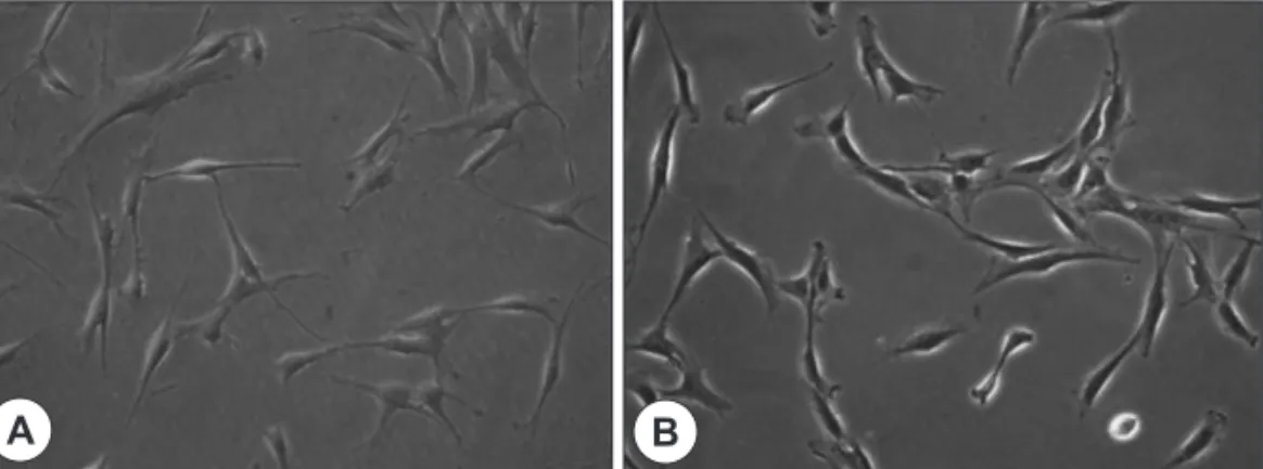
109 cells, with the majority being non adherent and, likely, of hematopoietic origin. Following repeated washes, adherent cells that had fibroblast-like spin- dle-shaped appearances were identified ; however, large round cells were also still present. After multiple trypsinization steps, an increased number of homoge- neous fibroblast-like cells with extended cytoplasmic processes was observed. The cells obtained from BM- MSCs were similar to those from T-MSCs (Fig. 1). The doubling time was higher in T-MSCs (73.5±4.2 h), compared with BM-MSCs (96.0±2.9 h). The prolif- eration profiles of BM-MSCs and T-MSCs were not significantly different. The cells per well did not limit
Fig. 1. Morphology of T-MSCs and BM-MSCs. The fibroblast-like spindle-shaped appearances of T-MSCs (A) is similar to BM-MSCs (B)(×200).
A B
the proliferation of MSCs during culture.
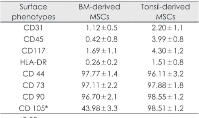
Phenotypes analysis of the various cell preparations The antigenic phenotypes of all cell preparations were investigated by flow cytometry. T MSCs and BM- MSCs were uniformly positive for CD44, CD73, and CD90. Higher levels of CD105 surface expression were observed in T-MSCs compared with BM MSCs (Table 1). CD31, CD45, CD117, and HLA-DR were negative in both MSCs. CD31 and CD45 negativity confirmed that the T-MSCs were of non-hematopoi- etic lineage. The expression of surface phenotypes of T-MSCs exhibited the similar cell surface phenotype as BM-MSCs. However, we observed significant dif- ferences concerning the expression of CD105.
Multi-lineage differentiation potential
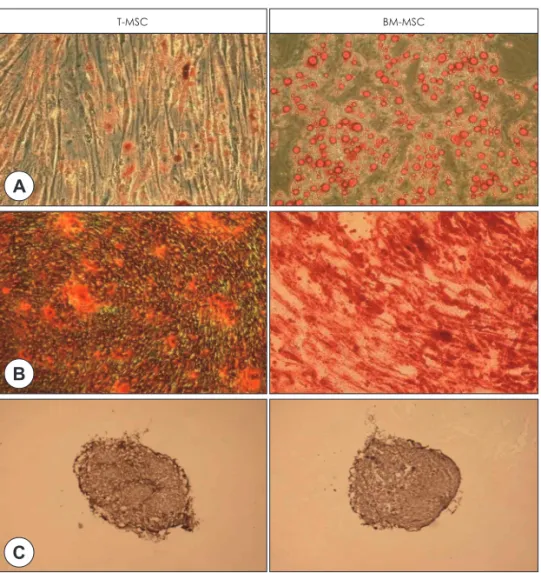
As previously described, when cultured in the appro- priate medium, T-MSCs and BM-MSCs were capable of in vitro differentiation into adipocytes, osteoblast, and chondrocytes.
Adipogenesis
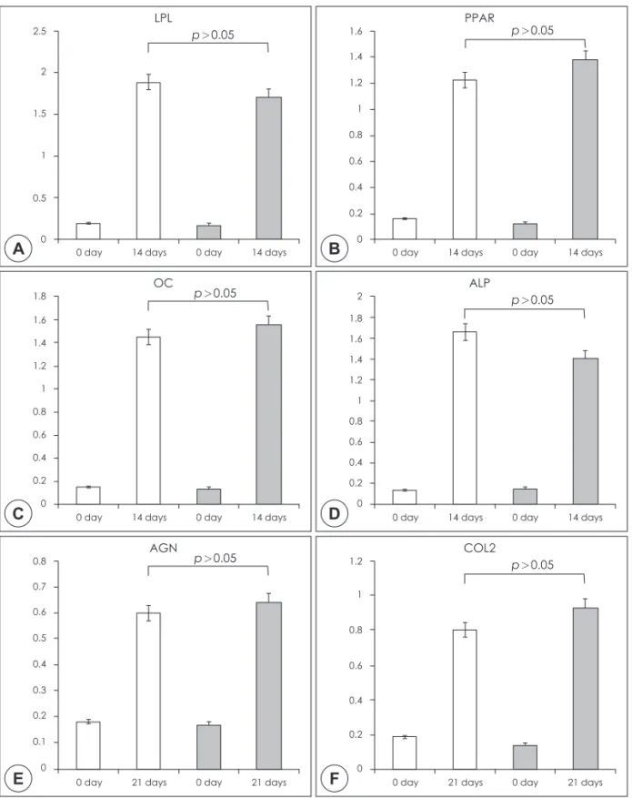
Following treatment of BM-MSCs and T-MSCs with adipogenic supplements, both MSCs were maintained in expansion medium. Multiple lipid droplets were ob- served in the cytoplasm of both MSCs, suggesting sim- ilar morphological changes (Fig. 2A). Quantitative RT- PCR analysis revealed a significant increase of LPL and PPARγ expression in both T-MSCs and BM-MSCs
(Fig. 3A, B)(p<0.05). The expression of adipogenic genes of T-MSCs and BM-MSCs were similar.
Osteogenesis
Osteogenic induction enabled both MSCs to be trans- formed from spindle-shaped to flattened and spread cells. Additionally, both osteoblast-like cells and ma- trix mineralization were observed (Fig. 2B). Quantita- tive RT-PCR analysis revealed a significant increase of OC and ALP expression in both T-MSCs and BM- MSCs (Fig. 3C, D)(p<0.05). The expression of osteo- genic genes of T-MSCs exhibited the similar to the find- ings of BM-MSCs.
Chondrogenesis
An accumulation of matrix sulfated proteoglycan was detected in both MSCs and the intensity of staining did not differ between the MSCs (Fig. 2C). Quantita- tive RT-PCR analysis revealed a significant increase of AGN and COL2 expression in both T-MSCs and BM- MSCs (Fig. 3E, F)(p<0.05). The expression of adipo- genic genes of T-MSCs and BM-MSCs were similar.
Effects of T-MSCs on MLR and mitogen proliferation assay
MLR and mitogen proliferation assays were per- formed to evaluate the immunomodulatory properties of both allogeneic MSCs. Two-way MLR between two mismatched HLA-DR PBMCs was suppressed using T-MSCs at varying cell densities. Changes in DNA syn- thesis profiles (cpm) in the absence or presence of var- ious concentrations of MSCs were also observed. Fig.
4A demonstrates that the lowest concentration of T- MSCs (T-MSCs to responder cell ratio of 1 : 5) pro- duced a marked decrease in lymphocyte proliferation (p<0.001). Similar effects were observed at higher concentrations of MSCs and the degree of inhibition did not differ between T-MSCs and BM-MSCs. In ad- dition to allogeneic stimuli, we evaluated the immuno- suppressive effect of both MSCs in mitogen (PHA)- induced proliferation assays. T-MSCs and BM-MSCs strongly inhibited the PHA-induced proliferation of
Table 1. Comparison of the expression of surface mark- ers of mesenchymal stem cells derived from bone mar- row and palatine tonsil as analyzed by flow cytometry
Surface
phenotypes BM-derived
MSCs Tonsil-derived MSCs CD31
CD45 CD117 HLA-DR CD 44 CD 73 CD 90 CD 105*
01.12±0.5 00.42±0.8 01.69±1.1 00.26±0.2 97.77±1.4 97.11±2.2 96.70±2.1 43.98±3.3
02.20±1.1 03.99±0.8 04.30±1.2 01.51±0.8 96.11±3.2 97.88±1.8 98.55±1.2 98.51±1.2
* : p<0.05
lymphocytes, to a similar extent (Fig. 4B) (p<0.05). In mitogen proliferation assays, the inhibition at the low- est concentration was less pronounced than in MLR, but was sustained at increasing concentrations. This was also evident when BM-MSCs were added to the reaction. These data demonstrate that both MSCs sup- press the proliferation of lymphocytes, to a similar de- gree, in a cell density-dependent manner.
Secretion of TNF-α, IFN-γ, and IL-10 on MLR and mitogen proliferation assays
Previous studies indicated that elicited soluble fac- tors participate in MSC-mediated immunosuppression.
To evaluate these effects, supernatants obtained from MLR and mitogen proliferation assays were assessed for changes in the levels of soluble factors by ELISA.
In MLR, both TNF-α and IFN-γ (which reflect T-lym- phocyte proliferation and inflammatory condition) de- creased significantly at the initial concentration of T- MSCs and BM-MSCS. At increasing concentrations of MSCs, significant changes were not evident. The levels of IL-10 showed no variation at any concentra- tion of MSCs (Fig. 5A). In the mitogen proliferation assays, similar results were observed, with graded re- sponses along the concentrations of MSCs evident.
IL-10 levels did not change significantly (Fig. 5B).
Fig. 2. Histological analysis of differentiation potential of T-MSCs and BM-MSCs. Representative micrscopic images views of the differentiation of T-MSCs compared to BM-MSCs into adipogenic (A), osteogenic (B) and chondrogenic (C) lineages (×200). Oil red O staining shows lipid vacuoles stained red, alizarlin red S staining shows deposition of calcium crystals stained orange to brown, and collagen type II staining shows cartilage-specific proteoglycan.
T-MSC BM-MSC
B A
C
Fig. 3. Gene expression analysis of differentiation potential of T-MSCs and BM-MSCs. Adipogenic genes are (A) lipopro- tein lipase (LPL) and (B) proliferator-activated receptor-gamma (PPARγ), osteogenic genes are (C) osteocalcin (OC) and (D) alkaline phosphatase (ALP), and chondrogenic genes are (E) aggrecan (AGH) and (F) collagen type II α1 (COL2). Quantitative RT-PCR analysis revealed a significant increase of LPL, PPARγ, OC, ALP, AGH, and COL2 expres- sion in both T-MSCs and BM-MSCs (p<0.05). The patterns of differentiation markers of T-MSCs and BM-MSCs were sim- ilar. Vertical axis indicates the relative gene expression level.
0 day 14 days 0 day 14 days
p>0.05 LPL
A
2.5 2 1.5 1 0.5
0
0 day 14 days 0 day 14 days
p>0.05 PPAR
B
1.6 1.4 1.2 1 0.8 0.6 0.4 0.2 0
0 day 14 days 0 day 14 days
p>0.05 OC
C
1.8 1.6 1.4 1.2 1 0.8 0.6 0.4 0.2 0
0 day 14 days 0 day 14 days
p>0.05 ALP
D
2 1.8 1.6 1.4 1.2 1 0.8 0.6 0.4 0.2 0
0 day 21 days 0 day 21 days
p>0.05 AGN
E
0.8 0.7 0.6 0.5 0.4 0.3 0.2 0.1 0
0 day 21 days 0 day 21 days
p>0.05 COL2
F
1.2 1 0.8 0.6 0.4 0.2 0
Expression of HLA-G5 in MLR and mitogen proliferation assays
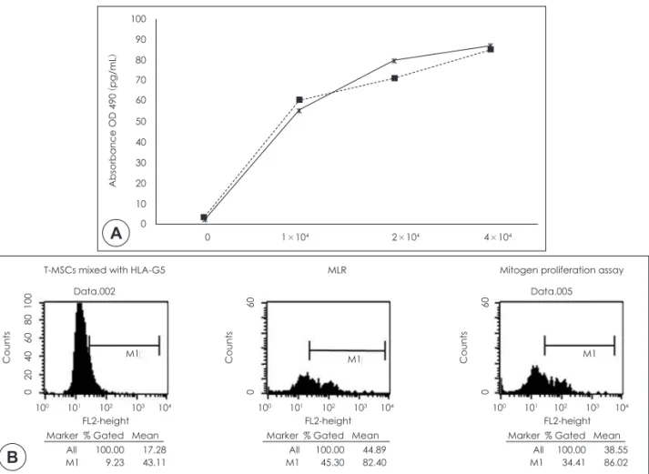
Both flow cytometry and ELISA were performed to evaluate the levels of HLA-G5 expression. Increases in HLA-G5 expression were observed following al- logeneic or mitogenic stimulation. HLA-G5 levels in- creased significantly in the MLR and mitogen prolif- eration assays as the concentration of T-MSCs increased (p<0.05 ; Fig. 6A). To validate that T-MSCs do not interact with HLA-G5, T-MSCs were mixed with HLA- G5 without allogeneic stimulus as the control group,
Fig. 4. Inhibition of lymphocyte proliferation in presence of T-MSCS and BM-MSCs. (A) MLR and (B) mitgen prolifera- tion assay showed immunomodulatory effects of both MSCs. T-MSCs and BM-MSCs inhibited proliferation of lympho- cytes in both the reactions.
0 1×1042×1044×104 0 1×1042×1044×104 Stem cells/well
A
90,000 80,000 70,000 60,000 50,000 40,000 30,000 20,000 10,000 0
cpm
0 1×1042×1044×104 0 1×1042×1044×104 Stem cells/well
B
120,000 100,000 80,000 60,000 40,000 20,000 0
cpm
where no change in HLA G5 expression was evident.
The increase in HLA-G5 expression was significant- ly higher in the mitogen proliferation assay than con- trol group (Fig. 6B).
Discussion
The isolation of MSCs from various other human adult and fetal tissues, including fat, bone, synovium, skin, thymus, periodontal ligament, placenta and am- niotic fluid, were reported.3-6) Recent study has report-
1×1042×1044×104
1×1042×1044×104
Fig. 5. Change of molecules participating immunomodulatory reaction of T-MSCs. (A) MLR and (B) mitogen prolifera- tion assay. In both reactions, TNF-α and IFN-γ decreased.
A
Absorbance OD 490 (pg/mL)
0 1×1042×1044×104 0 1×1042×1044×104
Stem cells/well TNF-α 500
450 400 350 300 250 200 150 100 50
0 0 1×1042×1044×104 0 1×1042×1044×104
Stem cells/well IFN-γ 700
600 500 400 300 200 100
0 0 1×1042×1044×104 0
Stem cells/well IL-10 400
350 300 250 200 150 100 50 0
B
Absorbance OD 490 (pg/mL)
0 1×1042×1044×104 0 1×1042×1044×104
Stem cells/well TNF-α 700
600 500 400 300 200 100
0 0 1×1042×1044×104 0 1×1042×1044×104
Stem cells/well IFN-γ 1000
900 800 700 600 500 400 300 200 1000
0 1×1042×1044×104 0
Stem cells/well IL-10 400
350 300 250 200 150 100 50 0
ed the palatine tonsil is a new source of MSCs.7) In our study, fibroblast-like cells (T-MSCs) were isolated from palatine tonsil and displayed a similar morphol- ogy to BM-MSCs. The proliferation profile of T-MSCs was not significantly different when compared to BM- MSCs. In flow cytometric analyses, T-MSCs were neg- ative for CD 31, CD45, CD117, HLA-DR and positive for CD44, CD73, CD90, and CD105. The expression of surface phenotypes of T-MSCs exhibited the simi- lar cell surface phenotype as BM-MSCs, specifically CD44, CD73, and CD90. However, we observed sig- nificant differences concerning the expression of CD105. It needs to be further investigated whether this molecule is important for the function and differentia- tion of stem cells.
The multilineage potential of T-MSCs was shown
based on their ability to differentiate into fat, bone, and cartilage. Adiopocytes displayed characteristic lipid droplets and expressed LPL and PPARγ. Analysis of the osteogenic condition showed areas of mineralization and osteoblasts, while OC and ALP were expressed as mRNA transcript markers. Chondrocytes displayed matrix accumulation of sulfated glycosaminoglycans in cell pellets and the expression of AGN and COL2 as mRNA transcript markers. These findings are con- sistent with Janjanin et al.7) However, they reported that the expression of OC, ALP, and COL2 in T-MSCs pel- lets were significantly lower than BM-MSCs. The low- er level expression of particular markers of differenti- ation could be the result of prior in vivo exposure of T-MSCs to high concentrations of inflammatory cy- tokines, which are characteristically present in donor
Fig. 6. Role of HLA-G5 was evaluated. HLA- G5 level changed as the concentration of T-MSCs was increased in both the MLR (solid line) and mitogen proliferation assay (dotted line) (A) and it was analyzed by ELISA. Significant change in the presence of T-MSCs was observed versus in the absence of T-MSCs (p<0.05). (B) Flow cytometry presenting in- creased HLA-G5 in MLR and mitogen proliferation assay compared with control group (T-MSCs mixed with HLA-G5).
Counts CountsCounts
0 20 40 60 80 100 0 60
0 60
100 101 102 103 104 100 101 102 103 104 100 101 102 103 104
Marker % Gated Mean Marker % Gated Mean Marker % Gated Mean
All 100.00 17.28
M1 9.23 43.11 All 100.00 38.55
M1 34.41 86.02 All 100.00 44.89
M1 45.30 82.40
M1 M1 M1
FL2-height FL2-height FL2-height
T-MSCs mixed with HLA-G5 MLR Mitogen proliferation assay
Data.002 Data.005
0 1×10⁴ 2×10⁴ 4×10⁴
A
100 90 80 70 60 50 40 30 20 10 0
Absorbance OD 490
(pg/mL
)
B
tissue of chronic tonsillitis.7) However, in this study, the levels of mRNA expressed from each differentiat- ed pellets did not differ significantly between T-MSCs and BM-MSCs. The main cause of tonsillectomy in our cases is a chronic tonsillar hypertrophy.
MSCs from allogenic donor could constitute a valu- able alternative source of stem cells for therapeutic application because there are some limitations of ob- taining sufficient autologous MSCs. MSCs-mediated immune modulation effect is a complex mechanism that involves changes in the maturation of antigen-pre- senting cells, the suppression of monocyte derived den- dritic cells, the cytokine secretion profile of dentritic cells, T cells, and natural killer cells.9,10) The molecular mechanism that medicates the immunosuppression effect of MSCs is not completely understood.
MSCs exert profound immunosuppression by in- hibiting T-cell proliferation and decreasing soluble factors in response to various stimuli in vitro.9,11) This suppressive effect of MSCs is mediated through sev- eral inducible soluble factors, such as transforming growth factor-β, hepatocyte growth factors, interleu- kin-10, prostaglandin E2, and indoleamine 2,3-dioxy- genase.12-14) Janjanin et al. reported that T-MSCs inhib- ited the proliferation of T cells stimulated by allergenic T cells or by non-specific mitogenic stimuli (PHA) and decrease the secretion of IFN-γ via an indoleamine 2,3-dioxygenase-dependent mechanism. These results are consistent with our findings that T-MSCs inhibits the proliferation of T cells stimulated by allergenic T cells in MLR study and by PHA stimuli in mitogen proliferation assay and also decreases the secretion of TNF-α and IFN-γ. Janjanin et al.7) reported that the immunomodulatory activity of T-MSCs was signifi- cantly less pronounced than that of BM-MSCs. How- ever, in our study, the immune suppressive effect of T- MSCs was not significantly different when compared to BM-MSCs.
MSCs modify the proinflammatory Th1 profile to- ward Th2 anti-inflammatory profile.12,13,15) In our study, T-MSCs inhibit the proinflammatory Th1 cyto- kines, such as TNF-α and IFN-γ, but do not show sig-
nificant decreasing secretion of anti-inflammatory Th2 cytokine, such as IL-10. These results are supported that T-MSCs can also modify the conversion from Th1 to Th2 lymphocytes.
One of immune suppression mechanism of MSCs is a histocompatibility locus antigen (HLA)-G mole- cules.16) Natural process allows fetal allografts to evade from rejection by the mother. This phenomenon is related with on HLA-G molecules.17) HLA-G is a non- classical major MHC class I, which is expressed in three types of membrane-bound and four types of soluble isoforms.18) HLA-G5 is a soluble form that me- diates an immunosuppressive effect by inducing apop- tosis of CD8+ T cells and own regulating CD4+ T-cell proliferation.18) HLA-G5 expressed and secreted by MSCs contributes to the suppression of the NK lytic ac- tivity and IFN-γ secretion, the direct inhibition of al- lergenic T-cell responses, and the increase of IL-10 concentration in the alloreaction microenvironment.19) In the ELISA and flow cytometry assay, T- MSCs that were stimulated by allergenic T cells or by non-specific mitogenic stimuli increased the secretion of HLA-G5.
These results are supported that the immune suppres- sion mechanism by T-MSCs are related with the in- crease of HLA-G5 level.
The current study demonstrated that T-MSCs do sat- isfy the phenotypical and functional definition of mes- echymal stem cells, as has previously been reported in BM-MSCs. T-MSCs also displayed immunomodula- tory effects that were associated with inhibiting T- cell proliferation, decreasing soluble factors, and in- creasing HLA-G5 secretion in response to various stimuli. These findings should contribute to improving clinical therapeutic trials using T-MSCs injection or in- fusion for immunomodulation purposes. Tonsil tissues that are harvested by otolaryngologist easily in the ENT field provide a new alternative source of MSCs. In fu- ture, T-MSCs will be useful and helpful for study using autologous MSCs in ENT field and may provide a good alternative source as allogeneic MSCs in therapeutic application.
Acknowledgments
This work was supported by a Korea Research Foundation Grant, funded by the Korean Government (KRF-E000394).
REFERENCES
1) Kolf CM, Cho E, Tuan RS. Mesenchymal stromal cells.
Biology of adult mesenchymal stem cells: regulation of niche, self-renewal and differentiation. Arthritis Res Ther 2007;(1)9:204.
2) Kuo CK, Li WJ, Mauck RL, Tuan RS. Cartilage tissue en-
gineering: its potential and uses. Curr Opin Rheumatol 2006;18(1):64-73.3) Pittenger MF, Mackay AM, Beck SC, Jaiswal RK, Doug- las R, Mosca JD, et al. Multilineage potential of adult human
mesenchymal stem cells. Science 1999;284(5411):143-7.4) In’t Anker PS, Scherjon SA, Kleijburg-van der Keur C, de Groot-Swings GM, Claas FH, Fibbe WE, et al. Isolation of
mesenchymal stem cells of fetal or maternal origin from hu- man placenta. Stem Cells 2004;22(7):1338-45.5) In’t Anker PS, Scherjon SA, Kleijburg-van der Keur C, Noort WA, Claas FH, Willemze R, et al. Amniotic fluid as
a novel source of mesenchymal stem cells for therapeutic transplantation. Blood 2003;102(4):1548-9.6) Zuk PA, Zhu M, Mizuno H, Huang J, Futrell JW, Katz AJ,
et al. Multilineage cells from human adipose tissue: implica- tions for cell-based therapies. Tissue Eng 2001;7(2):211-28.7) Janjanin S, Djouad F, Shanti RM, Baksh D, Gollapudi K, Prgomet D, et al. Human palatine tonsil: a new potential
tissue source of multipotent mesenchymal progenitor cells.Arthritis Res Ther 2008;10(4):R83.
8) Lee RH, Kim B, Choi I, Kim H, Choi HS, Suh K, et al.
Characterization and expression analysis of mesenchymal stem cells from human bone marrow and adipose tissue.
Cell Physiol Biochem 2004;14(4-6):311-24.
9) Nauta AJ, Fibbe WE. Immunomodulatory properties of
mesenchymal stromal cells. Blood 2007;110(10):3499-506.10) Uccelli A, Pistoia V, Moretta L. Mesenchymal stem cells:
a new strategy for immunosuppression? Trends Immunol
2007;28(5):219-26.
11) Gotherstrom C. Immunomodulation by multipotent mesen-
chymal stromal cells. Transplantation 2007;84(1 Suppl):S35-7.
12) Aggarwal S, Pittenger MF. Human mesenchymal stem cells
modulate allogeneic immune cell responses. Blood 2005;105(4):1815-22.
13) Beyth S, Borovsky Z, Mevorach D, Liebergall M, Gazit Z, Aslan H, et al. Human mesenchymal stem cells alter anti-
gen-presenting cell maturation and induce T-cell unrespon- siveness. Blood 2005;105(5):2214-9.14) Di Nicola M, Carlo-Stella C, Magni M, Milanesi M, Lon- goni PD, Matteucci P, et al. Human bone marrow stromal
cells suppress T-lymphocyte proliferation induced by cel- lular or nonspecific mitogenic stimuli. Blood 2002;99(10):3838-43.
15) Jiang XX, Zhang Y, Liu B, Zhang SX, Wu Y, Yu XD, et al.
Human mesenchymal stem cells inhibit differentiation and function of monocyte-derived dendritic cells. Blood 2005;
105(10):4120-6.
16) Nasef A, Mathieu N, Chapel A, Frick J, Francois S, Mazuri- er C, et al. Immunosuppressive effects of mesenchymal
stem cells: involvement of HLA-G. Transplantation 2007;84 (2):231-7.17) Rouas-Freiss N, LeMaoult J, Moreau P, Dausset J, Carosel- la ED. HLA-G in transplantation: a relevant molecule for
inhibition of graft rejection? Am J Transplant 2003;3(1):11-6.
18) Carosella ED, Moreau P, Le Maoult J, Le Discorde M, Dausset J, Rouas-Freiss N. HLA-G molecules: from ma-
ternal-fetal tolerance to tissue acceptance. Adv Immunol 2003;81:199-252.19) Selmani Z, Naji A, Zidi I, Favier B, Gaiffe E, Obert L, et al.
Human leukocyte antigen-G5 secretion by human mesen- chymal stem cells is required to suppress T lymphocyte and natural killer function and to induce CD4+CD25highFOXP3+
regulatory T cells. Stem Cells 2008;26(1):212-22.