J Korean Soc Coloproctol Vol. 19, No. 2, 2003
Effect of Lactacystin on the Sulindac-Indu- ced Apoptosis Mechanism s in HT-29 Cells
Jung-Min Kim, M.D., Ki-Jae Park, M.D., Sung-Heun Kim, M.D., Hong-Jo Choi, M.D., F.A.C.S.
Department of Surgery, Dong-A University College of Medicine, Busan, Korea
Purpose: One of possible mechanisms of the antineoplastic effect by nonsteroidal anti-inflammatory drugs (NSAIDs) is an induction of apoptosis. The NSAIDs-induced apoptosis appears to be caspase- and mitochondria-dependent. The ubiquitin-proteasome system, which is a fundamental non- lysosomal tool that cells use to process or degrade a variety of short-lived proteins, is known to be involved in apoptosis and to be located upstream of mitochondrial changes and caspase activation. The present study was conducted to explore the potential role of proteasome pathway in NSAIDs-induced apoptosis.
Methods: We employed sulindac as a NSAID, and the lactacystin as a proteasome inhibitor to investigate the extent of the apoptosis in colon cancer cell line, HT-29 cells. The proteasome activity and the amount of apoptosis were quantified after cells were treated with 1 mM sulindac, 1 M lactacystin or both.
Results: Sulindac treatment caused apoptosis of the HT-29 cells in a time-dependent manner with resultant changes in nuclear morphology. Western blots also showed caspase-3 activation and PARP cleavage after sulindac treatment. Not only single treatment with lactacystin decreased proteasome activity, but co-treatment with sulindac enhanced decrease in proteasome activity further (P<0.01). Treatment with lactacystin only did not induce apoptosis. However, lactacystin augmented the induction of sulindac-induced apoptosis (P<0.01). This synergistic effect was also proven by Western blot analyses, where co-treatment augmented the caspase-3 activation and PARP degradation.
Conclusions: The combination treatment of sulindac
with a proteasome inhibitor lactacystin is suggested to be a very effective strategy for the induction of cancer cell apoptosis. Elucidation of the mechanism underlying the regression of colon cancers by combination of sulindac and lactacystin seems to be an immediate challenge in the near future. J Korean Soc Coloproctol 2003;19:61-66
Key Words: ulindac, Apoptosis, Proteasome, Lactacystin, Colon cancer
Sulindac, 세포사멸 프로테아좀, , Lactacystin, 대장암
서 론
비스테로드성 항염증약물 이하( NSAIDs)은 대장암 의 발생빈도를 감소시킬 수 있는 것으로 현재 알려져 있다.1 NSAIDs에 의한 이러한 항종양효과의 기전 중 하나로서 세포사멸의 유도가 가장 많이 거론되고 있 는데, NSAIDs는 현재 사람의 대장암 세포주를 포함한 여러 형태의 세포들에서 세포사멸을 유도하는 것으로 알려져 있다.2-4 NSAIDs에 의해 유도되는 세포사멸 기 전은 caspase 및 미토콘드리아 의존성인 사실이 밝혀 지고 있다.5여러 실험 및 임상 연구를 통해 NSAIDs가 항종양약물로서의 이용 가능성을 암시하고는 있으나, 이들 약물이 암의 예방이나 치료를 위해 임상에서 이 용되기 위해서는 이들 약제의 안정성과 효능 적절한, 제제 그리고 약물의 작용기전에 대한 연구가 더 필요, 할 것으로 보인다.6Ubiquitin-proteasome system은 세포 들이 여러 단생(short-lived) 단백질의 생성과 파괴에 이용하는 중요한 비리소솜성(non-lysosomal) 기전으로 서 이것이 세포사멸에 관여하는 것으로 알려져 있다.
이 체계에 의한 세포사멸의 기작은 세포사멸 자극이 표적세포의 프로테아좀 활성을 억제함으로써 세포사 멸을 유도한다고 할 뿐만 아니라,7어떤 세포에서는 프 로테아좀 억제제 자체가 세포사멸을 유도한다고 한 다.8 일반적으로 프로테아좀에 의해 매개되는 세포사
대장암 세포주 HT-29 세포에서 Sulindac 에 의해 유도된 세포사멸 기전에 대한 Lactacystin 의 효과
동아대학교 의과대학 외과학교실
김정민・박기재・김성흔・최홍조
책임저자 최홍조 부산광역시 서구 동대신동 가 번지: , 3 1 동아대학교의료원 외과 우편번호( : 602-715) Tel: 051-240-5146, Fax: 051-247-9316 E-mail: [email protected]
본 논문의 요지는 2002년도 대한대장항문학회 추계학술대회 에서 포스터 발표되었음.
멸 과정은 미토콘드리아 변화 및 caspase 활성의 상위 과정으로서 NF-B, Bax 그리고 Bcl-2와 같은 다른 계통 에도 관여할 수 있다.9-12 현재 프로테아좀 억제제는 새 로운 항암제제로서 단독 혹은 다른 항암제제와 병용 으로 이용될 수 있을 것으로 기대되고 있다.13-18 비록 세포사멸에서의 NSAIDs의 기능에 대해서는 알려졌으 나 NSAIDs 유도성 세포사멸 과정에서 프로테아좀 및 프로테아좀 억제제의 역할에 대해서는 아직 명확히 규명되어 있지 않다 저자들은. NSAIDs유도성 세포사 멸 기전에서의 프로테아좀 억제제의 역할을 규명하고 자 NSAIDs로서 sulindac을 그리고 프로테아좀 억제제, 로서 lactacystin을 이용하여 대장암 세포주HT-29 세포 에서의 세포사멸의 정도를 평가하였다.
방 법
1) 시약
(1) 항체: Caspase의 활성과 PARP의 분해 정도를 평 가하기 위해 Rabbit polycolnal anti-human caspase-3 antibody (Santa Cruz Biotechnology, Santa Cruz, CA, USA)와 Rabbit polyclonal anti-human PARP antibody 를 각각 사용하였다 (Oncogene, Cambridge, MA, USA) .
(2) 배지 및 혈청: RPMI medium 1640및 fetal bovine 을 사용하 serum (FBS)(Gibco, Gaithersburg, MD, USA)
였다.
(3) 기타 프로테아좀 억제제인: lactacystin과 프로테 아좀 기질 III인 Suc-LLVY-AMC (Suc-Leu-Leu-Val- Tyr-aminomethylcoumarine)(Calbiochem, San Diego, 를 사용하였으며 그 외에 사용된 물질로서는 CA, USA)
Dimethyl sulfoxide (DMSO), RNase A, proteinase K, Poly-L-lysine, aprotinin, leupeptin, PMSF (Sigma, St.
Louis, MO, USA), 그리고 ECL western blotting de- tection reagents (Amersham International, Bucking ham-
가 포함되었다 shire, UK) .
2) 세포배양
대장암 세포주인 HT-29 (KCLB 30038)를 100 U/ml penicillin, 100 g/ml streptomycin 그리고 10% heat-
이 첨가된 배
inactivated fetal bovine serum (FBS) DMEM 지에 5% CO2를 포함한 37oC 배양기에서 배양하였다.
3) Sulindac 처치
세포를 시간 배양한 후 보존용액으로부터 HT-29 24
을 배지에 첨가하였다 보존용액은
sulindac . Sulindac
에 약제를 용해시켜 만들어 사용 시까지
DMSO -20°C
에서 보관하였다 다양한 농도의. sulindac으로 처리한 세포들을 배양한 후 trypan blue로 염색하여 hema 를 이용하여 그 수를 계산하였는데 이때
cytometer ,
처리 시간 후의 최대억제 용량
sulindac 72 1/2 (half-
은 이었기 때문에 세포사 maximal inhibition dose) 1 mM
멸에 대한 실험에서는 이 농도를 이용하였다.
세포사멸의 형태학적 평가 4)
세포 부유물을 cytocentrifuge를 이용하여 원심분리 한 후slide glass위에서 Hoechst 33342로 염색한 후 형 광현미경으로 관찰하였다 핵이 농축되거나 파편된 세. 포의 수를 실험의 정보를 모르는 판독자에 의해 실험 당 250~300의 세포 샘플에서 그 수를 계산하였다 이. 러한 과정의 동일한 실험을 4회 반복하였다.
5) Western blot 분석 으로 처리한
Sulindac 2×10⁶의 세포를 200 의l ice- cold solubilizing buffer [300 mM NaCl, 50 mM Tris-Cl (pH 7.6), 0.5% TritonX-100, 2 mM PMSF, 2 l/ml aprotinin and 2 l/ml leupeptin]에 다시 부유시킨 후4oC에서30분간 배 양하였다 그 후 용해질을. 4oC에서15분간14,000 rpm의 속도로 원심분리한 후 SDS와 Na-DOC (final con- 를 첨가하였다 용해질의 단 centration 0.2%, respectively) .
백질 농도는Bradford (Bio-Rad protein assay)방법에 의 해 분석한 후 동일한 양을7.5% SDS/PAGE에 첨가하였 다 겔을. NC막에 옮긴 후 caspase-3와 PARP 항체와 반응하게 하였다. 각 항체와의 면역염색은 ECL
를 이용하였으며
western blotting reagents LAS-1000PLUS (Fujifilm, Japan)에 의해 검색하였다.
프로테아좀 활성 분석 6)
으로 처리한 후 세포를 프로테아좀 완충액 Sulindac
[10 mM Tris-HCl, pH 7.5, 1 mM EDTA, 2 mM ATP, 에 용해시 20% glycerol, and 4 mM dithiothreitol (DTT)]
키고 그 후 4oC에서 약 10분간 13,000 rpm의 속도로 원심분리하였다 상층. (20 g of protein)을 37oC에서 프 로테아좀 활성 완충액 [0.05 M Tris-HCl, pH 8.0, 0.5 mM EDTA, 50 M Suc-LLVY-AMC]에 약 시간 배양하1 였다 각 용액의 형광 밀도는. modular fluorimetric 을 이용하여 측정하였 system (Spex Edison, NJ, USA)
다 모든 판독은 동일한 양의. AMC용액(50 M)의 형광 밀도를 이용하여 표준화하였다.
의 병용처리 7) lactacystin
세포를1 mM의 sulindac이 있는 경우와 없는 경우의 각각에서 1 M의 lactacystin으로 처리한 후 여러 시간 대에서 채취하였다 여기서 전술한 바와 같이 프로테. 아좀 활성도와 세포사멸의 정도를 분석하였다.
8) 통계분석
실험은 세 차례에 걸쳐 독립적으로 시행되었다 통. 계치는 각 실험에서 얻은 평균치를 기준으로 하여 평 균 표준편차로 나타내었다 실험군과 대조군에서의± . 결과에 대한 통계학적 유의성은 one-tailed Student's t 를 이용하여 검정하였으며 통계적 유의성 유무는
test ,
로 판단하였다 P<0.05 .
결 과
세포에서 에 의한 세포사멸
1) HT-29 Sulindac
세포를 의 과 함께 배양을 하였
HT-29 1 mM sulindac
더니 세포사멸을 나타내는 형태학적 변화를 관찰할 수 있었다 즉. Hoechst염색상 sulindac에 의한 세포핵 의 형태학적 변화를 관찰할 수 있었는데 정상 세포에, 서의 둥근 형태를 가진 핵에 비해(Fig. 1A) 사멸 세포 에서는 핵이 응축되고 파편이 된 소견을 확인할 수 있 었다(Fig 1B). 그리고 핵의 형태학적 변화로써 측정한 사멸세포의 수는 시간이 경과할수록 증가하는 양상을 보였다(Fig. 1C). Western blots상에서도 sulindac 처리 후 시간이 경과함에 따라 caspase-3의 활성과 PARP의 분해가 증가되는 소견을 확인할 수 있었다(Fig. 1D).
에 의해 유도된 세포사멸 기전에서의 2) Sulindac
Lactacystin의 효과
세포를 sulindac과 프로테아좀 억제제인 lactacystin 으로써 처리한 후 프로테아좀의 활성과 세포사멸의 정도를 관찰하였다. 1 M의 lactacystin만으로써 처리하 였을 경우에도 프로테아좀의 활성이 감소하는 소견을 확인할 수 있었지만, 1 M의 lactacystin과 1 mM의 을 함께 병용 처리하였을 경우에는 프로테아 sulindac
좀의 활성도가 더욱 더 감소하는 소견을 관찰할 수 있 었다(Fig. 2, P < 0.01).그리고 1 mM의 lactacystin만으 로써 처리하였을 경우 세포사멸은 관찰되지 않았으나, 동일한 용량을1 mM의 sulindac과 함께 처리를 하였더 니 sulindac을 단독 처리하였을 경우에 비해 세포사멸 의 특징인 핵 응축이 유의하게 더 증가하는 소견을 확
인하였다(Fig. 3A, P<0.01). Western blot상에서도 의 활성과 의 분해가 증가되는 소견을 caspase-3 PARP
통해lactacystin에 의한 sulindac유도성 세포사멸 기전 의 상승 효과를 확인할 수 있었다(Fig. 3B).
Fig. 1. Key manifestations of sulindac-induced apoptosis mechanisms in HT-29 cells (A & B) by Hoechst 33342 staining. Whereas the control cells had typical round nuclei (A), cells treated with 1 mM sulindac showed fragmented atypical nuclei (B). (C) Quantification of apoptotic cells after Hoechst staining. Four independent assays were performed and data shown were the mean±SD obtained from triplicates of each experiment. (D) Western blot showing caspase-3 activation (top) and PARP cleavage (bottom). Sulindac induced caspase-3 activation and PARP degradation, and produced the processed caspase-3 p20 and PARP p85 cleavage products.
D
32kd
20kd 17kd Caspase-3
PARP 32kd
20kd 0 8 16 24 48 72 96(h)
고 찰
여러 종양세포에서 NSAIDs가 세포사멸에 관여하는 것으로 알려져 있으나,1-3 대장암에서 NSAIDs의 일종 인 sulindac에 의해 유도되는 세포사멸의 기전에 대해 서는 아직 밝혀지지 않고 있다 프로테아좀의 활성 억. 제가 세포사멸을 유도한다는 사실은 여러 세포에서 확인되고 있다.7,8 자료가 제한적이기는 하지만 대장암 세포에서도 세포사멸에 대한 프로테아좀의 활성의 역 할에 대해서는 연구가 이루어져 왔다. Loda등19은 결 장암에서 프로테아좀의 활성이 증가된 경우에는 생물 학적 특성상 종양이 공격적이어서 예후가 불량하다고 하였다 다른 한 연구에 의하면 대장암 세포에서 프로. 테아좀이 억제되면 p53에 의한 세포사멸이 유도된다 고 하였다.20 그러나NSAIDs에 의해 유도되는 세포사 멸 기전에서 프로테아좀의 역할에 대해서는 본 연구 이전에는 아직까지 보고된 예는 없다 이 연구를 통해. 저자들은 대장암 세포주 HT-29에서 NSAIDs의 일종인 이 프로테아좀의 활성을 억제함으로써 세포사 sulindac
멸을 유도한다는 사실을 확인할 수 있었다.
프로테아좀 억제제가 여러 종양에서 항종양 효과를 가진다는 사실이 여러 연구들을 통해 밝혀짐에 따
라,13,14이 약제가 앞으로 새로운 계열의 항암제로서의
이용가능성을 제시하였다 종양학에서의 프로테아좀. 억제제는 직접적으로 세포사멸을 유도하는 작용을 할 뿐만 아니라 타 항암약제의 작용에 대한 종양세포의 감수성을 증가시킬 수 있다는 점 때문에 그 관심도가 더욱 높아지고 있다 즉 프로테아좀 억제제로써 종양. 세포에 대한 종양괴사인자(tumor necrosis factor) 그리 고 방사선 혹은 화학요법의 세포사멸의 감수성을 증 가시키는 것으로 알려져 있다.15,16 프로테아좀 억제제 가 세포사멸 자극에 대한 감수성을 증가시키는 기전 중 하나로서NF-B활성을 조절하는 작용과 관계 있을 것으로 생각되고 있다.16반면에 프로테아좀 억제제는 급성 전골수성백혈병(acute promyelocytic leukemia)에 서 백혈병유발 단백질인PML/RARalpha의 분해를 억 제하여 급성 전골수성백혈병 세포에 대한 retinoic acid 의 감수성을 회복시키는 것으로 알려져 있다.17 쥐의 암종에서 프로테아좀 억제제와 종양괴사인자를 C-26
Fig. 2. Time course proteasome activity assay in HT-29 cells. After incubation with proteasome activity buffer, the intensity of fluorescence was measured by a modular fluorimetric system. Data were expressed as the percent of control. Proteasome activity decreased after 1 mM sulindac treatment compared to the control (P<0.01, 8~96 h). The activity was lower in co-treated-group compared to sulindac or lactacystin only treated-group (P<0.01, 8~96 h). S = sulindac; L = lactacystin; S+L = sulindac+lactacystin.
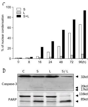
Fig. 3. Sulindac-induced apoptosis mechanisms were augmented by co-treatment of 1 M lactacystin. (A) The extent of apoptosis was augmented in co-treated-group compared to sulindac only treated-group (P < .01, 16~96 h). (B) Western blot showing caspase-3 activation and PARP cleavage. Co-treatment of lactacystin augmented sulindac- induced caspase-3 activation and PARP degradation, and production of the processed caspase-3 p20 and PARP p85 cleavage products. S = sulindac; L = lactacystin; S+L = sulindac+lactacystin.
A
B C S L S+L
Caspase-3
32kd
20kd 17kd 116kd PARP 85kd
동시에 투여하였더니 이들을 각각 투여했을 때와 비 교하여 항종양 효과가 매우 효과적으로 관찰되어 종 양의 성장이 지연되고 생존기간이 연장되었다고 하였 으며 이때 약제 투여에 따른 체중감소 설사 혹은 위, , , 장관 출혈과 같은 부작용은 전혀 관찰되지 않았다고 하였다.18 그러므로 프로테아좀 억제제와 특정 항암제 를 병용 투여할 경우 훨씬 적은 용량을 사용할 수 있음 으로 인해 약제의 투여에 따르는 부작용을 최소화하 면서 기대되는 적절한 항종양 효과를 가져 올 수 있을 것이다 저자들은 이 연구에서 프로테아좀 억제제인. 이 에 의해 유도된 세포사멸 기전에 lactacytrine sulindac
서 세포사멸의 정도를 더욱 증가시키는 사실을 확인 할 수 있었을 뿐만 아니라 본 연구에서는 나타나 있지, 않으나 1 M의 lactacystin과1 mM의 sulindac의 병용 투 여의 효과는 2.5 mM의 sulindac을 단독으로 투여했을 경우에서와 유사한 정도의 세포사멸을 유도함을 관찰 할 수 있었으며, 1 mM의 sulindac을 단독으로 투여하 였을 경우에 관찰되는 세포사멸의 정도는 1 M의
과 의 을 병용 투여했을 때 관
lactacystin 0.5 mM sulindac
찰되는 세포사멸의 정도와 같아 프로테아좀 억제제의, 병용 투여로 항종양제의 투여량을 줄일 수 있다는 장 점을 확인할 수 있었다.
결 론
저자들은 이 연구를 통해 대장암 세포주HT-29에서
의 일종인 과 프로테아좀 억제제인
NSAIDs sulindac
을 병용 투여하였을 경우 항암 효과가 더욱 lactacystin
더 증가하는 사실을 최초로 확인할 수 있었다 그러므. 로 종양세포의 세포사멸을 유도하는 데 있어서 sulin
과 의 병용 투여가 아주 효과적인 방법임
dac lactacystin
을 시사한다고 판단된다. Sulindac과 lactacystin의 병용 투여에 의한 대장암 세포의 소멸에 관여하는 기전에 대해서는 앞으로 보다 더 심층적인 연구가 뒤따라야 할 것으로 생각된다.
R E FE R E N C ES
1. Rosenberg L, Palmer JR, Zauber AG, Warshauer ME, Stolley PD, Shapiro S. A hypothesis: nonsteroidal anti- inflammatory drugs reduce the incidence of large-bowel cancer. J Natl Cancer Inst 1991;83:355-8.
2. Pasricha PJ, Bedi A, O'Connor K, Rashid A, Akhtar AJ, Zahurak ML, et al. The effects of sulindac on colorectal proliferation and apoptosis in familial adenomatous poly-
posis. Gastroenterology 1995;109:994-8.
3. Piazza GA, Rahm AK, Finn TS, Fryer BH, Li H, Stoumen AL, et al. Apoptosis primarily accounts for the growth- inhibitory properties of sulindac metabolites and involves a mechanism that is independent of cyclooxygenase inhibition, cell cycle arrest, and p53 induction. Cancer Res 1997;57:
2452-9.
4. Shiff SJ, Qiao L, Tsai LL, Rigas B. Sulindac sulfide, an aspirin-like compound, inhibits proliferation, causes cell cycle quiescence, and induces apoptosis in HT-29 colon adenocarcinoma cells. J Clin Invest 1995;96:491-503.
5. Pique M, Barragan M, Dalmau M, Bellosillo B, Pons G, Gil J. Aspirin induces apoptosis through mitochondrial cyto- chrome c release. FEBS Lett 2000;480:193-6.
6. Thun MJ, Henley SJ, Patrono C. Nonsteroidal anti-infla mmatory drugs as anticancer agents: mechanistic, pharma cologic, and clinical issues. J Natl Cancer Inst 2002;
94:252-66.
7. Meng L, Kwok BH, Sin N, Crews CM. Eponemycin exerts its antitumor effect through the inhibition of proteasome funtion. Cancer Res 1999;59:2798-801.
8. Drexler HC, Risau W, Konerding MA. Inhibition of proteasome function induces programmed cell death in proliferating endothelial cells. FASEB J 2000;14:65-77.
9. Orlowski RZ. The role of the ubiquitin-proteasome pathway in apoptosis. Cell Death Differ 1999;6:303-13.
10. Grimm LM, Goldberg AL, Poirier GG, Schwartz LM, Osborne BA. Proteasome play an essential role in thymocyte apoptosis. EMBO J 1996;15:3835-44.
11. Sadoul R, Fernandez PA, Quiquerez AL, Martinou I, Maki M, Schroter M, et al. Involvement of the proteasome in the programmed cell death of NGF-deprived sympathetic neu- rons. EMBO J 1996;15:3845-52.
12. Li B, Dou QP. Bax degradation by the ubiquitin/protea some-dependent pathway: involvement in tumor survival and progression. Pro Natl Acad Sci USA 2000;97:3850-5.
13. Orlowski RZ, Eswara JR, Lafond-Walker A, Grever MR, Orlowski M, Dang CV. Tumor growth inhibition induced in a murine model of human Burkitt's lymphoma by a proteasome inhibitor. Cancer Res 1998;58:4342-8.
14. Adams J, Palombella VJ, Sausville EA, Johnson J, Destree A, Lazarus DD, et al. Proteasome inhibitors: a novel class of potent and effective antitumor agents. Cancer Res 1999;
59:2615-22.
15. Chandra J, Niemer I, Gilbreath J, Kliche K-O, Andreeff M, Freireich EJ, et al. Proteasome inhibitors induce apoptosis in glucocorticoid-resistant chronic lymphocytic leukemic lymphocytes. Blood 1998;92:4220-9.
16. Delic J, Masdehors P, Omura S, Cosset JM, Dumont J, Binet JL, et al. The proteasome inhibitor lactacystin induces apoptosis and sensitizes chemo- and radioresistant human chronic lymphocytic leukaemia lymphocytes to TNF-alpha-
initiated apoptosis. Br J Cancer 1998;77:1103-7.
17. Fanelli M, Minucci S, Gelmetti V, Nervi C, Gambacorti- Passerini C, Pelicci PG. Constitutive degradation of PML/
RARalpha through the proteasome pathway mediates re- tinoic acid resistance. Blood 1999;93:477-81.
18. Golab J, Stoklosa T, Czajka A, Dabrowska A, Jakobisiak M, Zagozdzon R, et al. Synergistic antitumor effects of a selective proteasome inhibitor and TNF in mice. Anticancer Res 2000;20:1717-21.
19. Loda M, Cukor B, Tam SW, Lavin P, Fiorentino M, Draetta GF, et al. Increased proteasome-dependent degradation of the cyclin-dependent kinase inhibitor p27 in aggressive colorectal carcinomas. Nat Med 1997;3:231-4.
20. Chen F, Chang D, Goh M, Klibanov SA, Ljungman M. Role of p53 in cell cycle regulation and apoptosis following exposure to proteasome inhibitors. Cell Growth Differ 2000;
11:239-46.