Ⅰ. 서 론
치과 임플랜트는 결손 치아를 수복하기 위하여 널 리 사용되고 있으며, 많은 연구에서 높은 성공률을 보여주고 있다.1-3)임플랜트 수복을 위한 기존의 프로 토콜은 임플랜트를 식립하는 1차 수술시 임플랜트를 연조직 하방에 묻고 이후 3�6개월 간의 치유기간을 거친 후 2차 수술을 시행하는 것이었다. 이는 골유착 이 일어나는 시기에 임플랜트에 기능적 하중이 전달 됨으로써 미세동요에 의해 골유착이 방해받지 않게 하기 위해서였다.1) 그러나 기존 2회법을 이용한 임 플랜트 시술은 2번의 수술과정과 장기간의 치료기간 이라는 단점을 가진다. 최근에는 즉시하중이 시도되 어 그 영역을 넓혀가고 있으며,4) 특히 하악 완전 무 치악 환자에서 즉시하중을 가한 임플랜트 지지 고정 성 보철물은 높은 성공률을 보여주었다.5-7)
Henry 등5)은 5명의 환자에게 각각 6개의 Branemark 시스템 임플랜트를 식립하고 그 중 4개 의 임플랜트를 이용하여 즉시하중을 가하였으며 나 머지 2개의 임플랜트는 실패를 대비하여 골 속에 묻 어두었다. 2년 평가 결과 100%의 생존율을 보여주 었다. Randow 등6)은 27명의 환자에서 118개의 Branemark MK Ⅱ 임플랜트를 하악 무치악에 식립 하여 임플랜트 지지 고정성 보철물로 수복하였으며 이 중 16명의 환자, 88개의 임플랜트는 실험군으로
서 즉시하중을 시행하였고 11명의 환자, 30개의 임 플랜트는 대조군으로 일반적인 2회법을 적용하였다.
18개월 평가시 변연골 소실량은 실험군에서 0.4 ㎜, 대조군에서는 0.8 ㎜였으며 생존율은 두 군 모두에 서 100%였다. Testori 등7)도 하악무치악 환자 62명 에게 325개의 Osseotite 임플랜트를 식립하고 고정성 보철물을 제작하여 즉시하중을 가한 후 12�60개월 평가한 결과 99.4%의 성공률을 보고하였다.
기능적 하중이 전달되었을 때 임플랜트-골 계면에 서 150 ㎛ 이상의 미세동요가 발생하게 되면 임플랜 트의 골유착이 방해받게 되므로 즉시하중 시 이러한 사항을 잘 고려하여야 한다.8)이 등9)은 치밀골 양이 즉시하중 부담능과 임플랜트 안정성에 유의한 영향 을 미치며 치밀골이 두꺼워질수록 즉시하중 부담능 과 임플랜트 안정성이 증가한다고 보고한 바 있으 며, 하악 전방부는 이러한 면에서 구강내 다른 어떤 부위보다 더 나은 조건을 가지고 있다. 또한 하악 무 치악 증례에서 일반적으로 계획되는 임플랜트의 연 결고정은 즉시하중을 위한 더 나은 환경을 조성한 다. 임플랜트의 횡단 치열궁 연결고정(cross-arch splinting)시 임플랜트의 일차 안정성이 향상되며 더 유리한 하중 상태를 가진다는 것은 이미 알려진 사실이다.10-13)
임플랜트의 일차 안정성은 골유착을 위한 전제조 건3)일 뿐 아니라 골유착의 정도를 예견하기 때문에 대한치과보철학회지:Vol. 45, No. 2, 2007
하악 무치악에서 즉시하중과 지연하중간 임플랜트의 안정성 변화
강릉대학교 치과대학 보철학교실 및 구강과학연구소 정혜은ㆍ김대곤ㆍ박찬진ㆍ조리라ㆍ이양진
※ 본 연구는 식품의약품안전청의 용역연구개발사업(06112의기술285)의 지원으로 수행됨.
많은 임상가들은 임플랜트의 일차 안정성을 평가하 기 위해 타진 반응, 방사선 사진, Periotest�, 식립시 절삭력 측정 등 여러 가지 방법들을 이용하였다.14)
임플랜트의 안정성을 평가하는 방법 중 Meredith14) 에 의해 개발된 공진주파수분석법(resonance fre- quency analysis, RFA)은 임플랜트-골 계면의 견고 도(stiffness)를 휨(bending)에 의해 측정하는 방법으 로 비파괴적이고 간단하여 임상적용이 용이하다. 공 진주파수 측정치는 1에서 100까지의 ISQ (implant stability quotient) 수치와 진동파형으로 표시되며, 임플랜트의 안정성을 정량적으로 평가할 수 있다.
임플랜트-골 계면의 견고도 혹은 주위골의 견고도가 증가할수록 ISQ 수치는 증가하게 된다.14,15)이 등16)과 김 등17)은 공진주파수 분석법의 ISQ 수치가 임플랜 트 주위 골 변화를 인지할 수 있음을 보고하였으며 Glauser 등18)은 실패한 임플랜트에서 ISQ 수치가 1개월부터 특징적인 양상을 나타낸다고 하여 공진주 파수분석법이 임플랜트-골 계면의 견고도뿐 아니라 변연 골 수준의 변화를 나타낼 수 있어 임플랜트의 일차, 이차 안정성 평가 및 장기간 평가가 가능한 진 단적 방법임이 증명되었다.
본 연구의 목적은 하악 무치악 환자에서 임플랜트 지지 고정성 보철물로 수복시 통상적인 지연하중과 즉시하중을 가한 경우에 있어 시간이 지남에 따른 변연골 수준(marginal bone level)과 ISQ 수치의 변 화를 비교하고자 함이다. 또한 하악 이공 사이에 식 립한 임플랜트의 위치에 따라 이러한 수치가 어떤 차이를 보이는지 함께 살펴보고자 한다.
Ⅱ. 연구재료 및 방법 1. 실험군 선택
한 명의 치과의사에게 치료받은 16명의 환자(남자 9명, 여자 7명, 평균 나이 51.5세, Table I)가 포함되 었다.
연구에 포함된 환자들은 다음과 같은 기준을 만족 하였다.
1) 하악 완전 무치악
2) 가철성 보철물에 대해 거부감을 나타내며 임플 랜트를 이용한 고정성 보철물로 수복하는 것에
동의할 것
3) 하악 전방부에 길이 10 ㎜ 이상, 직경 3.75 ㎜ 이상의 임플랜트를 식립할 수 있는 가용골을 가 지며 수술시 부가적인 골 증대술이 필요하지 않 을 것
4) 전신적인 상태가 임플랜트 수술이 금기가 아니 며 수복과정을 견딜 수 있을 것
환자 선택시 대합치 상태는 고려하지 않았으며 흡 연가도 포함하였다. 흡연가들은 모두 4명이었으며 지연하중군에 속하였다. 흡연가들에게는 수술 전 1 주, 수술 후 8주간 금연을 지시하였다.
2. 임플랜트의 종류
식립된 모든 임플랜트는 TiUnite MKⅢ(Bra- nemark system�, Nobel Biocare AB, Goteborg, Sweden)였으며 직경은 3.75 ㎜ 또는 4.0 ㎜, 길이는 10 ㎜ 또는 13 ㎜였다(Table I).
3. 수술 및 보철 과정
하악 무치악에 총 90개의 임플랜트를 식립하였으 며 16명의 환자들을 하중 시기에 따라 2개의 군으로 분류하였다. 첫 번째 군(D군)은 지연 하중(delayed loading)군으로 통상적인 2회법으로 시술하여 임플 랜트 식립을 위한 1차 수술 후 일반적인 치유기간(3 개월)을 거쳐 2차 수술을 시행하였으며, 4주후 인상 을 채득하여 보철물을 제작하였다. 두 번째 군(I군) 은 즉시하중(immediately loading)군으로 임플랜 트 식립 후 24�48시간 내에 보철물을 제작, 장착하 여 즉시하중을 가하였다.
임플랜트 식립 전에 하악의 컴퓨터 단층 방사선 사 진을 촬영하여 임플랜트의 식립 부위를 평가하였다.
임플랜트 식립시 부가적인 골 증가술은 시행되지 않 았다. 두 군으로 분류된 환자의 상태는 군에 따라 큰 차이를 나타내지는 않았으며(Table I) 환자와 상의 후에 치료방법을 선택하였다.
하악의 악궁 형태에 따라 5�6개의 임플랜트를 식립 하였으며 U 형태의 악궁인 경우 양측 이공 사이에 6개 의 임플랜트를 식립하였고, V 형태의 악궁인 경우 양 측 이공 사이에 5개의 임플랜트를 식립하였다(Fig. 1).
임플랜트 식립시 골질과 골양을 평가하였으며 평 가기준은 Lekholm과 Zarb에 의한 악골의 골질과 골 양 분류법19)을 이용하였다(Table II).
즉시하중군은 기존 임시의치를 변경하여 수술 당 일 레진 임시보철물을 제작하였으며 의치의 파절을 예방하기 위하여 식립 위치를 예상하여 임시수복물 을 보강할 수 있는 금속 구조물을 미리 제작해 두었 다.20)임시수복물을 장착하여 기능한지 3�4개월 후 최종 보철물을 제작하였다.
4. 주기적인 평가 및 검사
임플랜트 식립시, 보철물 장착시, 그리고 매 6개월 마다 주기적인 평가를 시행하였다. 평가사항으로는 방사선 검사, 공진주파수 측정(ISQ 수치), 부작용들 을 포함하였다. 보철물 장착후 24개월까지 평가한 내용을 기록하였으며 ISQ 수치는 30개월까지 측정 하였다. 즉시하중군에서는 임플랜트 식립과 보철물 장착이 거의 동시에 이루어지므로 보철물 장착시의 Table I. Patients information and types of implants
Patient Sex Age Smoker Group Arch Bone Bone Implant
number shape quality quality type
1 M 57 Yes Delayed U-shaped 2 or 3 B �3.75×13
2 F 59 No Delayed U-shaped 2 or 3 B �4.00×13
3 F 43 No Delayed V-shaped 2 or 3 C �3.75×13
4 M 51 Yes Delayed V-shaped 1 or 2 D �3.75×10
5 M 54 Yes Delayed U-shaped 2 C �4.00×13
6 F 61 No Delayed V-shaped 2 C �3.75×13
7 M 46 No Delayed U-shaped 2 or 3 B �4.00×13
8 M 50 Yes Delayed V-shaped 1�3 C �3.75×10
9 F 52 No Immediate U-shaped 2 B �4.00×13
10 M 44 No Immediate V-shaped 3 B �3.75×13
11 M 45 No Immediate U-shaped 2 B �4.00×13
12 F 53 No Immediate U-shaped 2 or 3 B �4.00×13
13 M 49 No Immediate V-shaped 1 or 2 B �4.00×13
14 F 60 No Immediate U-shaped 1 or 2 B �4,00×13
15 F 52 No Immediate U-shaped 2 B �4.00×13
16 M 49 No Immediate U-shaped 2 or 3 B �4.00×13
Fig. 1. Implantation sites on anterior mandible according to arch shape. Left - U shape, Right - V shape.
측정값을 임플랜트 식립시의 측정값과 동일하게 설 정하였다.
방사선 검사는 평행 촬영법으로 개개 임플랜트의
치근단 사진(periapical x-ray)을 촬영하였으며, 방사 선 사진상에서 근심과 원심의 변연골 수준을 측정하 여 평균치를 구하였다. 기준 측정점은 임플랜트-지 대주 계면으로 설정하였다. 공진 주파수 측정시에는 상부 보철물을 제거한 후 개개 임플랜트의 안정성을 OsstellTM(Integration Diagnositc Ltd., Goteborg, Sweden)을 이용하여 평가하였다.
임플랜트 성공 기준은 Roos 등21)의 기준을 따랐으 며 이는 다음과 같다.
1) 개개 임플랜트의 안정성 평가시 동요도가 없어 야 한다.
2) 방사선 검사시 임플랜트 주위의 방사선 투과상 이 없어야 할 뿐 아니라 변연 골 소실량은 기능 첫 해에는 1.0 ㎜ 이내이며, 기능 첫해 이후에 는 매년 0.2 ㎜ 이하여야 한다.
3) 극심한 연조직 염증, 지속적인 동통, 감각 이상, 불편감 등의 증상 및 징후가 없어야 한다.
Table III. Marginal bone levels (mean ± standard deviation) in delayed loading and immediately load- ing groups
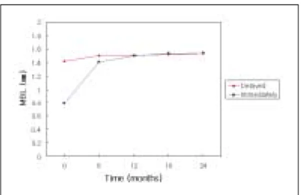
Prosthesis 6 Months 12 Months 18 Months 24 Months Delayed 1.42 ± 0.20 1.50 ± 0.13 1.51 ± 0.13 1.52 ± 0.12 1.54 ± 0.11 Immediately 0.79 ± 0.23 1.41 ± 0.17 1.50 ± 0.19 1.53 ± 0.18 1.54 ± 0.18 Total 1.10 ± 0.38 1.45 ± 0.16 1.51 ± 0.16 1.53 ± 0.15 1.54 ± 0.15 Repeated measured ANOVA P<0.001
Table II. Classification of bone quality and quantity by Lekholm and Zarb(1985) A. Bone quality
Type 1 : Almost the entire jaw is comprised of homogenous compact bone
Type 2 : A thick layer of compact bone surrounds a core of dense trabecular bone
Type 3 : A thin layer of cortical bone surrounds a core of dense trabedular bone of favorable strength Type 4 : A thin layer of cortical bone surrounds a core of low density trabedular bone
B. Bone quantity
Class A : Most of the alveolar ridge is present
Class B : Moderate residual ridge resorption has occurred
Class C : Advanced residual ridge resorption has occurred and only basal bone remains Class D : Some resorption of the basal bone has started
Class E : Extreme resorption of the basal bone has taken place
Fig. 2. Marginal bone level changes in delayed load- ing and immediately loading groups.
5. 통계 분석
95% 신뢰수준에서 분석하였다. 군간, 그리고 임플 랜트 위치간 시간에 따른 ISQ 수치 및 변연 골 수준 의 변화 양상 비교는 반복측정이 있는 일원분산분석
을 시행하였으며 임플랜트 위치별 비교는 Scheffe법 으로 사후검정하였다. 임플랜트 위치에 따른 측정값 비교시에 6개의 임플랜트를 식립한 경우에는 가운데 2개의 평균치를 5개 임플랜트를 식립한 경우의 중간 임플랜트의 측정값과 비교하였다.
Table IV. ISQ values (mean ± standard deviation) in delayed loading and immediately loading groups Installa- Prosth- 6 Months 12 Months 18 Months 24 Months 30 Months
tion esis
Delayed 75.1±4.8 73.8±3.7 73.3±3.5 73.0±3.3 73.0±3.1 72.8±3.2 72.8±3.2 Immediately 75.3±3.3 75.3±3.3 73.5±3.0 73.5±2.8 73.3±2.8 73.4±2.8 72.8±2.7 Total 75.2±4.1 74.5±3.6 73.4±3.2 73.3±3.1 73.2±3.0 73.1±3.0 72.8±3.0 Repeated measured ANOVA P=0.079
Fig. 3. ISQ value changes in delayed loading and immediately loading groups.
Fig. 4. Comparison of marginal bone level changes among implantation sites.
Table V. Comparison of marginal bone levels (mean ± standard deviation) among implantation sites Prosthesis 6 Months 12 Months 18 Months 24 Months
1 1.06±0.42 1.45±0.17 1.48±0.14 1.52±0.13 1.53±0.11
2 1.17±0.33 1.43±0.15 1.49±0.11 1.51±0.11 1.52±0.10
3 1.11±0.37 1.46±0.12 1.49±0.11 1.52±0.11 1.52±0.11
4 1.13±0.37 1.44±0.14 1.49±0.14 1.51±0.12 1.51±0.12
5 1.07±0.42 1.51±0.21 1.61±0.25 1.62±0.25 1.63±0.25
Repeated measured ANOVA P=0.604
Table VI. Comparison of ISQ values (mean ± standard deviation) among implantation sites
Installa- Prosth- 6 Months 12 Months 18 Months 24 Months 30 Months
tion esis
1 75.0±3.4 74.4±3.4 72.4±3.1 72.3±2.7 71.9±2.9 72.3±2.6 71.7±2.8 2 75.3±3.9 74.4±3.2 74.0±2.7 73.7±2.7 73.8±2.4 73.8±2.6 73.4±2.6 3 75.2±4.2 74.4±3.5 73.8±2.9 73.8±2.9 73.8±2.4 74.0±2.8 73.4±2.4 4 75.0±4.8 74.2±4.0 73.8±3.3 73.6±3.1 73.8±2.9 73.4±3.1 73.2±3.5 5 75.3±4.3 74.9±3.8 72.4±4.0 72.1±3.7 72.0±3.7 71.6±3.5 71.4±3.2 Repeated measured ANOVA P=0.047
Table VII. Comparison of ISQ values (mean ± standard deviation) among implantation sites in immediately loading group
Installa- Prosth- 6 Months 12 Months 18 Months 24 Months 30 Months
tion esis
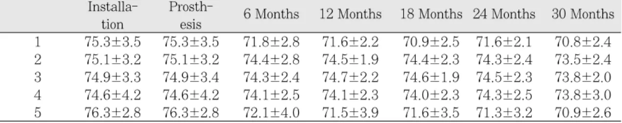
1 75.3±3.5 75.3±3.5 71.8±2.8 71.6±2.2 70.9±2.5 71.6±2.1 70.8±2.4 2 75.1±3.2 75.1±3.2 74.4±2.8 74.5±1.9 74.4±2.3 74.3±2.4 73.5±2.4 3 74.9±3.3 74.9±3.4 74.3±2.4 74.7±2.2 74.6±1.9 74.5±2.3 73.8±2.0 4 74.6±4.2 74.6±4.2 74.1±2.5 74.1±2.3 74.0±2.3 74.3±2.5 73.8±3.0 5 76.3±2.8 76.3±2.8 72.1±4.0 71.5±3.9 71.6±3.5 71.3±3.2 70.9±2.6 Repeated measured ANOVA P=0.001
Fig. 5. Comparison of ISQ value changes among implantation sites.
Fig. 6. Comparison of ISQ value changes among implantation sites in immediately loading group.
Ⅱ. 결 과 1. 임플랜트의 성공률
관찰기간 동안 두 군 모두에서 실패한 임플랜트는 없었으며 임플랜트 성공률은 100%였다. 즉시하중군
의 3명의 환자에서 각각 1개씩, 3개의 임플랜트에서 나사풀림이 관찰되어 다시 조여 주었으며 그 외의 부작용은 관찰되지 않았다. 24개월 평가시 평균 변 연골 수준은 1.54±0.15 ㎜였으며 30개월 평가시의 평균 ISQ 수치는 72.8±3.0으로 안정된 값을 나타내 었다.
2. 즉시하중군과 지연하중군간 시간에 따른 변연 골 수준과 ISQ 수치의 변화 비교
지연하중을 시행한 경우와 즉시하중을 가한 경우, 시간에 따른 변연골 수준의 변화 양상은 두 군에서 다르게 나타났다(P<0.001)(Table III, Fig. 2). ISQ 수치의 변화양상은 군간 의미있는 차이를 보이지 않 았으며 모든 군에 있어 안정된 값을 나타내었다 (P=0.079)(Table IV, Fig. 3). 두 군 모두 시간이 지 남에 따라 ISQ 수치는 감소하는 경향을 나타내었다.
3. 악궁에서 임플랜트 위치간 시간에 따른 변연골 수준과 ISQ 수치의 변화 비교
시간에 따른 변연골 수준의 변화양상은 임플랜트 위치에 따른 차이를 나타내지 않았으나(P=0.604), ISQ 수치는 임플랜트 위치에 따른 유의한 차이를 나 타내었다(P=0.047)(Table V, VI, Fig. 4, 5). 모든 임플랜트는 시간이 지남에 따라 ISQ 수치가 감소하 는 경향을 나타내었으며 특히 최후방(1, 5번 위치) 임플랜트의 ISQ 수치는 시간에 지남에 따라 다른 위 치의 임플랜트에 비해 더욱 감소하는 경향을 나타내 었다. 이러한 양상은 즉시하중군에서 더욱 두드러졌 으며 즉시하중군 내에서 시간에 따른 ISQ 수치의 변 화양상을 임플랜트 위치에 따라 비교하였을 때 통계 적으로 유의한 차이를 나타내었다(P=0.001) (Table VII, Fig. 6).
Ⅳ. 고 찰
본 연구에서는 16명의 하악 무치악 환자에게 90개 의 Bra�nemark TiUnite MKⅢ 임플랜트를 식립하여 임플랜트 지지 고정성 보철물로 수복하고 즉시하중 및 지연하중을 적용하여 30개월까지 추적연구하였 다. 관찰 기간동안 실패한 임플랜트는 없었으며 모 든 임플랜트의 성공률은 100%였다. 이러한 결과는 즉시하중을 가한 다른 연구 결과와 부합되는 높은 성공률이다. 앞선 연구들에서 하악 무치악 환자에게 즉시하중을 적용하여 1년에서 5년의 관찰기간동안 99.4�100%의 생존율을 보고한 바 있다.5-7)이러한 결과들은 하악 무치악 환자에서 즉시하중을 적용하
는 방법이 통상적인 지연하중과 견줄만한 예지성 높 은 치료라는 것을 뒷받침하고 있다.
본 연구에서 임플랜트 지지 보철물 장착후 첫 1년 간의 변연골 소실량은 두 군 모두에서 1 ㎜ 이하였으 며 2년째에는 0.04 ㎜였다. 이는 Roos 등21)의 성공 기준인 기능 첫해 이후의 골 소실량인 0.2 ㎜의 범위 내에 부합하였다. Lindquist 등22)은 하악 완전 무치악 에 임플랜트 지지 완전 고정성 보철물로 수복한 47 명의 환자를 12�15년간 평가한 결과 첫 해 이후부 터 연간 0.05 ㎜의 골 소실량을 보고한 바 있어 본 연구에서의 첫 해 이후 골 소실량이 안정된 수준임 을 알 수 있다. 그러나 지연하중군과 즉시하중군 간 에 시간에 따른 변연골 수준의 변화 양상을 반복측 정이 있는 일원분산분석으로 비교하였을 때 통계학 적으로 유의한 차이(P<0.001)를 보이고 있으며 보철 물 장착시의 변연골 수준이 지연하중군에서 1.42
㎜, 즉시하중군에서 0.79 ㎜로 큰 차이를 나타내고 있다. 이는 지연하중의 경우 보철물 장착시의 시점 이 치유 지대주를 연결하고 6�10주 후의 시점으로 치유 지대주를 연결한 후 어느 정도의 시간 경과가 있었으나 즉시하중의 경우 임플랜트 식립후 24�48 시간 내에 보철물이 장착되었기 때문일 것으로 생각 된다. Engquist 등23)은 Bra�nemark시스템이 임플랜 트 고정체 식립 직후에는 약간의 골 소실이 일어날 뿐이지만 치유 지대주를 연결하고 보철물이 장착되 는 사이의 기간 동안 많은 양의 골 소실이 일어남을 보고하였다.
본 연구에서 임플랜트 식립시의 ISQ 수치의 분포 는 63�84 사이의 값을 보였으며 평균값은 75였다.
이는 O¨stman 등24)이 보고한 하악에서의 평균인 71.4보다 더 큰 값으로, 식립된 임플랜트들의 높은 일차안정성을 보여준다. 30개월 평가시기의 ISQ 수 치도 65�79 사이의 값을 나타내었고 평균값은 72 로서 30개월 평가시기까지 안정된 값을 보여주어 모 든 임플랜트의 안정성이 우수함을 나타내었다. 지연 하중군과 즉시하중군의 ISQ 수치는 거의 동일하여 차이를 나타내지 않았으나 보철물 장착시의 ISQ 수 치는 지연 하중군에서 약간 낮게 나타났으며 이는 앞서 설명한 변연골 수준의 차이 때문인 것으로 생 각된다.
본 연구에서 ISQ 수치는 시간에 따라 감소하는 경
향을 나타내어 6개월 평가시기까지는 큰 폭으로 감 소하였으며 그 이후의 감소량은 미미하였다. 하지만 Glauser 등18)은 즉시하중 또는 조기하중을 가한 81개 의 임플랜트의 ISQ 수치 평가시 보철물 장착후 1, 2 개월까지는 ISQ 수치가 감소하였으나 이후에 안정 화되어 12개월 평가시에는 오히려 증가하는 경향이 있음을 보고하였다. 이에 반해 Nedir 등25)은 36명의 환자에게 106개의 ITI 임플랜트를 식립하여 임플랜 트 안정성을 평가하였는데, 식립시 ISQ 수치가 70 미만이었던 임플랜트는 3개월까지 ISQ 수치가 증가 하는 양상을 나타낸 반면, ISQ 수치가 70 이상인 임 플랜트는 3개월까지 ISQ 수치가 감소하는 경향을 나타낸다고 보고하였다. 본 연구에서는 ISQ 수치가 시간이 지남에 따라 감소하였으며 증가하는 양상은 나타내지 않았다. 앞선 연구들은 상악에서의 증례와 골 이식 증례를 포함하여, 식립시에는 불량한 골질 로 인해 ISQ 수치가 낮았으나 골유착을 이뤄가면서 ISQ 수치가 증가한 것으로 여겨지며 본 연구에서는 우수한 골질과 숙련된 수술방법으로 인해 식립시 70 이상의 높은 ISQ 수치를 보였으나 시간이 지남에 따 라 골 재형성을 거치며 골유착을 이뤄가는 과정에서 ISQ 수치가 감소한 것으로 생각된다.
하악 무치악 이공 사이 임플랜트 위치에 따른 임플 랜트의 안정성을 시간별로 비교하였을 때 본 연구에 서는 임플랜트 위치에 따른 ISQ 수치의 유의한 차이 (P=0.047)를 나타내었다. 모든 임플랜트들은 시간 이 지남에 따라 ISQ 수치가 감소하는 경향을 나타내 었으며 Fig. 5에 보이는 바와 같이 최후방 임플랜트 에서 감소경향이 더 뚜렷하였다. Fig. 5와 Fig. 6을 비교해 보면 이러한 최후방 임플랜트의 ISQ 수치 감 소경향은 즉시하중군에서 더욱 두드러져 나타난다.
최후방 임플랜트의 이러한 감소경향의 원인으로는 크게 두가지 요인을 추측해 볼 수 있다. 첫 번째는 하악 전방부에 임플랜트를 식립하여 완전 고정성 보 철물로 수복하는 경우 항상 고려되는 캔틸레버(can- tilever)로 인한 영향이다. Tashkandi 등26)은 우골에 3개의 13 ㎜ 임플랜트를 식립하고 6 ㎜ 폭과 5 ㎜ 두 께를 가지는 다양한 길이의 캔틸레버를 포함하는 보 철물을 연결하여 변형(strain)을 평가하였는데 최후 방 지대치의 경우 캔틸레버의 길이가 증가할수록 변 형의 강도가 다른 부위보다 크게 증가한다는 것을
보고하였다. Sertgo¨z 등27)은 하악의 삼차원 유한요소 분석법을 이용하여 임플랜트 지지 고정성 보철물에 서 응력 분석시 최후방 임플랜트에서 가장 큰 von Mises 응력을 나타낸다고 보고하였다. McAlar- ney 등28)은 55개의 임상 증례 자료를 컴퓨터에 입력하여 Skalak 모형으로 기계적 과하중(mechanical over- load)를 계산하였으며 그 결과 캔틸레버 부위에 가해 지는 교합력은 최후방 임플랜트에 2�3배의 하중을 발생시켰으며 5�6개의 임플랜트를 식립한 경우 최 후방 임플랜트의 질이 더 중요하다고 하였다.
Romeo 등29)은 부분 무치악에 캔틸레버를 포함하는 임플랜트 지지 고정성 보철물로 수복한 38명의 환자 를 평균 3.9년간 평가하였는데 가장 큰 변연골 소실 은 대부분 캔틸레버에 가장 가까운 고정체에서 발생 한다고 보고하였다. 본 연구에서는 5번 최후방 임플 랜트의 경우 24개월 평가시의 평균 변연골 수준은 1.63 ㎜이며 2, 3, 4번 임플랜트의 평균 변연골 수준 은 1.52 ㎜로 통계학적으로 유의하지는 않지만 차이 를 나타내었다. 그에 반해 1번 최후방 임플랜트의 평균 변연골 수준은 1.53 ㎜로 가운데 임플랜트들의 평균 변연골 수준과 별반 차이가 없어 최후방 임플 랜트들의 ISQ 수치의 감소가 변연골 수준에 의한 결 과만은 아닐 것으로 생각된다. 두 번째로 생각해 볼 수 있는 것은 하악의 굴곡(mandibular flexure)현상 으로 인한 영향이다. 여러 연구에서 크게 개구시 구 치부에서 0.03�0.7 ㎜의 하악 변형량을 보고하였
다.30-33) Fishchman34)은 하악에 고정성 연결고정
(fixed splint)이 존재할 때 하악의 변형량이 감소하며 이는 연결고정이 응력을 겪고 있다는 것을 의미하며 자연치에서는 치주상태에 따라 응력을 치주조직으 로 분산한다고 보고하였다. Zarone 등35)은 임플랜트 지지 하악 완전 고정성 보철물에서 하악굴곡과 응력 양상을 평가하였으며 모든 임플랜트를 연결고정한 경우 최후방 임플랜트에서 가장 큰 응력을 나타낸다 고 보고하였다. 이러한 결과들로 유추해볼 때 캔틸 레버를 포함하고 연결고정된 보철물에서 최후방 임 플랜트는 가운데 임플랜트보다 더 큰 응력 하에 있 으며 그것이 어떠한 방법으로 영향을 미치는지는 알 수 없지만 임플랜트-골 계면 사이의 견고도에 영향 을 미치는 것으로 사료된다.
이러한 연구 보고들과 본 연구의 결과로 추론해 보
면 무치악에 다수의 임플랜트를 식립하여 보철물을 제작할 경우 최후방 임플랜트의 충분한 길이와 직경 을 확보하여야 할 것이다. 특히 즉시하중과 같이 골 재형성에 의한 골유착이 일어나기 전에 하중을 가하 는 상황이라면 연결고정이 미세동요량을 줄여준다 하더라도 최후방 임플랜트 식립부위의 골 밀도 및 식립 가능한 임플랜트의 길이와 직경에 대해 더욱 신중히 평가하여야 하겠다.
본 연구는 16명의 환자를 30개월까지 추적연구하 여 하악 무치악 전방부에 5�6개의 임플랜트를 식립 하고 임플랜트 지지 고정성 보철물로 수복하였을 때 즉시하중을 적용한 경우에 있어서도 일반적인 지연 하중의 경우와 같은 높은 성공률과 임플랜트 안정성 을 보여줌을 확인하였다. 그러나 포함된 환자의 수 가 적고 다소 단기간의 연구라는 한계가 있어 추후 많은 환자를 대상으로 한 장기간의 연구가 필요할 것으로 사료된다.
Ⅴ. 결 론
16명의 하악 무치악 환자에게 90개의 Bra�nemark MKⅢ 임플랜트를 식립하여 임플랜트 지지 고정성 보철물로 수복하고 즉시하중 및 통상적 지연하중을 적용하여 30개월까지 추적연구하였다. 시간이 지남 에 따른 변연골 수준과 ISQ 수치의 변화를 비교하였 으며 하악 이공 사이 임플랜트의 위치에 따라 이러 한 수치의 변화량에 차이가 있는지를 평가하여 다음 과 같은 결론을 얻었다.
1. 관찰기간동안 실패한 임플랜트는 없었으며 모든 임플랜트의 변연골 수준과 ISQ 수치가 안정된 값 을 나타내었다.
2. 즉시하중군과 지연하중군간 시간에 따른 변연골 수준 변화 양상 비교시 유의성 있는 차이를 나타 내었다(P<0.001). 이는 보철물 장착 시점이 다르 기 때문이며 6개월 평가 시기 이후로는 두 군간의 차이를 보이지 않았으며 안정된 변연골 수준을 보여주었다.
3. 즉시하중군과 지연하중군간 시간에 따른 ISQ 수 치의 변화 양상 비교시 유의한 차이를 나타내지 않았다(P=0.079). 두 군 모두에서 시간이 지남에 따라 ISQ 수치가 감소하는 양상을 보였으며 30개
월 평가시에 ISQ 수치는 안정된 값을 나타내 었다.
4. 하악 이공 사이에 식립된 임플랜트 위치별 시간 에 따른 변연골 수준의 변화 양상 비교시 유의한 차이를 나타내지 않았다(P=0.604).
5. 하악 이공 사이에 식립된 임플랜트 위치별 시간 에 따른 ISQ 수치의 변화 양상 비교시 위치에 따 라 유의한 차이를 나타내었다(P=0.047). 최후 방 임플랜트의 ISQ수치는 다른 부위의 임플랜트보 다 더욱 감소하는 경향을 나타내었으며 이런 양 상은 즉시하중군에서 두드러졌으나 ISQ 수치는 안정된 값을 나타내었다.
참고문헌
1. Albrektsson T, Zarb G, Worthington P, Eriksson AR. The long-term efficacy of cur- rently used dental implants: A review and proposed criteria of success. Int J Oral Maxillofac Implants 1986;1:11-25.
2. Adell R, Eriksson B, Lekholm U, Bra�ne- mark PI, Jemt T. A long-term follow-up study of osseointegrated implants in the treatment of totally edentulous jaws. Int J Oral Maxillofac Implants 1990;4:347-59.
3. Adell R, Lekholm U, Rockler B, Bra�nemark PI. A 15-year study of osseointegrated im- plants in the treatment of the edentulous jaw. Int J Oral Surg 1981;10:387-416.
4. Gapski R, Wang HL, Mascarenhas P, Lang NP. Critical review of immediate im- plant loading. Clin Oral Impl Res 2003;
14:515-27.
5. Henry P, Rosenberg L. Single-stage surgery for rehabilitation of the edentulous mandible: Preliminary results. Pract Perio Aesth Dent 1994;6:15-22.
6. Randow K, Ericsson I, Nilner K, Petersson A, Glantz PO. Immediate functional load- ing of Bra�nemark dental implants. An 18- month clinical follow-up study. Clin Oral
Impl Res 1999;10:8-15.
7. Testori T, Meltzer A, Fabbro M, Zuffetti F, Troiano M, Francetti L, Weinstein RL. Immediate occlusal loading of Osseotitie implants in the lower edentulous jaw. A multicenter prospective study. Clin Oral Impl Res 2004;15:278-84.
8. Szmukler-Moncler S, Salama H, Reinge- wirtz Y, Dubruille JH. Timing of loading and effect of micromotion on bone-dental implant interface: Review of experimental literature. J Biomed Mater Res 1998;
43:192-203.
9. Yi YJ, Park CJ, Cho LR. An evaluation of the primary implant stability and the immediate load-bearing capacity accord- ing to the change of cortical bone thickness.
J Korean Acad Prosthodont 2005; 43:
248-57.
10. Glantz PO, Nyman S, Strandman E, Randow K. On functional strain in fixed mandibular reconstructions. Ⅱ. An in vi- vo study. Acta Odontol Scand 1984;5:
269-76.
11. Glantz PO, Strandman E, Svensson SA, Randow K. On functional strain in fixed mandibular reconstructions. Ⅰ. An in vitro study. Acta Odontol Scand 1984;
4:241-9.
12. Salama H, Rose LF, Salama M, Betts NJ. Immediate loading of bilaterally spli- nted titanium root-form implants in fixed prosthod- ontics - a technique reexamined:
two case reports. Int J Perio Res Dent 1995;15:344-61.
13. Tarnow DP, Emtiaz S, Classi A. Immediate loading of threaded implants at stage 1 surgery in edentulous arches: Ten con- secutive case reports with 1-to 5-year data. Int J Oral Maxillofac Implants 1997;12:319-24.
14. Meredith N. Assessment of implant stability as a prognostic determinant. Int J Prostho- dont 1998;11:491-501.
15. Meredith N, Alleyne D, Cawley P.
Quantitative determination of the sta- bility of the implant-tissue interface using resonance frequency analysis. Clin Oral Impl Res 1996;7:261-7.
16. Lee MR, Cho LR, Yi YJ, Choi HM, Park CJ. Correlation assessment between res- onance frequency analysis and radiogra- phic method according to peri-implant bone change. J Korean Acad Prosthodont 2005;43:736-44.
17. Kim SM, Park CJ, Yi YJ, Chang BS, Cho LR. A comparative analysis with resonance frequency according to vari- ous simulated bone defects. J Korean Acad Prosthodont 2005;43:487-97.
18. Glauser R, Sennerby L, Meredith N, Ree A, Lundgren A, Gottlow J, Hammerle CHF. Resonance frequency analysis of implants subjected to immediate or early functional occlusal loading. Successful vs.
failing implants. Clin Oral Impl Res 2004;
15:428-34.
19. Lekholm U, Zarb GA. Patient selection and preparation. In: Bra�nemark PI, Zarb GA, Albrektsson T. Tissue-integrated pros- theses: Osseointegration in clinical dentistry.
Chicago: Quintessence;1985. p. 199-209.
20. Park CJ, Kim DS, Yi YJ, Cho LR. Fabri- cation of a screw-retained, implantsup- ported, provisional prosthesis: A clinical report. J Prosthet Dent 2004;91:409-13.
21. Roos J, Sennerby L, Lekholm U, Jemt T, Grondahl K, Albrektsson T. A qualitative and quantitative method for evaluating im- plant success: A 5-year retrospective analysis of the Bra�nemark implant. Int J Oral Maxillofac implants 1997;12:504-14.
22. Lindquist LW, Carlsson GE, Jemt T. A prospective 15-year follow- up study of mandibular fixed prostheses supported by osseointegrat ed implants. Clinical re- sults and marginal bone loss. Clin Oral Impl Res 1996;7:329-36.
23. Engquist B, Astrand P. Dahlgren S, Engquist E, Feldmann H, Gron dahl K.
Marginal bone reaction to oral implants:
a prospective com parative study of Astra Tech and Bra�nemark System implants.
Clin Oral Impl Res 2002;13:30-7.
24. Ostman PO, Hellan M, Wendelhag I, Sennerby L. Resonance frequen cy analy- sis measurements of implants at placement surgery. Int J Prosthodont 2006;19:77-83.
25. Nedir R, Bischof M, Szmukler-Moncler S, Bernard JP, Samson J. Predicting os- seointegration by measn of implant primary stability. A resonance-frequency analysis study with delayed and immediately loaded ITI SLA implants. Clin Oral Impl Res 2004;15:520-8.
26. Tashkandi EA, Lang BR, Edge MJ.
Analysis of strain at selected bone sites of a cantilevered implant-supported pros- thesis. J Prosthet Dent 1996;76:158-64.
27. Sertgo¨z A, Guvener S. Finite element analysis of the effect of can- tilever and im- plant length on stress distribution in an implant- supported fixed prosthesis. J Prosthet Dent 1997;76:165-9.
28. McAlarney ME, Sc DE, Stavropoulos DN.
Theoretical cantilever lengths versus clin- ical variables in fifty-five clinical cases. J Prosthet Dent 2000;83:332-43.
29. Romeo E, Lops D, Margutti E, Ghisolfi M, Chiapasco M, Vogel G. Implant-support- ed fixed cantilever prostheses in partial- ly edentulous arches. A seven-year prospec- tive study. Clin Oral Impl Res 2003;14:
303-11.
30. Goodkind RJ, Heringlake CB. Mandibular flexure in opening and closing movement.
J Prosthet Dent 1973;30:134-8.
31. De Marco TJ, Paine S. Mandibular di- mensional change. J Prosthet Dent 1974;
31:482-5.
32. Omar R, Wise MD. Mandibular flexure as- sociated with muscle force applied in the retruded axis position. J Oral Rehabil 1981;8:209-21.
33. Fischman B. The rotational aspect of mandibular flexure. J Prosthet Dent 1990;
64:483-5.
34. Fischman BM. The influence of fixed splints on mandibular flexure. J Prosthet Dent 1976;35:643-7.
35. Zarone F, Apicella A, Nicolais L, Aversa R, Sorrentino R. Man dibular flexure and stress build-up in mandibular full-arch fixed prostheses supported by osseointegrated implants. Clin Oral Impl Res 2003;14:
103-14.
Reprint request to:
Lee-Ra Cho, D.D.S., M.S.D., Ph.D.
Dept. of Prosthodontics & Research Institute of Oral Science, College of Dentistry, Kangnung National University
Gangneung Daehangno 120, Gangneung, Gangwon-Do, 210-702, Korea [email protected]
Statement of problem: It was reported high success rate of implant-supported fixed prostheses using with 5�6 implants on anterior mandible. Recently, immediate loading pro- tocol was focused to overcome disadvantages of classic 2-stage delayed loading protocol.
Purpose: This clinical study was to evaluate stability changes with time of immediately loaded and delayed loaded implants in edentulous mandible and to compare stability changes with time according to implantation sites.
Materials and methods: Five or six implants were placed on anterior mandible depending on the arch shape. The immediately loading group was consisted of 8 patients received their prostheses within 24�48 hours after implantation. The delayed loading group was consisted of 8 patients received their definitive prostheses following classical prosthetic procedures after a healing period of 3 months.
All patients were recalled every 6 months for check-up. The evaluations of radiograph- ic examination, ISQ value measurement and recording of complication were done. To eval- uate marginal bone level, intraoral periapical radiographs were taken with long cone par- alleling technique. At every evaluation recall, all prostheses were removed and ISQ values were measured with OsstellTMon individual implants.
Results:
1. None of implants was failed. All implants showed stable marginal bone levels and ISQ values.
2. Marginal bone level changes with time showed statistically significant difference between immediately loading group and delayed loading group (P<0.001).
3. ISQ value changes with time did not show statistically significant difference between immediately loading group and delayed loading group (P=0.079). ISQ value decreased with time in both groups, however, all implants showed stable ISQ value at 30 months-recall evaluation.
STABILITY CHANGES OF IMMEDIATELY LOADED AND DELAYED LOADED IMPLANTS IN EDENTULOUS MANDIBLE
Hye-Eun Jung, D.D.S., Dae-Gon Kim, D.D.S., Chan-Jin Park, D.D.S., Ph.D., Lee-Ra Cho, D.D.S., Ph.D., Yang-Jin Yi, D.D.S., Ph.D.
Department of Prosthodontics and Research Institute of Oral Science, College of Dentistry, Graduate School, Kangnung National University
ABSTRACT
4. Marginal bone level changes with time did not show statistically significant differences among implantation sites (P=0.604).
5. ISQ value changes with time showed statistically significant differences among implantation sites (P=0.047). ISQ values of most posterior implants decreased with time comparing to other implants.
Conclusion: Although the marginal bone level of the terminal abutment didn’t differ- ent with the other implants, ISQ value of the terminal abutment was lower than that of the other implants. Therefore, further clinical evaluation would be needed in this point of view.
Key words : Immediately loading, Delayed loading, Marginal bone level, ISQ value