https://doi.org/10.4174/astr.2020.99.3.146 Annals of Surgical Treatment and Research
Surgical skin adhesive bond is safe and feasible wound closure method to reduce surgical site infection following minimally invasive colorectal cancer surgery
Chul Seung Lee, Seung-Rim Han, Bong-Hyeon Kye, Jung Hoon Bae, Wooree Koh, In Kyu Lee, Do-Sang Lee, Yoon Suk Lee
Division of Colorectal Surgery, Department of Surgery, Seoul St. Mary’s Hospital, College of Medicine, The Catholic University of Korea, Seoul, Korea
INTRODUCTION
Surgical site infections (SSIs) increase the risk of long-term morbidity among postoperative patients, with a higher rate of rehospitalization and postdischarge outpatient expenses [1]. In fact, SSIs are the most common cause of unplanned postoperative readmission [2]. Colorectal surgical procedures,
which account for a relatively high volume of gastrointestinal operations, pose a high risk for SSIs [3]. Several factors, such as age, the American Society of Anesthesiologists (ASA) physical status classification, diabetes mellitus, body mass index (BMI), smoking, and tumor location, are known to be independent risk factors of SSIs [3]. Other operative characteristics, including blood transfusion [4], emergency cases, and longer operative
Received March 10, 2020, Revised June 1, 2020, Accepted June 16, 2020 Corresponding Author: Yoon Suk Lee
Division of Colorectal Surgery, Department of Surgery, Seoul St. Mary’s Hospital, College of Medicine, The Catholic University of Korea, 222 Banpo-daero, Seocho-gu, Seoul 06591, Korea
Tel: +82-2-2258-6095, Fax: +82-2-595-2282 E-mail: yslee@catholic.ac.kr
ORCID: https://orcid.org/0000-0002-1849-2774
Copyright ⓒ 2020, the Korean Surgical Society
cc Annals of Surgical Treatment and Research is an Open Access Journal. All articles are distributed under the terms of the Creative Commons Attribution Non- Commercial License (http://creativecommons.org/licenses/by-nc/4.0/) which permits unrestricted non-commercial use, distribution, and reproduction in any medium, provided the original work is properly cited.
Purpose: Minimally invasive colorectal surgery had reduced the rate of surgical site infection. The use of surgical skin adhesive bond (2-octyl cyanoacrylate) for wound closure reduces postoperative pain and provides better cosmetic effect compared to conventional sutures or staples. But role of surgical skin adhesive bond for reducing surgical site infection is unclear. Our objective in this study was to evaluate the role of surgical skin adhesive bond in reducing surgical site infection following minimally invasive colorectal surgery.
Methods: We performed a retrospective analysis of 492 patients treated using minimally invasive surgery for colorectal cancer at Seoul St. Mary’s Hospital, the Catholic University of Korea. Of these, surgical skin adhesive bond was used for wound closure in 284 cases and skin stapling in 208. The rate of surgical site infection including deep or organ/space level infections was compared between the 2 groups.
Results: The rate of superficial surgical site infection was significantly lower in the group using skin adhesive (P = 0.024), and total costs for wound care were significantly lower in the skin adhesive group (P < 0.001).
Conclusion: This study showed that surgical skin adhesive bond reduced surgical site infection and total cost for wound care following minimally invasive colorectal cancer surgery compared to conventional skin stapler technique. Surgical skin adhesive bond is a safe and feasible alternative surgical wound closure technique following minimally invasive colorectal cancer surgery.
[Ann Surg Treat Res 2020;99(3):146-152]
Key Words: Cyanoacrylates, Octyl 2-cyanoacrylate, Surgical stapling, Surgical wound infection, Tissue adhesives
time [5], have also been associated with an increased risk for SSIs. A clearly modifiable factor is the type of surgical wound closure used, which is also known to be associated with the risk for SSIs [6].
Skin stapling provides surgeons with a convenient and quick technique for surgical wound closure. However, skin staples can increase postoperative pain and carry a risk for scarring.
Surgical skin adhesives have been developed to overcome these shortcomings of skin stapling. Surgical skin adhesives create a strong polymeric bond across the edges of the wound, promoting natural healing, with the synthetic flexible microbial barrier providing in-vitro protection against penetration of organisms [7]. A prospective randomized study further reported a trend to better cosmetic outcomes with the use of skin adhesives compared to staples for the closure of surgical wounds in open elective colectomies for benign or malignant indications [8]. However, there has been less comparative study evaluating the effect of surgical skin adhesives compared to that of skin stapling on the risk of SSIs after elective colorectal cancer surgery. In fact, although the effects of skin stapling on the risk of SSIs have been evaluated in clean surgeries, such as spinal surgery [9], the risk for SSIs with the use of skin stapling in clean-contaminated surgery, such as colorectal surgery, has not been evaluated. Considering the decreasing rate of SSIs with the increasing use of minimally invasive surgical (MIS) techniques for colorectal surgery, there is a need to evaluate the effect of different surgical wound closures on the risk of SSIs for colorectal MIS. The objective of this study was, therefore, to comparatively evaluate the role of surgical skin adhesives with that of skin stapling in reducing the rate of SSIs following MIS for colorectal cancer surgery.
METHODS
Patients
Our study was approved by the Institutional Review Board (IRB) of The Catholic University of Korea (KC19RESI0247).
Informed consent from patients to be included in this study was omitted according to the policy of our IRB. We reviewed the medical records of patients who underwent MIS (robotic or laparoscopic colorectal surgery) for clinical TNM stage I–
IV colorectal cancer at Seoul St. Mary’s Hospital, the Catholic University of Korea, between January 2017 and February 2019.
From January 2017 to May 2018, surgeons have used only skin stapler (Visistat Skin Stapler, Teleflex, Wayne, PA, USA) for surgical wound closure. From August 2018 to February 2019, surgeons have used only surgical skin adhesive bond (Dermabond Advanced Topical Skin Adhesive, Ethicon, Cincinnati, OH, USA). We excluded patients who underwent concomitant surgeries, emergency surgery, conversion to an open approach, or abdominoperineal resection, as well as those
with a history of open abdominal surgery. A total of 492 eligible patients were identified, with surgical adhesive closure used in 284 (57.7%, group I) and skin stapling in 208 patients (42.3%, group II).
Perioperative care and intraoperative treatment were similar for the 2 groups, adhering to the preventive SSI bundle for colorectal surgery [10], which includes hair removal with a clipper before surgery, use of 2% chlorhexidine gluconate–70% isopropyl alcohol solution for skin disinfection [11], administration of prophylactic antibiotic treatment within 30 minutes prior to skin incision, maintenance of an adequate circulating volume, and incisional wound irrigation using povidone-iodine solution before closure. The following precautions were also adhered to during surgery; use of a wound protector for specimen removal, limiting operating room traffic to essential personnel, and close attention by the anesthesiologist to maintain normothermia and euglycemia.
Other pre- and postoperative managements adhered to the enhanced recovery after surgery (ERAS) protocol at our institution [12], including intravenous pain control medication (900-mg fentanyl citrate and 180-mg ketorolac tromethamine).
The incidence of SSIs was defined using the Centers for Disease Control and Prevention criteria [13], with a maximum follow-up for wound care of 90 days after surgery (superficial or deep incisional SSI, 30 days; organ/space SSI, 90 days).
Monitoring for SSIs was conducted under the Korean National Healthcare-associated Infections Surveillance System (KONIS) program, using the standard methods implemented by the KONIS network for the surveillance of healthcare-associated infection [14]. Superficial incisional SSIs were treated mainly with wound dressing care, whereas deep incisional or organ/
space SSIs were treated with additional drainage of any discharge or pus. Antibiotics were chosen to cover enteric gram- negative and facultative/anaerobic bacilli [15].
After colon or rectal resection using a laparoscope or robot, extension of the umbilical incision within 5.0–7.0-cm long was performed for removal of the specimen through the wound protector (SurgiTractor, Surgicore, Gwangju, Korea).
An extracorporeal anastomosis was preferred for right-sided colon cancers and an intracorporeal anastomosis for left- sided colon cancers. The daVinci Xi Surgical System (Intuitive Surgical, Sunnyvale, CA, USA) was used for all robot-assisted procedures. Antibiotic-coated sutures (Coated VICRYL Plus Antibacterial [polyglactin 910] Suture, PDS Plus Antibacterial [polydioxanone] Suture; Ethicon) were used for closure of the fascia and subcutaneous layers in both groups. In surgical skin adhesive group, skin bond was applied after subcuticular layer was approximated using 4-0 vicryl sutures. When using a skin stapler, skin stapler was applied just after subcutaneous suture.
Surgical wound dressing was performed at postoperative 1st, 2nd, and 4th day in surgical skin stapler group and skin staplers
were removed at postoperative 7th to 10th days at outpatient’s clinic. Routine surgical wound dressing was not performed in surgical skin adhesive group.
Costs were measured in US dollar (USD). One USD was calculated at 1,137 Korean Won. The total costs for wound care included the cost of the surgical material (skin adhesive bond, skin stapler, and dressing material) and dressing charge during hospital stay and outpatient’s clinic.
Statistical analysis
Differences between groups were evaluated using Student t-test and the chi-squared test for continuous and categorical variables, respectively. Statistical analysis was performed using IBM SPSS Statistics software (ver. 24.0, IBM Corp., Armonk,
NY, USA). Significant associations obtained on univariate analysis were used in a multivariate logistic regression analysis to identify independent predictors of SSIs. These variables included age, diabetes mellitus, ASA physical status, BMI, operation time, tumor location, stoma formation, and operation method. A P-value of <0.05 was considered significant.
RESULTS
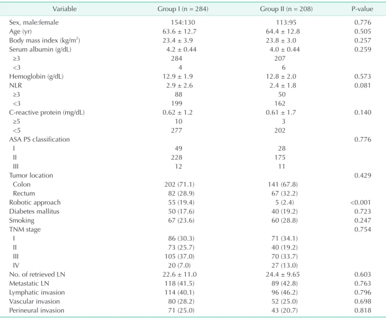
The characteristics for the 284 patients in group I (surgical skin adhesive bond) and the 208 in group II (skin stapler) are shown in Table 1. There were no differences in patients’
characteristics between the 2 groups, including patient-related factors (age, BMI, ASA physical status, and underlying disease),
Table 1. Patient characteristics
Variable Group I (n = 284) Group II (n = 208) P-value
Sex, male:female 154:130 113:95 0.776
Age (yr) 63.6 ± 12.7 64.4 ± 12.8 0.505
Body mass index (kg/m2) 23.4 ± 3.9 23.8 ± 3.0 0.257
Serum albumin (g/dL) 4.2 ± 0.44 4.0 ± 0.44 0.259
≥3 284 207
<3 4 6
Hemoglobin (g/dL) 12.9 ± 1.9 12.8 ± 2.0 0.573
NLR 2.9 ± 2.6 2.4 ± 1.8 0.081
≥3 88 50
<3 199 162
C-reactive protein (mg/dL) 0.62 ± 1.2 0.61 ± 1.7 0.140
≥5 10 3
<5 277 202
ASA PS classification 0.776
I 49 28
II 228 175
III 12 11
Tumor location 0.429
Colon 202 (71.1) 141 (67.8)
Rectum 82 (28.9) 67 (32.2)
Robotic approach 55 (19.4) 5 (2.4) <0.001
Diabetes mallitus 50 (17.6) 40 (19.2) 0.723
Smoking 67 (23.6) 60 (28.8) 0.247
TNM stage 0.754
I 86 (30.3) 71 (34.1)
II 73 (25.7) 40 (19.2)
III 105 (37.0) 70 (33.7)
IV 20 (7.0) 27 (13.0)
No. of retrieved LN 22.6 ± 11.0 24.4 ± 9.65 0.603
Metastatic LN 118 (41.5) 89 (42.8) 0.763
Lymphatic invasion 114 (40.1) 96 (46.2) 0.796
Vascular invasion 80 (28.2) 52 (25.0) 0.698
Perineural invasion 71 (25.0) 43 (20.7) 0.818
Values are presented as number only, mean ± standard deviation, or number (%).
Group I, patients for whom surgical skin adhesive bond closure was used (Dermabond Advanced Topical Skin Adhesive, Ethicon, Cincinnati, OH, USA); group II, patients for whom skin stapling was used for closure (Visistat Skin Stapler, Teleflex, Wayne, PA, USA).
ASA, American Society of Anesthesiologists; PS, physical status; NLR, neutrophil-lymphocyte ratio; LN, lymph node.
systemic inflammation factors (neutrophil-lymphocyte ratio, C-reactive protein level), and nutritional status (albumin). The mean ages of patients in group I and group II were 63.6 ± 12.7 years and 64.4 ± 12.8 years, respectively (P = 0.505). The mean BMI in group I and group II was 23.4 ± 3.9 kg/m2 and 23.8 ± 3.0 kg/m2, respectively (P = 0.791). The distribution of location of the cancer was also not different between the groups. Similarly, the prevalence of diabetes mellitus (50 cases vs. 40 cases, P = 0.723) and smoking history (67 cases vs. 60 cases, P = 0.247) was not different between the groups. The robotic approach was more in group I (55 cases vs. 5 cases, P < 0.001).
With regard to perioperative outcomes (Table 2), the estimated intraoperative blood loss and rate of intraoperative transfusion were not different between the groups. However, the operative time was significantly longer in group I (222.5
± 72.2 minutes) than in group II (145.4 ± 63.8 minutes) (P <
0.01). Superficial SSIs were observed more frequently in group II (5/208 cases) than in group I (0/284 cases) (P = 0.024). The rate of deep and organ/space SSIs was not significantly different between the groups (deep SSIs: 1/284 vs. 1/208, P > 0.990; and organ: 3/284 vs. 7/208, P = 0.703). The mean visual analogue pain score was also not different between group I and group II on postoperative day (POD) 1 (4.1 ± 1.8 vs. 4.0 ± 2.0, P = 0.429) and POD 3 (3.7 ± 1.6 vs. 3.77 ± 1.8, P = 0.732). Postoperative hospital stay was not significantly different between group I (5.8 ± 4.6 days) and group II (6.1 ± 5.6 days) (P = 0.504). The average total cost for wound care was significantly lower in group I (48.0 ± 5.3 vs. 68.7 ± 14.3 USD, P < 0.001).
On univariate analyses, age > 75 years, tumor location, stoma formation, and method of surgical wound closure were
Table 2. Perioperative outcomes
Variable Group I (n = 284) Group II (n = 208) P-value
Operative time (min) 222.5 ± 72.2 145.4 ± 63.8 0.001
Estimated blood loss (mL) 71.2 ± 78.2 70.8 ± 147.1 0.969
Intraoperative transfusion 3 (1.1) 6 (2.9) 0.381
Surgical site infection 3 (1.1) 12 (5.8) 0.003
Superficial 0 (0) 5 (2.4) 0.024
Deep 1 (0.4) 1 (0.5) >0.990
Organ space 3 (1.1) 7 (3.4) 0.703
VAS
POD 1 4.1 ± 1.8 4.0 ± 2.0 0.429
POD 3 3.7 ± 1.6 3.77 ± 1.8 0.732
Length of postoperative hospital stay (day) 5.8 ± 4.6 6.1 ± 5.6 0.504
Total costa) (USD) 48.0 ± 5.3 68.7 ± 14.2 <0.001
Material cost (USD) 47.0 ± 2.2 46.2 ± 13.1 0.311
Wound dressing fee (USD) 1 ± 4.6 22.5 ± 4.1 <0.001
Values are presented as mean ± standard deviation or number (%).
Group I, patients for whom surgical skin adhesive bond closure was used (Dermabond Advanced Topical Skin Adhesive, Ethicon, Cincinnati, OH, USA); group II, patients for whom skin stapling was used for closure (Visistat Skin Stapler, Teleflex, Wayne, PA, USA).
VAS, visual analogue scale for pain; POD, postoperative day; USD, US dollar.
a)Calculating the cost of the surgical procedure including skin adhesive bond, skin stapler, dressing, and suture.
Table 3. Predictors of surgical site infection after colorectal minimally invasive surgery identified using univariate and multivariate logistic regression analysis
Variable Univariate analysis Multivariable analysis
HR (95% CI) P-value HR (95% CI) P-value
Age, >75 yr 3.766 (1.149–12.348) 0.029 3.791 (1.095–13.123) 0.035
Diabetes mellitus 0.311 (0.040–2.399) 0.263 - -
ASA PS classification, I vs. II + III + IV 1.088 (0.617–1.921) 0.770 - -
Body mass index, >25 kg/m2 0.679 (0.352–1.312) 0.250 - -
Operation time, >200 min 0.651 (0.344–1.232) 0.187 - -
Operation method, laparoscopic vs. robotic 0.701 (0.415–1.180) 0.183 - -
Tumor location, rectum vs. colon 2.723 (0.969–7.653) 0.057 - -
Stoma formation 4.690 (1.651–13.321) 0.004 4.388 (1.502–12.817) 0.007
Wound closure using skin adhesive glue 0.179 (0.050–0.644) 0.008 0.183 (0.050–0.667) 0.010 HR, hazard ratio; CI, confidence interval; ASA, American Society of Anesthesiologists; PS, physical status.
associated with SSIs. On multivariate analysis, age > 75 years, stoma formation, and the method of surgical wound closure was retained as an independent risk factor of SSIs (Table 3).
DISCUSSION
According to our findings, surgical skin adhesive bond significantly lowered the incidence of superficial SSIs compared to skin stapling. Moreover, despite the longer operative time in the surgical skin adhesive bond group, due to more robot- assisted MIS, which is usually considered as one of the risk factors of SSIs [16], the rate of SSIs was significantly low compared to that in the skin stapler group.
Generally, the use of surgical skin adhesive bond for wound closure reduces postoperative pain compared to conventional staples. However, in this study, there was not significantly different between the 2 groups. Since 2017, our institute applied ERAS protocol including postoperative pain management and reported less postoperative pain in ERAS group [12]. Due to the effective postoperative pain management program, we could not find significant difference in postoperative pain between the groups.
In this study, we find that the benefit of surgical skin adhesive bond, over skin staples, for reducing superficial SSI is clinically valuable. The benefits of tissue surgical skin adhesive bond for skin closure have previously been reported.
For cesarean delivery, the use of n-butyl-2-cyanoacrylate skin closure provides more favorable cosmetic results than skin sutures, with no increase in the rate of wound complication [17].
Another recent study reported that cyanoacrylate glue serves better cosmetic results and patient satisfaction than skin suture for wound closure after brain surgery [18]. One randomized controlled trial reported that n-butyl-2-cyanoacrylate yielded better results than nylon sutures in terms of wound infection and dehiscence and necrosis of tissue edges, for extra oral maxillofacial wound closure [19].
To date, surgical skin adhesives have not been commonly used in practice and the clinical role of adhesives, including in lowering the risk of SSIs, in colorectal cancer surgery has not been previously evaluated. To our knowledge, our study is the first to describe the clinical impact of adhesives in patients who have undergone MIS for colorectal cancer.
Operation time is now considered as one of the risk factors of SSI, and operative time was significantly longer for the surgical skin adhesive bond group than for the skin stapler group because of more robotic surgeries were in skin adhesive bond group (group I, 55 [19.4 %] vs. group II, 5 (2.4 %); P < 0.001).
Previous studies have identified increased operative time as a specific disadvantage of robot-assisted colorectal surgery [20,21].
But it was so interesting that the benefit of skin adhesives bond over skin staples in terms of reducing SSI was maintained even
for longer operative times.
Surgical skin adhesive bond closure methods have already been proven to provide a number of advantages that include fast and painless application, reduction in total wound closure time, antibacterial and waterproof properties that allow patients to take a shower, and unnecessary application of stitches or clip removal [22]. However, the cost of surgical skin adhesive bond is still expensive compared to that of traditional suture or skin stapling techniques. However, the higher cost of adhesives can be balanced with fewer dressings and fewer visits on an outpatient basis. When we calculated the average of total cost for wound care which included surgical adhesive, skin stapler, dressing material, and dressing charge, the total cost for wound care in surgical adhesive bond group was significantly lower than skin stapler group, because routine surgical wound dressing was not necessary after applying surgical adhesive bond. Moreover, considering the time and costs needed to manage SSIs, adhesive might be more economical overall.
The limitations of our study included the following. First, because of the retrospective design of the study, an effect of selection bias cannot be denied. Second, the study sample was not large, and the rate of SSIs was quite low, which makes it difficult to clearly differentiate the risk factors for SSIs. A large-scale multi-institutional prospective validation study is necessary to confirm our findings. Third, bacterial cultures were not used; therefore, the mechanism of SSIs could not be confirmed. Finally, other risk factors of postoperative pain, such as surgical incision length, could not be measured exactly.
Despite these limitations, all procedures were performed by experienced colorectal surgeons and perioperative care and intraoperative treatment including fascia and subcutaneous layers closure technique were same.
In conclusion, our study indicated that surgical skin adhesive bond is a safe and feasible technique for wound closure in reducing superficial SSI following MIS for colorectal surgery.
ACKNOWLEDGEMENTS
We thank Joon Ho Son, Soo Ji Park, and other colleagues who helped with data collection in Division of Colorectal Surgery, Department of Surgery, Seoul St. Mary’s Hospital, College of Medicine, The Catholic University of Korea.
Conflict of Interest
No potential conflict of interest relevant to this article was reported.
ORCID iD
Chul Seung Lee: https://orcid.org/0000-0002-4859-3015 Seung-Rim Han: https://orcid.org/0000-0002-7362-3888 Bong-Hyeon Kye: https://orcid.org/0000-0002-5251-990X
Jung Hoon Bae: https://orcid.org/0000-0002-7598-2825 Wooree Koh: https://orcid.org/0000-0002-7960-7918 In Kyu Lee: https://orcid.org/0000-0001-9074-5214 Do-Sang Lee: https://orcid.org/0000-0002-1528-2317 Yoon Suk Lee: https://orcid.org/0000-0002-1849-2774
Author Contribution
Conceptualization: YSL
Data Curation and Formal Analysis: SRH, JHB, WK Investigation: IKL, DSL
Resources: IKL, BHK, DSL Software: CSL
Supervision: YSL, IKL, BHK, DSL
Writing – Original Draft: CSL, SRH, BHK, JHB, WK Writing – Review & Editing: IKL, DSL, YSL
REFERENCES
1. Anderson DJ, Podgorny K, Berrios-Torres SI, Bratzler DW, Dellinger EP, Greene L, et al. Strategies to prevent surgical site infections in acute care hospitals: 2014 update. Infect Control Hosp Epidemiol 2014;35 Suppl 2:S66-88.
2. Merkow RP, Ju MH, Chung JW, Hall BL, Cohen ME, Williams MV, et al.
Underlying reasons associated with hospital readmission following surgery in the United States. JAMA 2015;313:483-95.
3. Hedrick TL, Sawyer RG, Friel CM, Stukenborg GJ. A method for estimating the risk of surgical site infection in patients with abdominal colorectal procedures. Dis Colon Rectum 2013;56:627-37.
4. Young H, Bliss R, Carey JC, Price CS.
Beyond core measures: identifying modifiable risk factors for prevention of surgical site infection after elective total abdominal hysterectomy. Surg Infect (Larchmt) 2011;12:491-6.
5. Bekelis K, Coy S, Simmons N. Operative duration and risk of surgical site infection in neurosurgery. World Neurosurg 2016;94:551-5.
6. Pommerening MJ, Kao LS, Sowards KJ, Wade CE, Holcomb JB, Cotton BA.
Primary skin closure after damage control laparotomy. Br J Surg 2015;102:67-75.
7. Tong AY, Gupta PK, Kim T. Wound closure and tissue adhesives in clear corneal incision cataract surgery. Curr Opin Ophthalmol 2018;29:14-8.
8. Ong J, Ho KS, Chew MH, Eu KW. Prospec- tive randomised study to evaluate the use of DERMABOND ProPen (2-octylcyanoa-
crylate) in the closure of abdominal wounds versus closure with skin staples in patients undergoing elective colectomy.
Int J Colorectal Dis 2010;25:899-905.
9. Ando M, Tamaki T, Yoshida M, Sasaki S, Toge Y, Matsumoto T, et al. Surgical site infection in spinal surgery: a comparative study between 2-octyl-cyanoacrylate and staples for wound closure. Eur Spine J 2014;23:854-62.
10. Keenan JE, Speicher PJ, Thacker JK, Walter M, Kuchibhatla M, Mantyh CR. The preventive surgical site infection bundle in colorectal surgery: an effective approach to surgical site infection reduction and health care cost savings. JAMA Surg 2014;149:1045-52.
11. Darouiche RO, Wall MJ Jr, Itani KM, Otterson MF, Webb AL, Carrick MM, et al.
Chlorhexidine-alcohol versus povidone- iodine for surgical-site antisepsis. N Engl J Med 2010;362:18-26.
12. Kim MK, Kim JG, Lee G, Won DD, Lee YS, Kye BH, et al. Comparison of the effects of an ERAS program and a single-port laparoscopic surgery on postoperative outcomes of colon cancer patients. Sci Rep 2019;9:11998.
13. Allegranzi B, Bischoff P, de Jonge S, Kubilay NZ, Zayed B, Gomes SM, et al. New WHO recommendations on preoperative measures for surgical site infection prevention: an evidence-based global perspective. Lancet Infect Dis 2016;16:e276-87.
14. Kim ES, Kim HB, Song KH, Kim YK, Kim HH, Jin HY, et al. Prospective nationwide
surveillance of surgical site infections after gastric surgery and risk factor analysis in the Korean Nosocomial Infec- tions Surveillance System (KONIS). Infect Control Hosp Epidemiol 2012;33:572-80.
15. Solomkin JS, Mazuski JE, Bradley JS, Rodvold KA, Goldstein EJ, Baron EJ, et al. Diagnosis and management of complicated intra-abdominal infection in adults and children: guidelines by the Surgical Infection Society and the Infectious Diseases Society of America.
Surg Infect (Larchmt) 2010;11:79-109.
16. Campbell DA Jr, Henderson WG, Englesbe MJ, Hall BL, O’Reilly M, Bratzler D, et al.
Surgical site infection prevention: the importance of operative duration and blood transfusion: results of the first American College of Surgeons-National Surgical Quality Improvement Program Best Practices Initiative. J Am Coll Surg 2008;207:810-20.
17. Kwon JY, Yun HG, Park IY. n-Butyl-2- cyanoacrylate tissue adhesive (Histoacryl) vs. subcuticular sutures for skin closure of Pfannenstiel incisions following cesarean delivery. PLoS One 2018;13:e0202074.
18. Tacconi L, Spinelli R, Signorelli F. Skin glue for wounds closure in brain surgery:
our updated experience. World Neurosurg 2019;121:e940-6.
19. Sahu S, Mishra S, Lenka S, Banerjee R, Pachisia S, Ghosh S. Comparison between N-butyl cyanoacrylate tissue adhesive and Ethilon nylon sutures in extraoral maxillofacial incisions: a randomized prospective study. J Oral Biol Craniofac
Res 2019;9:173-8.
20. Kim MJ, Park SC, Park JW, Chang HJ, Kim DY, Nam BH, et al. Robot-assisted versus laparoscopic surgery for rectal cancer: a phase II open label prospective randomized controlled trial. Ann Surg
2018;267:243-51.
21. Trastulli S, Cirocchi R, Desiderio J, Coratti A, Guarino S, Renzi C, et al. Robotic versus laparoscopic approach in colonic resections for cancer and benign diseases:
systematic review and meta-analysis.
PLoS One 2015;10:e0134062.
22. Singer AJ, Hollander JE, Quinn JV.
Evaluation and management of traumatic lacerations. N Engl J Med 1997;337:1142-8.