기록부위에 따른 정중신경 체성감각유발전위의 P22와 P27 발현 양상
박병규∙신용범∙이현충∙안영현
부산대학교 의과대학 재활의학교실 – Abstract –
Changes of P22 and P27 of Median Nerve Somatosensory Evoked Potentials Related to Recording Locations
Byung Kyu Park, M.D., Yong Beom Shin, M.D., Hyun Choong Lee, M.D., Young Hyun Ahn, M.D.
Department of Rehabilitation Medicine, Pusan National University College of Medicine
Objectives:To investigate variability of P22 and P27 following N20 of somatosensory evoked potentials (SEPs) according to different recording derivations.
Methods:Twenty-one healthy subjects underwent SEPs assessments of both median nerves. SEPs were recorded over 9 sites over both hemispheres. In middle array, one electrode was placed over C3’/C4’and two electrodes were placed 2 cm medial and lateral to C3’/C4’, respectively. In the anterior and posterior arrays 3 cm apart (anterior and posterior, respectively) from middle array, three electrodes were designat- ed in same manner. Absolute latencies of the N20 and following positive peaks and amplitudes identified as N20-P22, N20-P27, or N20-P (one positive peak) were measured.
Results:In the anterior array, P22 was more frequently evoked at position A of the dominant hemisphere and at all sites of non-dominant side, compared to P27. N20-P22 amplitude was smaller at position B comparing to position C on the non-dominant hemisphere, however, it was not changed on dominant side.
In the posterior array, P27 was more frequent than P22 at all positions of the dominant hemisphere but it was similarly evoked on non-dominant side.
Conclusion: P22 may be affected by P27, and therefore P27 is more dominant than P22 on the both hemispheres, suggesting asymmetry of neural generators distribution.
Key Words: Somatosensory evoked potential, P22, P27
Address reprint requests to Young Hyun Ahn, M.D.
Department of Rehabilitation Medicine, College of Medicine, Pusan National University College of Medicine 1-10 Seu-gu, Ami-dong, Busan, 602-739, Korea
TEL: 82-51-240-7485, FAX: 82-51-247-7485, E-mail: [email protected]
서 론
뇌졸중에 의한 운동기능의 회복은 뇌의 기능적 재구 성에 의하여 가능하다.
1,2이러한 과정에 구심성 신경계 가 중요한 역할을 담당하며 이에 대한 평가를 위하여 체성감각유발전위 검사가 뇌졸중 환자의 예후 평가나
뇌 병변 부위와의 상관관계를 규명하고자 시도되기도
하였다.
3-5그러나 정상인을 대상으로 체성감각유발전위
를 측정한 결과 진폭의 심각한 변이성이 보고되어 정량
적인 평가에 어려움이 있어 전위의 잠시가 평가에 주로
이용되고 있다.
6잠시는 정량적으로 평가하기에 한계가
있을 뿐 아니라 뇌졸중 이후 만성적인 상태에 이르면
구심성 신경계의 전달경로가 안정화되어 정상에 가까워
질 수도 있다. 또한, 체성감각유발전위를 발생시키는 뇌 조직 내의 발생장치의 기능은 전위의 진폭에 더 잘 반영된다. 그러므로 진폭의 변이성을 줄인다면 임상적 인 상관관계가 높은 정량적인 평가가 가능할 것으로 기 대된다.
통상적인 방법에 의하여 유발된 상지 체성감각유발전 위는 N20, P22 또는 P27, N30 등으로 구성된다.
7일 반적으로 진폭이라 함은 N20과 바로 다음에 발생되는 양성 파형의 정점간의 수직거리를 의미한다. 이러한 양 성 파형은 P22 또는 P27로 구성되는데, P22는 시상- 피질투사(thalamocortical projection) 섬유를 통하여 전두엽의 운동영역에서 유발되고 P27은 감각피질의 다 양한 체성감각 수용체들의 상호작용으로 발생되는 것으 로 보고된 바 있다.
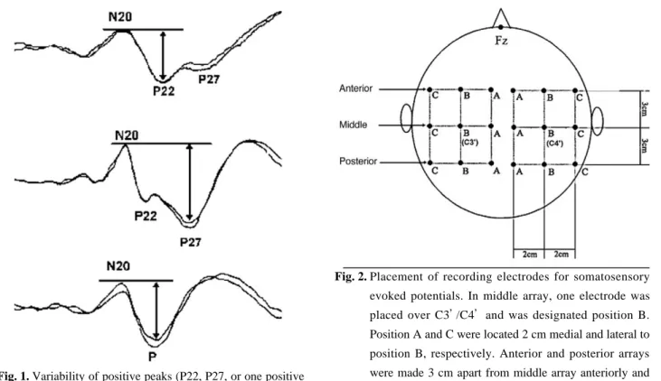
8,9통상적인 방법으로 기록되는 체성 감각유발전위에서는 P22와 P27 전위가 독립적으로 구 분되어 발생되기도 하지만 서로 융합되어 하나의 양성 파형(P)으로 나타나는 예도 적지 않다(Fig. 1). 이러 한 양성 파형의 다양성 때문에 진폭의 변이성이 증가할 수도 있다고 생각된다.
본 연구에서는 정상인을 대상으로 N20 이후에 발생 되는 양성 파형을 관찰하여 통상적인 기록부위를 기준 으로 전후방 및 내외측방 기록부위에 따른 P22와 P27 의 발현 빈도를 조사하고 P22 및 P27 진폭의 양측간 변이성을 비교하고자 한다.
연구 대상 및 방법
23세에서 29세까지(평균 25.8±2.2세)의 건강한 성 인 21명으로 남자 16명, 여자 5명이었고 신장은 평균 172.3±7.6(160~185)cm이었다.
검사실의 온도를 22~24℃로 일정하게 유지하면서 Synergy (Oxford-Medelec, UK) 근전도기기를 사용 하였고 양측 정중신경을 각각 자극하여 유발된 체성감 각유발전위를 기록하였다. 자극의 강도는 단무지의 경 미한 움직임이 육안으로 보이기 시작하는 정도로 하였 고, 빈도는 초당 3회, 자극지속시간은 0.1 msec, 주파 수 폭은 3 Hz~3 KHz, 기록속도는 5 msec/divi- sion, 기록감응도는 5 μ V/division으로 설정하였다.
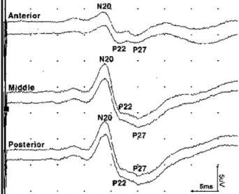
기록전극은 1 cm 길이의 표피 침전극을 이용하여 국제 뇌파 10-20 시스템에 입각한 Fz 지점에 참고기록전극 을 삽입하였다. 활성기록전극은 통상적 부위 즉, C3’
/C4’ 에 삽입하였고 이 부위를 B 지점으로 정의하였다.
이를 기준으로 내측 및 외측 2 cm 부위에 각각 삽입하 고 각각 A 및 C 지점으로 정의하였다. 이러한 A, B, C 지점의 배열은 중간부로 정의하고 이를 기준으로 전 방 및 후방 3 cm 부위에 같은 방식으로 각각 A, B, C 부위에 활성기록전극을 삽입하여 일측의 정중신경을 자 극하여 발생되는 체성감각유발전위가 반대측 두피의 총 9 부위에서 동시에 기록되도록 배치하였다(Fig. 2).
Fig. 1. Variability of positive peaks (P22, P27, or one positive waveform) following N20 in median nerve somatosen- sory evoked potential.
Fig. 2. Placement of recording electrodes for somatosensory evoked potentials. In middle array, one electrode was placed over C3’/C4’and was designated position B.
Position A and C were located 2 cm medial and lateral to position B, respectively. Anterior and posterior arrays were made 3 cm apart from middle array anteriorly and posteriorly, respectively. Three electrodes were also des- ignated in same manner. Common reference electrode was placed over Fz.
400회 반복 자극 후 통합∙평균하는 과정을 2회 시행 하여 얻어진 체성감각유발전위의 N20과 P22, P27 전 위를 조사하여 기록부위에 따른 잠시 및 진폭의 변화를 분석하였다. P22와 P27 전위의 구분은 각 기록부위의 발현양상을 기준으로 하였고 모든 부위에서 하나의 양 성 파형만 유발될 경우에는 P22와 P27 전위를 구분할 수 없어 P 전위로 정의하였다(Fig. 1, 3). 각 파형의 잠시는 정점발생 시점으로 측정하였고 진폭은 N20 정 점을 기준으로 그 이후에 유발되는 양성파형들의 정점 까지의 수직거리로 계산하였다. 양측 진폭의 변이성은 크기가 큰 진폭에 대한 양측의 진폭 차의 비로 정하여 측정하였다.
10통계학적인 검정을 위하여 마이크로소프 트 윈도우용 SPSS 10.0에 의한 비모수적인 분석방법 으로 Wilcoxon 부호순위합 검정을 시행하여 P값이 0.05 미만이면 통계학적으로 의미 있다고 정하였다.
결 과
모든 기록 부위에서 하나의 양성 파형만 유발되어 P22 또는 P27 전위로 구분할 수 없었던 P 전위가 발 견된 경우는 우성대뇌반구에서 9명이었고 비우성대뇌반 구에서 8명이었다. N20 이후의 양성 전위 즉, P22, P27, P의 발현 양상은 전후방 및 내외측 기록부위에 따라 통계학적으로 의미 있는 차이를 보였다(Table 1). 전방부의 경우 P22는 우성대뇌반구의 전방부 A 지 점과 비우성대뇌반구 A, B, C 지점에서 그 발현빈도가 P27보다 유의하게 높았다(p<0.05). 후방부의 P27은 우성대뇌반구에서는 A, B, C 지점에서 P22보다 발현
빈도가 통계학적으로 의미 있게 높았으나(p<0.05) 비우 성대뇌반구에서는 유의한 차이를 보이지 않았다 (p>0.05). 통상적인 기록부위를 포함한 중간부의 경우 P22와 P27의 발현 양상은 의미 있는 차이를 보이지 않 았다(p>0.05).
전방부에서 기록된 P22 진폭은 우성대뇌반구의 경우
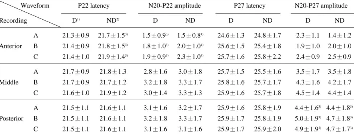
Fig. 3. Waveform identification of each peak of median nerve somatosensory evoked potentials. P22 is apparent and P27 is equivocal in anterior array. However, P22 is obliterated and P27 becomes to be prominent in middle and posterior arrays. Identification of P27 in anterior array can be referenced to definite waveform of posi- tive peak in middle or posterior array. Determination of P22 in middle and posterior arrays can be also based on distinct waveform following N20 in anterior array.
Table 1. Distribution of Positive Peaks According to Recording Locations
Waveform P22 P27 P
Recording D1) ND2) D ND D ND
A �12 �13 6 6 9 8
Anterior B 12 �13 12 9 9 8
C 12 �13 12 7 9 8
A 10 12 12 13 9 8
Middle B 10 12 12 13 9 8
C 10 11 12 13 9 8
A 7 11 *12 13 9 8
Posterior B 7 11 *12 13 9 8
C 7 11 *12 13 9 8
1. D: Dominant hemisphere 2. ND: Non-dominant hemisphere
*p<0.05 with comparing to the value of P27 at same recording site of the identical hemisphere
�p<0.05 with comparing to the value of P22 at same recording site of the identical hemisphere
A 지점에서 통계학적으로 의미 있게 작았고(p<0.05) B와 C 지점에서는 서로 의미 있는 차이를 보이지 않았 다(p>0.05)(Table 2). 비우성대뇌반구에서는 진폭의 크기가 A, B, C 지점의 순으로 증가하였다(p<0.05).
P22의 잠시는 우성대뇌반구의 경우 A, B, C 지점간 유의한 차이를 보이지 않았으나(p>0.05) 비우성대뇌반 구에서는 B와 C 지점보다 A 지점에서 유의하게 짧았 다(p<0.05). 후방부의 P27 진폭은 A 지점에 비하여 B 와 C지점에서 유의하게 증가하였으나(p<0.05) 양측대 뇌 반 구 간 의 미 있 는 차 이 를 보 이 지 는 않 았 다
(p>0.05)(Table 2).
각 기록부위에서 N20-P22 및 N20-P27의 진폭의 양 측간 변동계수를 측정하였는데, N20-P22 진폭 변동계 수의 평균값이 N20-P27에 비하여 작았으나 통계학적 으로 유의한 차이를 규명할 수는 없었다(p>0.05).
고 찰
체성감각유발전위는 말초신경의 전기자극에 의해 말 초신경, 척수, 대뇌피질 등에서 기록될 수 있고 감각신 경계의 기능을 개괄적으로 반영하는 파형 중의 하나로 간주되고 있다.
6그러므로 운동단위를 침범하는 질환의 진단 및 중추신경계의 구심성 감각섬유의 기능을 평가 하기 위하여 체성감각유발전위가 임상에서 많이 이용되 고 있지만 명확하게 표준화된 방법은 설정되지 못한 실 정이다. 이는 전위의 잠시뿐만 아니라 진폭의 개체간 변이성 때문인 것으로 생각되는데 특히 진폭의 경우 80%의 차이가 정상인들을 대상으로 한 연구에서 보고 된 바 있다.
10,11이러한 진폭의 양측간 변이성은 통상적 인 기록방법의 경우 N20 이후에 유발되는 양성 파형의 다양성 때문인 것으로 생각된다. 이러한 양성 파형의 양상은 P22와 P27 존재의 유무 및 두 파형간의 상호작 용에 의하여 영향을 받을 것으로 생각된다.
Desmedt와 Bourguet
8, Desmedt 등
9은 두정엽과 전두엽에서 기록되는 체성감각유발전위 양상을 칼라 영 상으로 재조합하여 지도화하였다. 그 결과 P22는 시상- 피질투사(thalamocortical projection) 섬유를 통하여
Table 2. Latencies and Amplitudes of P22 and P27 at Different Recording SitesWaveform P22 latency N20-P22 amplitude P27 latency N20-P27 amplitude
Recording D1) ND2) D ND D ND D ND
A 21.3±0.9 21.7±1.53) 1.5±0.93) 1.5±0.84) 24.6±1.3 24.8±1.7 2.3±1.1 1.4±1.2 Anterior B 21.4±0.9 21.8±1.53) 1.8±1.03) 2.0±1.04) 25.6±1.5 25.4±1.8 1.9±1.0 2.0±1.0 C 21.4±1.0 21.9±1.43) 1.9±0.93) 2.3±1.04) 25.7±1.6 25.8±2.2 2.4±0.9 2.5±0.9
A 21.7±0.9 21.8±1.3 2.8±1.6 3.0±1.8 25.7±1.5 25.5±1.6 3.5±1.7 3.5±1.8
Middle B 21.7±0.9 21.7±1.2 3.2±1.8 3.3±1.7 25.8±1.6 25.7±1.7 4.3±1.6 4.2±1.7
C 21.6±1.0 21.9±1.2 3.0±1.4 3.3±1.3 25.9±1.6 25.7±1.8 4.5±1.4 4.4±1.4
A 21.5±1.1 21.6±1.1 3.1±1.6 3.2±1.7 25.9±1.6 25.8±1.9 4.4±1.63) 4.4±1.83) Posterior B 21.5±1.1 21.6±1.1 3.2±1.8 3.3±1.7 25.9±1.7 25.8±1.9 5.0±1.93) 4.7±1.83) C 21.5±1.1 21.6±1.1 3.1±1.6 3.1±1.6 25.9±1.7 25.9±2.0 4.9±1.93) 4.7±1.73) Values are mean±S.D.
1. D: Dominant hemisphere 2. ND: Non-dominant hemisphere
3. Value of position A is significantly smaller than that of position B and C (p<0.05)(A<B=C) 4. Value is lowest in position A and highest in position C (p<0.05)(A<B<C)
Table 3. Comparison of Amplitude Variability of P22 and P27 Amplitude
N20-P22 (case) N20-P27 (case) Recording
A 0.27±0.16 (10) 0.41±0.32 (4) Anterior B 0.19±0.16 (10) 0.31±0.11 (8) C 0.24±0.17 (10) 0.26±0.21 (6) A 0.27±0.19 (8) 0.32±0.16 (10) Middle B 0.22±0.14 (8) 0.26±0.12 (10) C 0.17±0.15 (8) 0.21±0.12 (10) A 0.16±0.14 (7) 0.24±0.13 (10) Posterior B 0.14±0.14 (7) 0.23±0.12 (10) C 0.14±0.14 (7) 0.21±0.12 (10) Values are mean±S.D.
p>0.05 by comparing of N20-P22 amplitude variability with N20-P27
전두엽의 제4 운동영역 및 보조운동영역에서 유발되고 P27은 감각피질의 다양한 체성감각 수용체들의 상호작 용으로 발생되는 것으로 보고된 바 있다. 본 연구에서 도 P22는 전방부에서, P27은 후방부에서 그 발현빈도 가 높아 이러한 연구보고와 유사한 결과가 확인되었다.
P27은 우성대뇌반구의 경우 전방부의 내측 기록부위를 제외한 지점에서 P22와 발현빈도가 유사하였으나 비우 성대뇌반구의 경우 전방부의 모든 부위에서 P22보다 그 발현빈도가 낮았다. 이러한 결과를 통하여 P27 전위 의 영향이 우성대뇌반구에서 더 우세하다고 생각할 수 있다. 한편, P27의 호발부위로 생각되는 후방부의 경우 P22의 발현빈도가 비우성대뇌반구에서는 P27과 유사 하였으나 우성대뇌반구에서는 유의하게 낮았다. 이는 P27 전위가 P22의 발생에 영향을 주기 때문인 것으로 생각된다.
본 연구에서 전방부 P22의 진폭은 비우성대뇌반구의 경우 내측에서 외측 방향의 순으로 증가하였으나 우성 대뇌반구에서는 내측부에서만 작았다. 이러한 결과도 P27 전위의 영향 때문인 것으로 설명할 수 있다. 비우 성대뇌반구의 전방부에서는 P27의 영향이 가장 적고, 내측 방향에 가까울수록 참고기록전극의 기록부위인 Fz 지점과 가까워져 진폭이 감소할 것으로 가정할 수 있 다. 비우성대뇌반구에서는 Fz 지점과 멀어질수록 진폭 이 증가할 수 있으나 우성대뇌반구의 경우 P27의 영향 이 커지므로 이러한 진폭 상승의 효과는 상쇄될 수 있 을 것으로 생각된다. 한편, 후방부 P27 진폭의 변화가 양측대뇌반구간 차이를 보이지 않았는데 이는 P22에 의한 영향이 P27보다 미약하기 때문인 것으로 추정된 다.
체성감각유발전위의 진폭은 N20 전위 및 그 다음으 로 이어지는 P22 전위의 정점들간의 수직거리로 계산 하는 것이 실제 신호발생기의 기능을 더 잘 반영할 것 으로 생각된다. 본 연구에서 P22가 발현된 경우만을 대 상으로 측정된 진폭의 양측간 변이성은 P27 진폭과 비 교하여 통계학적으로 유의한 차이를 보이지 않았다. 이 는 통계량이 충분하지 못한 이유뿐만 아니라 우성대뇌 반구에서 유의하게 크게 기록되는 P27 전위의 영향을 극복할 수 없을 가능성도 제시한다고 생각된다. 그러나 P22 전위는 N20 바로 다음으로 이어지기 때문에 진정 한 의미의 진폭을 반영할 수 있을 뿐 아니라 운동영역 의 기능과도 연관될 수 있기 때문에 중추신경계의 기능 평가나 운동기능과의 상관성 연구에 있어 통상적인 방 법보다 그 유용성이 더 높을 것으로 기대된다.
뇌졸중 이후 운동기능의 회복은 감각운동피질의 재배 치와 깊은 연관성이 있다고 보고되고 있다.
1,2,12운동기 능은 구심성 신경계와 원심성 신경계 사이의 복잡한 상 호작용을 통하여 발현되기 때문에 동작을 실행하는 동 안 체성감각피질 및 운동피질의 활동성이 함께 촉진된
다.
13,14그러므로 뇌졸중 이후의 운동기능 회복은 감각
기능, 즉 체성감각기능과 상관성이 있다고 생각되고 있 다. 이러한 기능을 전기신경생리학적으로 평가하는 체 성감각유발전위를 이용하여 뇌졸중 환자의 예후와 상관 성을 조사한 결과들이 보고된 바 있다.
15-17이러한 연구보고에서 체성감각유발전위는 잠시와 진 폭의 양상을 등급화 하여 임상적인 점수와 연관하여 평 가되고 있다. 그러나 만성적인 뇌졸중 환자에서 체성감 각유발전위가 유발되는 경우 잠시는 대개 정상범위일 가능성이 높다. 이는 전위가 유발될 정도로 신경 전달 경로가 안정화되기 때문에 다소 지연된다고 하더라도 그 차이를 등급화하기에는 어려움이 있기 때문이다. 그 리고 체성감각유발전위를 발생시키는 신호발생기의 기 능은 전위의 진폭에 더 잘 반영될 수 있기 때문에 진폭 을 정확하게 측정하려는 노력이 필요하다고 생각된다.
그러나 대뇌반구간 신호발생기의 분포가 확연히 다르다 면 양측을 비교하여 평가하는 방법으로는 한계가 있을 것으로 판단된다. 향후 동일한 쪽의 대뇌반구손상 환자 들만을 대상으로 임상적 상관성 연구가 필요할 것으로 생각한다.
결 론
본 연구결과를 통하여 P27은 우성대뇌반구에서 우세 하게 작용하고 P22에 영향을 줄 수 있다고 생각된다.
그러므로 체성감각유발전위 신호발생기의 분포는 양측 대뇌반구간 비대칭적일 것으로 판단된다.
참고문헌
01. Nudo RJ, Milliken GW: Reorganization of movement rep- resentations in primary motor cortex following focal ischemic infarcts in adult squirrel monkeys. J Neurophysi- ol 1996: 75: 2144-2149
02. Nudo RJ, Wise BM, SiFuentes F, Milliken GW: Neural substrates for the effects of rehabilitative training on motor recovery after ischemic infarct. Science 1996: 272: 1791- 1794
03. Feys H, Van Hees J, Bruyninckx F, Mercelis R, De Weerdt W: Value of somatosensory and motor evoked potentials in predicting arm recovery after a stroke. J Neu- rol Neurosurg Psychiatry 2000: 68: 323-331
04. Gott PS, Karnaze DS, Fisher M: Assessment of median nerve somatosensory evoked potentials in cerebral ischemia. Stroke 1990: 21: 1167-1171
05. Vang C, Dunbabin D, Kilpatrick D: Correlation between
functional and electrophysiological recovery in acute ischemic stroke. Stroke 1999: 30: 2126-2130
06. Dumitru D. Electrodiagnostic medicine, 1st ed, Philadel- phia: Hanley and Belfus, 1995: pp281-337
07. Aminoff MJ, Eisen AA: AAEM minimonograph 19:
somatosensory evoked potentials. Muscle Nerve 1998: 21:
277-290
08. Desmedt JE, Bourguet M: Color imaging of parietal and frontal somatosensory potential fields evoked by stimula- tion of median or posterior tibial nerve in man. Electroen- cephalogr Clin Neurophysiol 1985: 62: 1-17
09. Desmedt JE, Nguyen TH, Bourguet M: Bit-mapped color imaging of human evoked potentials with reference to the N20, P22, P27 and N30 somatosensory responses. Elec- troencephalogr Clin Neurophysiol 1987: 68: 1-19
10. Dumitru D, Newton BY, Dreyfuss P: Segmental v der- matomal somatosensory-evoked potentials: normal inter- trial variation and side-to-side comparison. Am J Phys Med Rehabil 1993: 72: 75-83
11. Tsuji S, Shibasaki H, Kato M, Kuroiwa Y, Shima F: Sub- cortical and cortical somatosensory potentials evoked by posterior tibial nerve stimulation: normative values. Elec-
troencephalogr Clin Neurophysiol 1984: 59: 214-228 12. Kusoffsky A, Wadell I, Nilsson BY: The relationship
between sensory impairment and motor recovery in patients with hemiplegia. Scand J Rehabil Med 1982: 14:
27-32
13. Nudo RJ, Friel KM, Delia SW: Role of sensory deficits in motor impairments after injury to primary motor cortex.
Neuropharmacology 2000: 39: 733-742
14. Tach WT: Neural basis of motor control: an overview.
Phys Med Rehabil Clin N Am 1993: 4: 615-622
15. Minamide H, Onishi H, Yamashita J, Ikeda K: Reversibili- ty of transient focal cerebral ischemia evaluated by somatosensory evoked potentials in cats. Surg Neurol 1994: 42: 138-147
16. Steinberg GK, Gelb AW, Lam AM, Manninen PH, Peer- less SJ, Rassi Neto A, et al: Correlation between somatosensory evoked potentials and neuronal ischemic changes following middle cerebral artery occlusion.
Stroke 1986: 17: 1193-1197
17. Zeman BD, Yiannikas C: Functional prognosis in stroke:
use of somatosensory evoked potentials. J Neurol Neuro- surg Psychiatry 1989: 52: 242-247