Original Article Urogenit Tract Infect 2017;12(2):89-94
http://crossmark.crossref.org/dialog/?doi=10.14777/uti.2017.12.2.89&domain=pdf&date_stamp=2017-08-25
Type Distribution of Human Papillomavirus in Genital Warts of Korean Men
Kyoung Ho Ryu, Jeong Ho Cho, Min Chong Lee, Tae Young Jung
1Goldman Urology Clinic, Seoul,
1Department of Urology, VHS Medical Center, Seoul, Korea
Purpose: To analyze the distribution of human papillomavirus (HPV) types and the characteristics of genital condyloma in Korean men.
Materials and Methods: Between January 2015 and December 2015, we reviewed the medical charts of 435 male patients diagnosed with genital condyloma. A total of 441 samples were identified. The detection rate of each HPV type and its associated characteristics (age, number of HPV types, low-risk and/or high-risk types, number of lesions) were analyzed. Our sample population was divided into two groups: The non-urethral condyloma group and the urethral condyloma group.
In addition, subgroup analysis was also performed.
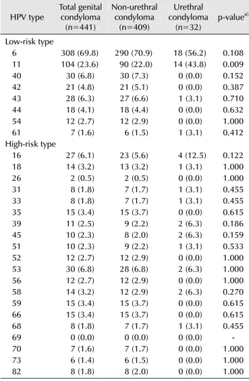
Results: Among the total 441 specimens, 409 (92.7%) were non-urethral condyloma and 32 (7.3%) were urethral condyloma. Single-type infection was observed in 56.7% and multiple-type infection was seen in 43.3%. HPV type 6 and type 11 were the most common types in total genital condyloma and subgroups.
HPV type 11, which was detected in 43.8% of those in the urethral condyloma group and in 22.0% of those in the non-urethral condyloma group (p=0.009), showed a statistically significant difference with respect to the type-specific detection rate.
Conclusions: As in previous studies, our study also showed that HPV type 6 was the most prevalent type among all genital condylomas, followed by HPV type 11.
A subgroup analysis also showed the same result.
Keywords: Human papilloma virus; Genital wart; Condyloma acuminata
Copyright 2017, Korean Association of Urogenital Tract Infection and Inflammation. All rights reserved.
This is an open access article distributed under the terms of the Creative Commons Attribution Non-Commercial License (http://creativecommons.org/licenses/by-nc/4.0) which permits unrestricted non-commercial use, distribution, and reproduction in any medium, provided the original work is properly cited.
Received: 18 April, 2017 Revised: 16 June, 2017 Accepted: 20 July, 2017
Correspondence to: Tae Young Jung
http://orcid.org/0000-0002-4634-3370 Department of Urology, VHS Medical Center, 53 Jinhwangdo-ro 61-gil, Gangdong-gu, Seoul 05368, Korea
Tel: +82-2-2225-1739, Fax: +82-2-484-4604 E-mail: [email protected]
INTRODUCTION
Human papillomavirus (HPV) infection is one of the most prevalent sexually transmitted infections (STIs) [1]. More than 100 types of HPV have been identified to date, and about 40 of these have been associated with genital disease [2,3]. Most HPV infections are naturally resolved within 2 years, but infection of several HPV types can persist and affect cell transformation. These types have oncogenic potentials and cause malignant diseases, such as cervical, penile, anal, and vulva cancer. Therefore, these types are classified as high-risk (HR) types [3,4]. A recent study
reported a moderate relationship between the occurrence
of bladder tumor and HPV infection [5]. However the most
commonly occurring disease related to HPV in men is
condyloma acuminate (genital condyloma), which is a
nonmalignant disease [6]. Most of the previous studies
regarding genital condyloma have been about penile,
scrotal, anal and urethral condyloma, showing that urethral
and meatal condyloma accounted for 20% of genital
condyloma [7-11]. Unlike other genital warts, urethral warts
require invasive procedures, such as urethro-cystoscopy
for diagnosis and treatment [12]. In cases of urethral wart
that are not confirmed with gross inspection or have no
obvious symptoms, appropriate treatment can be missed or postponed. Moreover, urethra and other genital areas have anatomical differences. Urethral orifice has a keratinized stratified squamous epithelium, and it abruptly changes to a non-keratinizing stratified squamous epithelium that lines the fossa navicularis. After that, it continues in the form of pseudostratified columnar epithelium in the penile urethra [13]. HPV also has various affinity for different anatomical sites, having different epithelial tropisms depending on the type [14]. Despite these differences and characteristics of HPV, studies of subgroup analysis in genital condyloma, especially in urethral condyloma, are very limited. Therefore, we analyzed the HPV type distribution in genital condyloma and performed a subgroup analysis between urethral condyloma and other genital condyloma.
MATERIALS AND METHODS
Between January 2015 and December 2015, we retrospectively reviewed the electronic charts of 438 patients diagnosed with genital condyloma via a histologic examination in the Goldman Urology Clinic (Gangnam-gu, Seoul, Korea). Three samples from three patients diagnosed with HIV or syphilis were excluded from the study; finally, 441 samples from 435 patients were included in this study.
The detection rate of HPV types and characteristics (age, number of HPV types, low-risk (LR) and/or HR types, number of lesions) of genital condyloma were compared.
For a subgroup analysis, total genital condyloma samples were divided into two groups. Regardless of whether other genital warts were accompanied or not, the samples of urethral condyloma were classified as the urethral condyloma group. Only urethral condyloma was analyzed for histologic examination and HPV typing test in urethral condyloma group. The samples of genital condyloma, which were not accompanied by urethral condyloma, were classified as the non-urethral condyloma group. Of the samples of multiple condyloma in the genital condyloma group, histological examination and HPV typing test were performed on the mass with the largest size.
Specimens were obtained by an excisional biopsy, and remaining wart tissues were removed by a laser therapy.
Electrocauterization was performed when bleeding control was required after an excisional biopsy. The specimens
from the non-urethral condyloma group were obtained from a penile shaft, penile glans, penile base, coronal sulcus, prepuce, pubic area, inguinal area, and anus. We conducted a histological examination only for urethral specimen in the urethral condyloma group. In the case of a meatal wart, an excisional biopsy was performed after the meatal eversion. Thereafter, urethrocystoscopy was performed to confirm additional intraurethral warts.
The HPV genotyping tests were performed with all samples using a multiplex real-time polymerase chain reaction (PCR) test, Anyplex
TMII HPV 28 Detection system (Seegene, Seoul, Korea). Twenty eight different HPV types, including 20 HR types (16, 18, 26, 31, 33, 35, 39, 45, 51, 52, 53, 56, 58, 59, 66, 68, 69, 70, 73, 82) and 8 LR types (6, 11, 40, 42, 43, 44, 54, 61), were identified by this multiplex real-time PCR test.
The detection rate of HPV type in each group was calculated by dividing the number of each HPV type by the number of total specimens in each group. Subgroup analysis and a comparison of HPV type-specific detection rate in each group were performed using Pearson's chi-square test and Fisher’s exact test. All statistical analyses were performed using IBM SPSS Statistics ver. 20.0 (IBM Co., Armonk, NY, USA). A p-value of less than 0.05 was considered to be statistically significant.
RESULTS
A total of 441 genital condyloma samples were analyzed;
of them, 409 (92.7%) were non-urethral condyloma and 32 (7.3%) were urethral condyloma.
Table 1 shows the characteristics of the total genital condyloma group and each subgroup. The mean age of patients in the total genital condyloma group was 32.1±6.9 years (range, 18.1-66.0 years), and the samples from patients aged 30.1 to 40.0 years showed the highest distribution rate (47.8%). Single-type infection was observed in 56.7%
of all genital condylomas, and among these, 64.4% had only LR type. Samples of multiple lesions were identified in 76.2% of all genital condylomas. According to a subgroup analysis, there was no statistically significant difference in the distribution rates of single and multiple HPV type infections, LR and/or HR types and number of lesions.
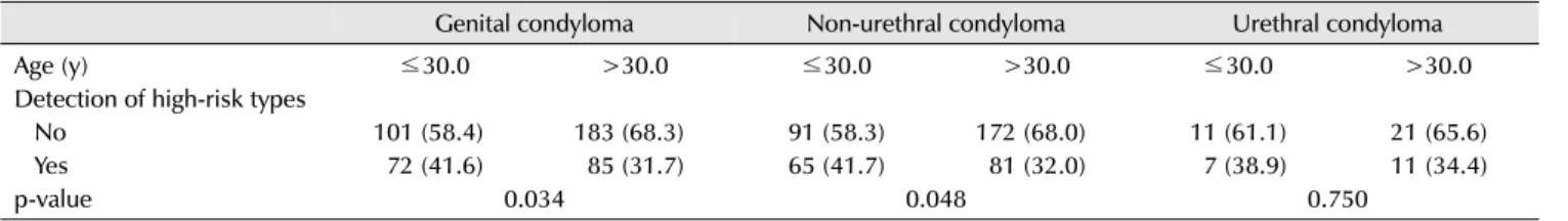
Approximately 38.1% of the non-urethral condyloma group
and 56.3% of the urethral condyloma group were younger
Table 1. The characteristics of specimens in the non-urethral and urethral condyloma groups
Characteristic
Total genital condyloma
(n=441)
Non-urethral condyloma
(n=409)
Urethral condyloma
(n=32) p-value
a)Mean age (y) 32.1±6.9 31.0±9.0 32.2±6.7 0.283
Age (y) 0.120
≤30.0 174 (39.5) 156 (38.1) 18 (56.3) 30.1-40.0 211 (47.8) 200 (48.9) 11 (34.4)
≥40.1 56 (12.7) 53 (13.0) 3 (9.4)
Number of HPV types 0.356
Single type 250 (56.7) 229 (56.0) 21 (65.6) Multiple type 191 (43.3) 180 (44.0) 11 (34.4)
Low- and/or high-risk type 0.914
Low 284 (64.4) 263 (64.3) 21 (65.6)
High 12 (2.7) 12 (2.9) 0 (0.0)
High & low 145 (32.9) 134 (32.8) 11 (34.4)
Number of lesions 0.059
Single 105 (23.8) 93 (22.7) 12 (37.5) Multiple 336 (76.2) 316 (77.3) 20 (62.5) Values are presented as mean±standard deviation or number (%).
HPV: human papillomavirus.
a)
Statistical analysis was performed between non-urethral condyloma group and urethral condyloma group.
Table 2. Distribution rate of HPV types in genital condyloma HPV type Total genital
condyloma (n=441)
Non-urethral condyloma
(n=409)
Urethral condyloma
(n=32) p-value
a)Low-risk type
6 308 (69.8) 290 (70.9) 18 (56.2) 0.108 11 104 (23.6) 90 (22.0) 14 (43.8) 0.009
40 30 (6.8) 30 (7.3) 0 (0.0) 0.152
42 21 (4.8) 21 (5.1) 0 (0.0) 0.387
43 28 (6.3) 27 (6.6) 1 (3.1) 0.710
44 18 (4.1) 18 (4.4) 0 (0.0) 0.632
54 12 (2.7) 12 (2.9) 0 (0.0) 1.000
61 7 (1.6) 6 (1.5) 1 (3.1) 0.412
High-risk type
16 27 (6.1) 23 (5.6) 4 (12.5) 0.122
18 14 (3.2) 13 (3.2) 1 (3.1) 1.000
26 2 (0.5) 2 (0.5) 0 (0.0) 1.000
31 8 (1.8) 7 (1.7) 1 (3.1) 0.455
33 8 (1.8) 7 (1.7) 1 (3.1) 0.455
35 15 (3.4) 15 (3.7) 0 (0.0) 0.615
39 11 (2.5) 9 (2.2) 2 (6.3) 0.186
45 10 (2.3) 8 (2.0) 2 (6.3) 0.159
51 10 (2.3) 9 (2.2) 1 (3.1) 0.533
52 12 (2.7) 12 (2.9) 0 (0.0) 1.000
53 30 (6.8) 28 (6.8) 2 (6.3) 1.000
56 12 (2.7) 12 (2.9) 0 (0.0) 1.000
58 14 (3.2) 12 (2.9) 2 (6.3) 0.270
59 15 (3.4) 15 (3.7) 0 (0.0) 0.615
66 15 (3.4) 15 (3.7) 0 (0.0) 0.615
68 8 (1.8) 7 (1.7) 1 (3.1) 0.455
69 0 (0.0) 0 (0.0) 0 (0.0) -
70 7 (1.6) 7 (1.7) 0 (0.0) 1.000
73 6 (1.4) 6 (1.5) 0 (0.0) 1.000
82 8 (1.8) 8 (2.0) 0 (0.0) 1.000
Values are presented as number (%).
HPV: human papillomavirus.
a)