• 검색 결과가 없습니다.

Surgical Resection of Thoracic Duct LymphangiomaJin San Bok, M.D., Jae Hyun Jun, M.D., Hyun Joo Lee, M.D., Ph.D., In Kyu Park, M.D., Ph.D., Chang Hyun Kang, M.D., Ph.D., Young Tae Kim, M.D., Ph.D.

N/A
N/A
Protected

Academic year: 2021

Share "Surgical Resection of Thoracic Duct LymphangiomaJin San Bok, M.D., Jae Hyun Jun, M.D., Hyun Joo Lee, M.D., Ph.D., In Kyu Park, M.D., Ph.D., Chang Hyun Kang, M.D., Ph.D., Young Tae Kim, M.D., Ph.D."

Copied!
4
0
0

로드 중.... (전체 텍스트 보기)

전체 글

(1)

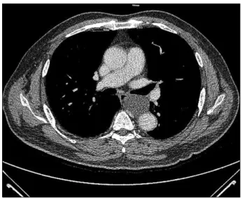
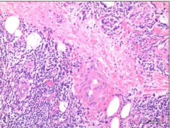
Korean J Thorac Cardiovasc Surg 2014;47:423-426 □ Case Report □ http://dx.doi.org/10.5090/kjtcs.2014.47.4.423 ISSN: 2233-601X (Print) ISSN: 2093-6516 (Online)

− 423 −

Department of Thoracic and Cardiovascular Surgery, Seoul National University Hospital

Received: October 23, 2013, Revised: November 25, 2013, Accepted: November 26, 2013, Published online: August 5, 2014

Corresponding author: Young Tae Kim, Department of Thoracic and Cardiovascular Surgery, Seoul National University Hospital, 101 Daehak-ro, Jongno-gu, Seoul 110-744, Korea

(Tel) 82-2-2072-3161 (Fax) 82-2-764-3664 (E-mail) [email protected]

C

The Korean Society for Thoracic and Cardiovascular Surgery. 2014. All right reserved.

CC

This is an open access article distributed under the terms of the Creative Commons Attribution Non-Commercial License (http://creative- commons.org/licenses/by-nc/3.0) which permits unrestricted non-commercial use, distribution, and reproduction in any medium, provided the original work is properly cited.

Fig. 1. Case 1. Chest computed tomography shows 4-cm sized mediastinal cystic mass adjacent to left side of the esophagus.

The mass was suspected to be an esophageal duplication cyst or a bronchogenic cyst.

Surgical Resection of Thoracic Duct Lymphangioma

Jin San Bok, M.D., Jae Hyun Jun, M.D., Hyun Joo Lee, M.D., Ph.D., In Kyu Park, M.D., Ph.D., Chang Hyun Kang, M.D., Ph.D., Young Tae Kim, M.D., Ph.D.

A 67-year-old male patient came to the hospital due to lung cancer and mediastinal cystic mass which was sus- pected to be esophageal duplication cyst. Video-assisted thoracoscopic surgery (VATS) was performed and in- tra-operative finding suggested it as a cystic mass along the thoracic duct. Thoracic duct was ligated and the cyst was completely resected. A 48-year-old female patient visited the hospital for dysphagia. Mediastinal cystic mass was suspected to be an esophageal duplication cyst. Intraoperative finding suggest a thoracic duct lymphangioma.

After thoracic duct ligation, the mass was completely resected with VATS. Postoperative chylothorax did not devel- op in both cases.

Key words: 1. Thoracic duct 2. Lymphangioma 3. Chylothorax

CASE REPORTS 1) Case 1

A 67-year-old male patient presented to the hospital with a right lower lobe nodule and a paraesophageal cystic mass, which were discovered by regular chest computed tomog- raphy (CT). The 2.5-cm right lower lobe nodule was sus- pected to be a lung cancer. The 4-cm paraesophageal cystic mass was located on the left side of the esophagus (Fig. 1).

The mass was suspected to be an esophageal duplication cyst or a bronchogenic cyst.

Video-assisted thoracoscopic surgery (VATS) was perfor- med. The right lower lobe nodule was proven to be ad- enocarcinoma by a frozen biopsy, and a right lower lobec- tomy and mediastinal lymph node dissection were carried out.

After further dissection through the mediastinum, the media-

stinal mass was located on the left side of the esophagus.

(2)

Jin San Bok, et al

− 424 − Fig. 2. Case 2. Chest computed tomography shows 5.5-cm sized mediastinal cystic mass which was located on the posterior to the lower esophagus. It was suspected to be an esophageal duplica- tion cyst or a bronchogenic cyst.

Fig. 3. Case 2. Video-assisted thoracoscopic surgery image shows division of thoracic duct after clipping.

Fig. 4. Case 2. Specimen of 4.5×4.0×1.0-cm sized thoracic duct lymphangioma. The cystic mass was filled with milky fluid and the probe is located in the thoracic duct.

However, rather than originating from the esophagus, it seemed that the mass was connected to the thoracic duct. On the basis of this finding, we suspected that the mass was a lymphangioma originating from the thoracic duct. To prevent chylothorax, the thoracic duct was carefully ligated with met- al clips and the mass was resected. A 32-French chest tube was inserted into the right pleural cavity.

There was no sign of chylothorax, and the chest tube was removed on the 4th postoperative day. The patient was dis- charged from the hospital on the 5th postoperative day with- out complication. The mass was confirmed to be a lym- phangioma by a pathologic examination and by immunohis- tochemistry, on which the mass was positive for CD31, CD34, factor VIII, and D2-40.

2) Case 2

A 48-year-old female patient presented to the hospital for dysphagia that had begun 1 month prior. Chest CT showed a 5.5-cm mediastinal cystic mass, which was located posterior to the lower esophagus (Fig. 2). It was suspected to be an esophageal duplication cyst or a bronchogenic cyst. Endosco- pic ultrasonography indicated that it may be a bronchogenic tumor or an esophageal duplication cyst.

An operation was performed by right-side VATS with the

patient in a semi-prone position. The mass seemed to origi- nate from the thoracic duct; therefore, the thoracic duct was clipped, and the mass was excised (Fig. 3). When the mass was dissected on a table, a milky fluid was found (Fig. 4). A 20-French chest tube was inserted into the patient’s pleural cavity.

Chylothorax did not occur postoperatively. The chest tube

was removed on the 2nd postoperative day. The patient was

discharged from the hospital on the 5th postoperative day. In

the pathologic examination, the mass was confirmed to be a

4.5×4.0×1.0 cm lymphangioma (Fig. 5). By immunohistoche-

mistry, the mass was demonstrated to be positive for CD31

and negative for D2-40 (Figs. 6, 7).

(3)

Thoracic Duct Lymphangioma

− 425 − Fig. 5. Case 2. Lymphoid aggregates are observed (H&E, ×200).

Fig. 6. Case 2. Lymphatic epithelial cells are stained with CD31 antibody (immunohistochemistry with CD31, ×200).

Fig. 7. Case 2. The cystic mass was negative for D2-40 (immunohistochemistry with D2-40, ×200).

DISCUSSION

The thoracic duct originates in the abdomen at the level of the diaphragm, penetrates through the posterior and superior mediastinum, and exits the thorax through the thoracic aper- ture, which is usually posterior to the left innominate vein and left of the common carotid artery [1]. Cysts of the thora- cic duct can occur either above or below the diaphragm.

Supradiaphragmatic thoracic duct cysts are typically found in the neck. However, mediastinal thoracic duct cysts are quite uncommon [2].

The cysts are often symptomatic because of the pressure they exert on the adjacent structures. Symptoms such as

coughing, dyspnea, and chest discomfort may be present.

Symptoms of dysphagia are often associated with the in- gestion of fatty foods. Furthermore, acute respiratory in- sufficiency after the ingestion of a fatty meal is also observed in some patients [1]. In case 2, the patient experienced dys- phagia, which may caused by the cystic mass.

Bronchogenic and esophageal duplication cysts account for a majority of the mediastinal cysts, and thoracic duct cysts are rarely included in the differential diagnosis. Magnetic res- onance imaging is not routinely required for the management of mediastinal cysts but may be able to demonstrate the com- munication of the cyst with the thoracic duct or the cisterna chyli. The high signal intensity of the T2-weighted images is attributed to the high lipid and protein content in the cyst.

Surgical resection of mediastinal cysts, including thoracic duct cysts, is indicated once they are discovered. These cysts are removed to relieve the symptoms and to prevent further growth, infection, and possible malignant transformation [2].

CD-31 and D2-40 are the known markers of lymphangio- ma [3]. In both cases, the mass was positive for CD-31, which is also called PECAM-1 and is an adhesion molecule that is expressed in the monocytes, granulocytes, and T-cell subsets [4]. D2-40, a monoclonal antibody, which binds lym- phatic epithelial cells, stained positive only in the mass from case 1 [5].

Surgical treatment for lymphangioma consists of the re-

moval of the cyst and the ligation of all the lymphatics con-

(4)

Jin San Bok, et al

− 426 − nected to it [1]. Kwak and Bae [6] reported that they ligated the thoracic duct and that the chylothorax was not observed thereafter. In the case of a mediastinal cystic mass that is ad- jacent to the esophagus, the mass may originate from the thoracic duct. As such, when removing the mass, the operator should note its relationship with the thoracic duct. If the mass seems to originate from the thoracic duct, it should be ligated to prevent chylothorax.

Here, we report two cases of thoracic duct lymphangioma.

Initially, these cases were thought to be esophageal duplica- tion cysts; however, they were found to be thoracic duct lymphangiomas. To prevent postoperative chylothorax, it is important to suspect thoracic duct lymphangioma if a cystic mass is present along the course of the thoracic duct.

CONFLICT OF INTEREST

No potential conflict of interest relevant to this article was reported.

REFERENCES

1. Turkyilmaz A, Eroglu A. A giant thoracic duct cyst: an un- usual cause of dysphagia. J Thorac Cardiovasc Surg 2007;

134:1082-3.

2. Mortman KD. Mediastinal thoracic duct cyst. Ann Thorac Surg 2009;88:2006-8.

3. Bhawan J, Silva C, Taungjaruwinai WM. Inconsistent im- munohistochemical expression of lymphatic and blood endo- thelial cell markers in cutaneous lymphangiomas. J Cutan Pathol 2013;40:801-6.

4. Janeway CA Jr. Immunobiology: the immune system in health and disease. 6th ed. New York: Garland Science;

2005.

5. Kahn HJ, Bailey D, Marks A. Monoclonal antibody D2-40, a new marker of lymphatic endothelium, reacts with Kaposi's sarcoma and a subset of angiosarcomas. Mod Pathol 2002;15:434-40.

6. Kwak MY, Bae CH. Thoracic duct cyst in mediastinum: a

case report. Korean J Thorac Cardiovasc Surg 2011;44:83-5.

수치

Fig. 1. Case 1. Chest computed tomography shows 4-cm sized  mediastinal cystic mass adjacent to left side of the esophagus
Fig. 4. Case 2. Specimen of 4.5×4.0×1.0-cm sized thoracic duct  lymphangioma. The cystic mass was filled with milky fluid and the  probe is located in the thoracic duct.
Fig. 6. Case 2. Lymphatic epithelial cells are stained with CD31  antibody (immunohistochemistry with CD31, ×200).

참조

관련 문서

#define 에서 인자 사용 예제 compoundassign.c... 관계와

▪ 실세계의 객체에서 불필요한 부분을 제거하여 필요한 부분만을 간결하고 이해하기 쉬운 클래스 로 만드는 작업. ▪

블록체인 기술은 보안, 금융, 의료 분야 등 여러 산업에서 활용이 가능해 4차 산업혁명의 핵심기술로 거론되지만 법으로 열거한 신성장 R&D 기술에 해당되지 않아

Scale Min 과 Scale Max 아래의 각 채널에 Disable 로 표시 되면 기본값으로 설정 되어 있는 상태 입니다.. 스케일 설정 값은 EEPROM에 저장되어 전원

위의 창에서 마우스로 해당 채널을 더블클릭 하거나 채널 선택 후 채널설정(S) 을 클릭 하면 아래와 같이 채널 설정 창이 나타납니다.. 8.1 채널

One might try to solve the problem by copying the first d elements of x to a temporary array, moving the remaining n − d elements left d places, and then copying the first d

(바) “액체 밀봉드럼 (Liquid seal drum)”이라 함은 플레어스택의 화염이 플레어시 스템으로 거꾸로 전파되는 것을 방지하거나 또는 플레어헤더에

• 글로벌 거버넌스란 국제사회에서 과거 주권국가들이 유읷핚 행위자로서 그들의 관계로 형성되었던 국제관계가 다양핚 다른 행위자들이 등장하고 새로욲 행위자들이 국가들과