대 한 생 식 의 학 회 지 : 제 37 권 제 3 호 2010
Growth Rate of Dominant Follicles
During Controlled Ovarian Hyperstimulation
Eun Ju Hwang1, Seung-Yup Ku1,2*, Yong Jin Kim1,2, Byung Chul Jee1,2, Chang Suk Suh1,2, Seok Hyun Kim1,2, Young Min Choi1,2, Jung Gu Kim1, Shin Yong Moon1,2
1Department of Obstetrics and Gynecology, Seoul National University College of Medicine,
2Institute of Reproductive Medicine and Population, Medical Research Center, Seoul National University, Seoul, Korea
과배란유도 여성에서 우성난포의 성장속도
서울대학교 의과대학 산부인과학교실1, 서울대학교 의학연구원 인구의학연구소2
황은주1·구승엽1,2*·김용진1,2·지병철1,2·서창석1,2·김석현1,2·최영민1,2·김정구1·문신용1,2
목 적: 과배란유도 환자에서의 우성난포의 성장속도와 임상인자들과의 연관성을 평가하고자 하였다.
연구방법: 체외수정시술을 위한 과배란유도 313주기를 대상으로 혈중 호르몬 농도를 측정하고 과배란유도 주기 중
난포 직경의 변화를 초음파를 이용하여 연속적으로 측정하였다. 우성난포의 성장 속도를 계산하고, 임상지표와의 연 관성을 분석하였다.
결 과: Gonadotropin releasing hormone 작용제와 길항제 주기 사이에 우성난포의 성장속도의 유의한 차이는 관찰되지 아니하였다. 우성난포의 성장속도와 환자의 연령, 체질량 지수, 생리 시작 3일의 follicle stimulating hormone, luteinizing hormone, 에스트라디올, 획득 난자의 수, 수정률 등의 임상지표는 유의한 연관성이 없었다.
결 론: 본 연구 결과는 우성난포의 성장속도는 난소 반응을 포함한 임상지표와 연관성이 없는 독립적인 변수임
을 시사한다. [Korean. J. Reprod. Med. 2010; 37(3): 253-259.]
중심단어: 난포성장, 과배란유도
The advent of ultrasonography provided a non- invasive means of visualizing human ovaries. Use of ultrasonographic evaluation for follicle development and ovulation has been well documented during natural cycles.1~7 Ultrasonographic evaluations of follicle growth
are used to complement hormonal estimations of ovarian status in women undergoing ovulation induction for the treatment of infertility.8~11
Follicles have been reported to grow in a linear fashion during a menstrual cycle,12 and the growth rates varying from one to four millimeter per day have been documented, with some researchers reporting increases or decreases in the growth rates of ovulatory follicles during the few days leading up to ovulation.1,2,7,8,13~15
There are few data on the relationship between the rates of growth of ovarian follicles and clinical char- acteristics or outcome during ovulation induction for in
접 수 일: 2010년 8월 17일, 수정일: 2010년 9월 22일 게재확정일: 2010년 9월 25일
Corresponding author: Seung-Yup Ku, MD, PhD, Department of Obstetrics and Gynecology, Seoul National University College of Medicine, 28 Yeongeon-dong, Jongno-gu, Seoul 110-744, Korea.
Tel: (02) 2072-1971, Fax: (02) 762-3599 e-mail: [email protected]
*This study was supported by a grant of the Korea Health 21 R&D Project, Ministry of Health & Welfare, Republic of Korea (01-PJ10-PG6-01GN13 -0002).
vitro fertilization (IVF). The objective of this study was to elucidate the correlation of g the rowth rates of the dominant follicles with possibly associated factors in women undergoing controlled ovarian hyperstimulation (COH).
MATERIALS AND METHODS
1. PatientsA total of 216 patients underwent 313 cycles of COH for IVF at Seoul National University Hospital from 2006 to 2009. Two hundred fifteen cycles in 144 patients underwent gonadotropin releasing hormone (GnRH) agonist long protocols, and 98 cycles in 72 patients GnRH antagonist protocols. Follicle stimulating hormone (FSH), luteinizing hormone (LH) and estradiol (E2) were measured on the day 3 of menstrual cycle. Serial ultrasonographic measurement of the diameter of growing follicles was performed.
2. COH protocols
For the GnRH angonist long protocol, GnRH agonist triptorelin (Decapeptyl, 0.1 mg/d; Ferring, Malmo, Sweden) was started in the mid-luteal phase of the previous cycle. After pituitary down-regulation, the triptorelin dose was reduced to 0.05 mg/d, and recom- binant FSH (Gonal-F; Serono, Geneva, Switzerland) was added until either the leading follicle reached a mean diameter of 18 mm or two or more follicles reached a diameter of 17 mm. For the GnRH antagonist multiple- dose flexible protocol, recombinant FSH (Gonal-F, Serono) was started on the 2nd or 3rd menstrual-cycle day without previous oral contraceptive pretreatment.
The GnRH antagonist cetrolelix (Cetrotide, 0.25 mg;
Serono) was added daily, starting when the leading follicle reached a diameter of 14 mm and until either the leading follicle reached a mean diameter of 18 mm or two or more follicles reached a diameter of 17 mm.
For both protocols, urinary hCG (Pregnyl, 10,000 IU, IM; Organon, Oss, the Netherlands) was administered 36 hours before transvaginal oocyte retrieval. Follicle growth rate was determined by calculating the slope of the change in leading follicular diameter (in millimeters) from stimulation day 5 or 6 to hCG day: Follicle growth (mm/day) = [Leading follicle diameter at hCG day- Leading follicle diameter at stimulation day 5 or 6]/
Number of days from stimulation day 5 or 6 to hCG day.
3. IVF/Intracytoplasmic sperm injection (ICSI)
Retrieved oocytes were cultured for 4 to 6 hours until insemination. In the cases of ICSI, after cumulus cells were removed by hyaluronidase (Sigma, St. Louis, MO, USA), the oocytes were evaluated for their maturity using an inverted microscope (Hoffman modulation, TE2000, Nikon, Tokyo, Japan). Only metaphase II oocytes, from which the first polar body was extruded, were used for ICSI. Semen samples obtained by ejaculation in the morning of the oocytes retrival day were liquefied at room temperature for 30 minutes, and centrifuged with SpermGrad (Vitrolife, Kungsbacka, Sweden) made of two gradient (45%/90%) at 1,500 rpm for 20 minutes. After removal of supernatant, we layered 2 mL of Universal IVF medium over the sperm pellet to centrifuge again at 1,000 rpm for 10 minutes.
After washing and swim-up procedure, the only sperm pellet in the supernatant were aspirated and used for insemination. Fertilization was determined by the presence of 2 pronuclei (2PN) using an inverted microscope on the first day after insemination. Zygotes with 2PN were cultured individually in microdrops of 25 μL of growth medium, G-1TM v5 (Vitrolife) overlaid with 8 mL of mineral oil (Sigma, USA) in Falcon 1007 culture dishes (Becton Dickinson Labware, Franklin Lakes, New Zealand) at 37℃ under 6% CO2.
4. Embryo transfer (ET)
ET was performed 3 days after oocytes retrieval.
Embryos were graded, according to their morphologies and cleavage rates. Embryos were graded from one to five (I, II, III, IV, V), based on number and uniformity of blastomere, and percentage of fragmentation, according to Veeck's classification system.16 We defined top- quality embryos as those of morphologic grade I/V.
After embryo grading, up to 4 embryos were selected and transferred into the uterus. The luteal phase was supported daily with progesterone in oil (Progest, 50 mg; Samil, Seoul, Korea) or with 8% progesterone gel (Crinone, Serono), initially for 14 days, starting on the
day of oocyte retrieval, and continuing for another 6~8 weeks in cases in which a pregnancy was achieved.
Clinical pregnancy was defined by the presence of an intrauterine gestational sac with pulsating fetal heartbeats at 3 to 4 weeks after oocyte retrieval. Fertilization rate was defined as number of 2PN divided number of retrieved oocytes.
5. Statistical analysis
Correlations between growth rates of the dominant follicles and possibly associated factors were analyzed.
Correlations between different parameters were deter- mined by bivariate correlation analysis and are expressed as Pearson's correlation coefficients. The statistical
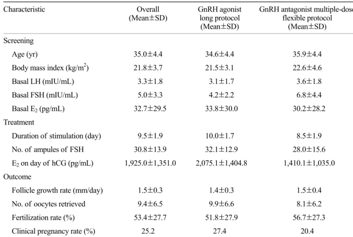
Table 1. Characteristics of patients
Characteristic Overall
(Mean±SD) GnRH agonist long protocol
(Mean±SD)
GnRH antagonist multiple-dose flexible protocol
(Mean±SD) Screening
Age (yr) 35.0±4.4 34.6±4.4 35.9±4.4
Body mass index (kg/m2) 21.8±3.7 21.5±3.1 22.6±4.6
Basal LH (mIU/mL) 3.3±1.8 3.1±1.7 3.6±1.8
Basal FSH (mIU/mL) 5.0±3.3 4.2±2.2 6.8±4.4
Basal E2 (pg/mL) 32.7±29.5 33.8±30.0 30.2±28.2
Treatment
Duration of stimulation (day) 9.5±1.9 10.0±1.7 8.5±1.9
No. of ampules of FSH 30.8±13.9 32.1±12.9 28.0±15.6
E2 on day of hCG (pg/mL) 1,925.0±1,351.0 2,075.1±1,404.8 1,410.1±1,035.0 Outcome
Follicle growth rate (mm/day) 1.5±0.3 1.4±0.3 1.5±0.4
No. of oocytes retrieved 9.4±6.5 9.9±6.6 8.1±6.2
Fertilization rate (%) 53.4±27.7 51.8±27.9 56.7±27.3
Clinical pregnancy rate (%) 25.2 27.4 20.4
GnRH, gonadotropin releasing hormone; SD, standard deviation; LH, luteinizing hormone; FSH, follicle stimulating hormone; E2, estradiol; hCG, human chorionic gonadotropin.
Eun Ju Hwang. Growth Rate of Dominant Follicles During Controlled Ovarian Hyperstimulation. Korean J Reprod Med 2010.
software package SPSS ver. 18.0 (SPSS Inc., Chicago, IL, USA) was used for the statistical analysis, and results were considered statistically significant at p-values of
<0.05.
RESULTS
Mean age of the patients was 35.0±4.4 years. Mean body mass index (BMI) was 21.8±3.7 kg/m2. Mean basal serum LH, FSH, and E2 levels were 3.3±1.8 mIU/mL, 5.0±3.3 mIU/mL, and 32.7±29.5 pg/mL, respectively.
There were no significant differences in these variables between two protocols. Mean duration and total dosage of gonadotropin used were 9.5±1.9 days, 30.8±13.9 ampoules, respectively. In GnRH agonist long protocol, mean duration and total dosage of gonadotropin used were 10.0±1.7 days and 32.1±12.9 ampoules, and in GnRH antagonist protocol, 8.5±1.9 days and 28.0±
15.6 ampoules, respectively. Mean E2 on the day of hCG was 1,867.0±1,334.9 pg/mL in total, 2,075.1±
1,404.8 pg/mL in GnRH agonist long protocol, and 1,410.1±1,035.0 pg/mL in GnRH antagonist protocol,
respectively. The mean growth rate of dominant follicles was 1.5±0.3 mm/day in total, 1.4±0.3 mm/day in GnRH agonist long protocol, and 1.5±0.4 mm/day in GnRH antagonist protocol, respectively. The mean number of oocytes retrieved was 9.4±6.5 in overall, 9.9±6.6 in GnRH agonist long protocol and 8.1±6.2 in GnRH antagonist protocol. The fertilization rate and clinical pre- gnancy rate were 53.4±27.7% and 25.2%, respectively (Table 1).
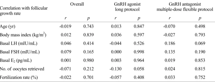
No significant correlation was found between growth rates of the dominant follicles and age (r=-0.019, p=0.743), BMI (r=0.012, p=0.839), Basal LH (r=0.046, p=0.414), FSH (r=0.079, p=0.165), and E2 (r=0.001, p=0.980), retrieved oocytes (r=-0.071, p=0.212), and fertilization rates (r=-0.022, p=0.701), and neither in GnRH agonist or GnRH antagonist protocol (Table 2).
DISCUSSION
Ovarian follicles comprise oocytes surrounded by granulosa cells. Follicular growth is achieved by a small increase in oocyte volume and predominantly by the
Table 2. Correlation between follicular growth rate and other factors
Overall GnRH agonist long protocol
GnRH antagonist multiple-dose flexible protocol Correlation with follicular
growth rate
r p r p r p
Age (yr) -0.019 0.743 0.013 0.847 -0.070 0.498
Body mass index (kg/m2) 0.012 0.839 0.036 0.597 -0.027 0.793
Basal LH (mIU/mL) 0.046 0.414 -0.044 0.526 0.186 0.069
Basal FSH (mIU/mL) 0.079 0.165 0.000 0.998 0.135 0.190
Basal E2 (pg/mL) 0.001 0.980 0.003 0.964 0.019 0.853
No. of oocytes retrieved -0.071 0.212 -0.130 0.058 0.024 0.815 Fertilization rate (%) -0.022 0.701 -0.057 0.408 0.033 0.752 GnRH, gonadotropin releasing hormone; LH, luteinizing hormone; FSH, follicle stimulating hormone; E2, estradiol.
*r=partial correlation coefficient adjusted by duration of stimulation and number of ampoules of FSH.
Eun Ju Hwang. Growth Rate of Dominant Follicles During Controlled Ovarian Hyperstimulation. Korean J Reprod Med 2010.
proliferation of surrounding gonadotropin-responsive granulosa cells and the expansion of antral cavity.17 A small proportion of follicles develop into healthy antral follicles. These follicles are highly sensitive and respon- sive to FSH.18,19 Majority of recruited follicles enter into atresia. During a spontaneous menstrual cycle, a single antral follicle is selected and becomes dominant, whereas the growth of multiple follicles is supported by exogenous gonadotropins in assisted reproduction cycles.
The follicular diameter increases to approximately 20 mm before ovulation.17
The occurrence of elevated FSH levels in women of older reproductive age may lead to advancement of normal dominant follicle growth, as in older ovulatory women who showed shorter follicular phase and overall cycle length. Previous studies suggested that this shorter length in old subjects results from earlier dominant follicle selection, independent of hormonal influences.
20,21 Our present study demonstrates that no significant correlations between age of patients and follicular growth rate. These results suggest that age has no significant effect in regard to follicular growth rate in COH cycle with pituitary suppression.
The negative impact of obesity in the outcome of assisted reproductive technology has been suggested by multiple reports. Obese patients undergoing COH are known to have increased FSH requirement, fewer colleted oocytes, frequent cycle cancellation, lower pregnancy rate, and increased miscarriage rate.22~24 However, they did not address the correlation between the BMI and follicular growth rate. While being obese, one can speculate, might have associated with ovarian follicle growth, our data showed that the follicular growth rate was not affected by BMI.
We intended to search for any correlation between follicular growth rate and basal hormone level, ovarian response or fertilization rate, however, no significant correlation was observed. Although our results suggest
that the follicular growth rate has no correlation with them in COH cycles, these factors may have significance in natural cycle, which is subject to future investigations.
In conclusion, our retrospective study observed that the growth rates of dominant follicles have no correlation with clinical characteristics or outcome variables in COH cycles using GnRH agonist or GnRH antagonist.
Acknowledgement
The authors would like to express our deepest gratitude to Sun Kyung Oh, Hee Sun Kim, Moon Ju Kang, Sung Ah Kim for their laboratory support of this study.
REFERENCES
1. Bomsel-Helmreich O. Ultrasound and the preovulatory human follicle. Oxf Rev Reprod Biol 1985; 7: 1-72.
2. Hackeloer BJ, Fleming R, Robinson HP, Adam AH, Coutts JR.
Correlation of ultrasonic and endocrinologic assessment of human follicular development. Am J Obstet Gynecol 1979;
135: 122-8.
3. Hackeloer BJ, Robinson HP. Ultrasound examination of the growing ovarian follicle and of the corpus luteum during the normal physiologie menstrual cycle (author's transl).
Geburtshilfe Frauenheilkd 1978; 38: 163-8.
4. Lenz S. Ultrasonic study of follicular maturation, ovulation and development of corpus luteum during normal menstrual cycles. Acta Obstet Gynecol Scand 1985; 64: 15-9.
5.O'Herlihy C, De Crespigny LJ, Robinson HP. Monitoring ovarian follicular development with real-time ultrasound. Br J Obstet Gynaecol 1980; 87: 613-8.
6. Queenan JT, O'Brien GD, Bains LM, Simpson J, Collins WP, Campbell S. Ultrasound scanning of ovaries to detect ovulation in women. Fertil Steril 1980; 34: 99-105.
7.Renaud RL, Macler J, Dervain I, Ehret MC, Aron C, Plas-Roser S, et al. Echographic study of follicular maturation and ovulation during the normal menstrual cycle. Fertil Steril 1980; 33: 272-6.
8.Dornbluth NC, Potter JL, Shepard MK, Balmacedo JP, Siler-Khodr TM. Assessment of follicular development by
ultrasonography and total serum estrogen in human meno- pausal gonadotropin-stimulated cycles. J Ultrasound Med 1983; 2: 407-12.
9. Kemeter P, Feichtinger W. Ultrasound monitoring of follicle growth in IVF. Wien Med Wochenschr 1991; 141: 9-13.
10. Leerentveld RA, van Gent I, van der Stoep M, Wladimiroff JW. Ultrasonographic assessment of Graafian follicle growth under monofollicular and multifollicular conditions in clomiphene citrate-stimulated cycles. Fertil Steril 1985; 43:
565-9.
11.Ritchie WG. Ultrasound in the evaluation of normal and induced ovulation. Fertil Steril 1985; 43: 167-81.
12.Doody MC, Gibbons WE, Zamah NM. Linear regression analysis of ultrasound follicular growth series: statistical relationship of growth rate and calculated date of growth onset to total growth period. Fertil Steril 1987; 47: 436-40.
13. Gougeon A, Lefevre B. Evolution of the diameters of the largest healthy and atretic follicles during the human menstrual cycle. J Reprod Fertil 1983; 69: 497-502.
14. Pache TD, Wladimiroff JW, de Jong FH, Hop WC, Fauser BC. Growth patterns of nondominant ovarian follicles during the normal menstrual cycle. Fertil Steril 1990; 54: 638-42.
15. Rossavik IK, Gibbons WE. Variability of ovarian follicular growth in natural menstrual cycles. Fertil Steril 1985; 44:
195-9.
16.Veeck LL. Fertilization and early embryonic development.
Curr Opin Obstet Gynecol 1992; 4: 702-11.
17. Broekmans FJ, de Ziegler D, Howles CM, Gougeon A, Trew
G, Olivennes F. The antral follicle count: practical recommen- dations for better standardization. Fertil Steril 2010; 94: 1044 -51.
18. Gougeon A. Regulation of ovarian follicular development in primates: facts and hypotheses. Endocr Rev 1996; 17: 121-55.
19.Jayaprakasan K, Hilwah N, Kendall NR, Hopkisson JF, Campbell BK, Johnson IR, et al. Does 3D ultrasound offer any advantage in the pretreatment assessment of ovarian reserve and prediction of outcome after assisted reproduction treatment? Hum Reprod 2007; 22: 1932-41.
20. Hansen KR, Thyer AC, Sluss PM, Bremner WJ, Soules MR, Klein NA. Reproductive ageing and ovarian function: is the early follicular phase FSH rise necessary to maintain adequate secretory function in older ovulatory women? Hum Reprod 2005; 20: 89-95.
21. Klein NA, Harper AJ, Houmard BS, Sluss PM, Soules MR. Is the short follicular phase in older women secondary to advanced or accelerated dominant follicle development? J Clin Endocrinol Metab 2002; 87: 5746-50.
22. Awartani KA, Nahas S, Al Hassan SH, Al Deery MA, Coskun S. Infertility treatment outcome in sub groups of obese population. Reprod Biol Endocrinol 2009; 7: 52.
23.Orvieto R, Meltcer S, Nahum R, Rabinson J, Anteby EY, Ashkenazi J. The influence of body mass index on in vitro fertilization outcome. Int J Gynaecol Obstet 2009; 104: 53-5.
24. Sneed ML, Uhler ML, Grotjan HE, Rapisarda JJ, Lederer KJ, Beltsos AN. Body mass index: impact on IVF success appears age-related. Hum Reprod 2008; 23: 1835-9.
= Abstract =
Objective: To evaluate if there is any correlation between the growth rate of dominant follicles and clinical characteristics or outcome variables in women undergoing controlled ovarian hyperstimulation (COH).
Methods: This study was performed in 313 in vitro fertilization (IVF) cycles. Follicle stimulating hormone (FSH), luteinizing hormone (LH) and estradiol (E2) were measured on day 3 of menstrual cycle, and serial ultrasonographic measurement of the diameter of growing follicles was performed. The growth rates of dominant follicles calculated by diameter difference divided by days were correlated with clinical characteristics and outcome variables.
Results: There was no significant difference in the growth rate of the dominant follicles between gonadotropin releasing hormone (GnRH) agonist and antagonist cycles. No significant correlation was found between the growth rates and evaluated factors such as age, body mass index, LH, FSH, E2, retrieved oocytes and fertilization rate.
Conclusion: The Growth rate of dominant follicles seems to show an independent feature of basal characteristics and ovarian response.
Key Words: Follicle growth, Controlled ovarian hyperstimulation