ISSN: 2233-601X (Print) ISSN: 2093-6516 (Online)
− 170 −
Received: September 6, 2018, Revised: November 14, 2018, Accepted: November 21, 2018, Published online: June 5, 2019
Corresponding author: Chun Wu, Department of Cardiothoracic Surgery, Children’s Hospital of Chongqing Medical University, No.136 Zhongshan Second Road,Yuzhong District, Chongqing 400014, China
(Tel) 86-18883883009 (Fax) 86-18883883009 (E-mail) wuchun007@sina.com
© The Korean Society for Thoracic and Cardiovascular Surgery. 2019. All right reserved.
This is an open access article distributed under the terms of the Creative Commons Attribution Non-Commercial License (http://creativecommons.org/
licenses/by-nc/4.0) which permits unrestricted non-commercial use, distribution, and reproduction in any medium, provided the original work is properly cited.
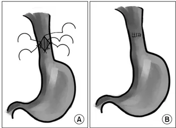
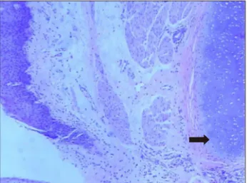
Congenital Esophageal Atresia Associated with a Tracheobronchial Remnant
Yuhao Wu, M.D. 1,2,3,4 , Chun Wu, M.D. 1,2,3,4
1
Department of Cardiothoracic Surgery, Children’s Hospital of Chongqing Medical University;
2