• 검색 결과가 없습니다.

Placement of a Retro-fitted Single Crown Restoration Under an Existing Denture by Employing Digital Technology: A Case Report

N/A
N/A
Protected

Academic year: 2021

Share "Placement of a Retro-fitted Single Crown Restoration Under an Existing Denture by Employing Digital Technology: A Case Report"

Copied!
6
0
0

로드 중.... (전체 텍스트 보기)

전체 글

(1)

pISSN : 1229-5418

Implantology 2018; 22(2): 100-105

https://doi.org/10.32542/implantology.20180009

Received: June 15, 2018 Revised: June 29, 2018 Accepted: June 29, 2018

Copyright © 2018. The Korean Academy of Oral &

Maxillofacial Implantology

This is an Open Access article distributed under the terms of the Creative Commons Attribution Non-Commercial License (http://creativecommons.

org/licenses/by-nc/4.0/) which permits unrestricted non-commercial use, distribution, and reproduction in

OPEN ACCESS

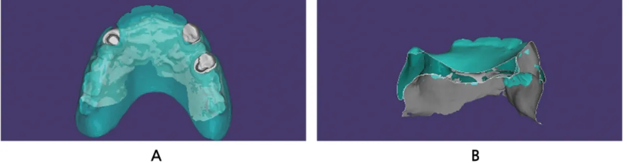
This article demonstrates a case that successfully placed a retro-fitted crown restoration under an existing denture by employing the characteristics of digital technology. Scanned data of the arch that involves edentulous area, and of the old denture itself were obtained and superimposed on each other.

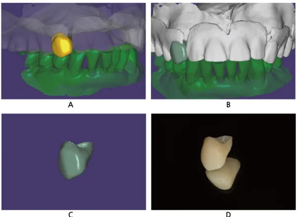
A virtual crown was modeled in computer-aided design software with marginally larger than the ideal size. The old denture was regarded as an adjacent tooth in the setting menu so that the portion of the virtual crown that extends over to the old denture could be automatically deleted by selecting the “Cut intersections” option. This retro-fitted crown was milled with a polymethylmethacrylate disc after a minor adjustment of its contour. The crown and the old denture were placed in the patient’s mouth, and resulted in favorable esthetic outcome and occlusion, showing proper relationship with the old denture.

Keywords: Digital technique, Retro-fitted crown restoration, Existing denture, Computer-aided design

Abstract

Restoration Under an Existing Denture by Employing Digital Technology: A Case Report

*Corresponding author: June-Sung Shim, [email protected] Kyung Chul Oh

1

, June-Sung Shim

2*

1

Clinical research assistant professor, Department of Prosthodontics, College of Dentistry, Yonsei University, Seoul, Korea

2

Professor, Department of Prosthodontics, College of Dentistry, Yonsei University, Seoul, Korea

Ⅰ. Introduction

Fabricating a well-fitted single crown restoration related to an existing denture is a chal- lenging procedure; in many cases, the denture may not fit appropriately on the retro-fitted crown. A few ideas have been suggested to resolve this limitation to the date. In the past, direct-indirect approaches were suggested to fit the crown with regard to the existing denture 1-5 .

With the advancement of digital technologies, several authors reported methods to fab-

ricate retro-fitted crowns based on the computer-aided design / computer-aided manu-

facturing (CAD/CAM) techniques 6-9 . A variety of CAD technologies have been devel-

oped in dental fields, impacting on all aspects of dentistry; however, in reality, their func-

tions are not fully utilized 10,11 . The present case described herein successfully placed a

(2)

A 73-year-old male patient presented to the Department of Prosthodontics at Yonsei University Den- tal Hospital for fabrication of a single crown restoration under an existing denture. Digital intraoral scans were obtained in occlusion without wearing the existing denture by using an intraoral scanner (Trios 3;

3Shape) (Fig. 1). Then, the entire surfaces of the existing denture were scanned with a tabletop scanner (T500; Medit Co) (Fig. 2). The maxillary digital intraoral scan and the scanned old denture were super- imposed by best-fit matching algorithm after selecting three pairs of points for matching in CAD soft- ware (Exocad DentalCAD; Exocad GmbH) (Fig. 3). The superimposed maxillary data were saved in the standard tessellation file (STL) format while maintaining their relationship with the mandibular data. Prop- er fit of the denture on the maxillary arch was evaluated in the software by referring to the sectional view.

Fig. 1. Digital intraoral scans and inter-arch relationship obtained without wearing the existing denture by using an intraoral scanner (Trios 3; 3Shape). A: Digital intraoral scan of maxillary arch. B: Digital intraoral scan of mandibular arch. C: Maxillary and mandibular arches in occlusion.

Kyung Chul Oh et al. : Placement of a Retro-fitted Single Crown Restoration under an Existing Denture by Employing Digital Technology: A Case Report. Implantology 2018

A B C

Fig. 2. Scanned data of the old denture obtained with a tabletop scanner (T500; Medit Co). Both polished and intaglio surfaces were scanned.

Kyung Chul Oh et al. : Placement of a Retro-fitted Single Crown Restoration under an Existing Denture by Employing Digital Technology: A Case

Report. Implantology 2018

(3)

An “Anatomic crown” module was selected for the tooth to be restored, “Adjacent tooth” modules were chosen for the adjacent teeth, and “Antagonist” modules were selected for the antagonists in a new project file. The margins were defined and the shape, contour, and occlusion of the virtual crown were adjusted (Fig. 4). The crown was designed in a slightly larger than the ideal size.

An “Approx.” option in the “Select type of adaptation:” menu was chosen followed by the “Cut inter- sections” option, in order to adapt the virtual crown to the existing denture. The denture is regarded as an adjacent tooth and the software automatically cut the portion of the virtual tooth that extended over the denture (Fig. 5). A minor adjustment was performed to the virtual crown, and the crown was fabricated with a polymethylmethacrylate (PMMA) disc and a milling machine (Fig. 6).

The fabricated PMMA crown and the old denture were placed in the patient’s mouth, and the relation- ship of the crown with the old denture was evaluated to examine if the crown is too tight or too loose with regard to the denture. After checking the esthetics and occlusion, the crown was cemented with a Fig. 3. Superimposition of the maxillary digital intraoral scan and the scanned old denture data sets. A:

Superimposition of the maxillary digital intraoral scan and the scanned old denture data sets by best-fit matching algorithm. B: Sectional view to confirm the fit of the denture on the maxillary arch.

Kyung Chul Oh et al. : Placement of a Retro-fitted Single Crown Restoration under an Existing Denture by Employing Digital Technology: A Case Report. Implantology 2018

A B

Fig. 4. Designing and adjusting the shape, contour, and occlusion of the virtual crown. A: Defining margin. B: Before occlusal adjustment. C: After occlusal adjustment.

Kyung Chul Oh et al. : Placement of a Retro-fitted Single Crown Restoration under an Existing Denture by Employing Digital Technology: A Case Report. Implantology 2018

A B C

(4)

Fig. 5. Adaptation procedures of the virtual crown. A: Clicking of “Approx.” option in “Select type of adaptation:” menu followed by “Cut intersections” option. B: Before adaptation of the virtual crown to the old denture in sectional view. Yellow-colored area indicates portion of the virtual crown. C: After adaptation of the virtual crown to the old denture in sectional view. Yellow-colored area indicates portion of the virtual crown.

Kyung Chul Oh et al. : Placement of a Retro-fitted Single Crown Restoration under an Existing Denture by Employing Digital Technology: A Case Report. Implantology 2018

A B C

Fig. 6. Finalized retro-fitted crown restoration. A: Final design of the virtual crown in lateral view. B:

Final design of the virtual crown in frontal view. C: Final design of the virtual crown. D: PMMA crown fabricated with a milling machine. PMMA, polymethylmethacrylate.

Kyung Chul Oh et al. : Placement of a Retro-fitted Single Crown Restoration under an Existing Denture by Employing Digital Technology: A Case Report. Implantology 2018

A

C

B

D

(5)

III. Discussion

The retro-fitted crown restoration was successfully placed in the patient’s mouth by automatically cutting the portion of the virtual crown that extended over the existing denture area in 3D aspects. The procedure was possible because the old denture was set to be regarded as an adjacent tooth. The virtual crown was adapted to the “adjacent area”, which refers to the old denture, on selecting the “Cut intersec- tions” option.

This approach could broaden its application range to a variety of materials because the values related to the amount of adaptation can be adjusted. Specifically, fabrication of retro-fitted surveyed crowns that are placed on the metal frameworks of removable dental prostheses would be valuable in the future, although the process involves difficulties of scanning metal structures such as clasps, occlusal rests, and minor connectors. It is the clinicians’ role to investigate the most appropriate values according to the materials or the computer-aided manufacturing machines they possess by trial and error to apply the present workflow.

The present case significantly reduced the chairtime to adjust the crown to fit appropriately with the existing denture. It may further reduce a clinical situation that might require remaking the denture. Ac- cordingly, patients can use their accustomed old denture for an extended period. Further in vitro studies assessing the accuracy of this retro-fitted crown with regard to the existing denture may also be worth investigating.

Fig. 7. Intraoral placement of the retro-fitted crown restoration. A: Frontal view. B: Occlusal view.

Kyung Chul Oh et al. : Placement of a Retro-fitted Single Crown Restoration under an Existing Denture by Employing Digital Technology: A Case Report. Implantology 2018

A B

(6)

intersections” option. The patient was satisfied to use his accustomed old denture for an extended period.

Further studies assessing the applicability to other prosthodontic materials would be of a great value.

References

1. Elledge DA, Schorr BL. A provisional and new crown to fit into a clasp of an existing removable partial denture. J Prosthet Dent. 1990; 63(5): 541-4.

2. Macpherson JR, Evans DB. Fabricating crowns to fit existing removable partial dentures: an illus- tration of two techniques. J Prosthodont. 1993; 2(3): 199-205.

3. Hansen CA, Russell MM. Making a crown to fit accurately under an existing removable partial denture clasp assembly. J Prosthet Dent. 1994; 71(2): 206-8.

4. Jackson AD, Butler CJ. Fabrication of a new crown and provisional to an existing removable par- tial denture. J Prosthodont. 1995; 4(3): 200-4.

5. Helvey GA. Retrofitting crowns to an existing removable partial denture clasp: a simple technique.

J Prosthet Dent. 2002; 87(4): 399-402.

6. Marchack BW, Chen LB, Marchack CB, et al. Fabrication of an all-ceramic abutment crown under an existing removable partial denture using CAD/CAM technology. J Prosthet Dent. 2007; 98(6):

478-82.

7. Yoon TH, Chang WG. The fabrication of a CAD/CAM ceramic crown to fit an existing partial removable dental prosthesis: a clinical report. J Prosthet Dent. 2012; 108(3): 143-6.

8. Ozawa D, Suzuki Y, Kawamura N, et al. Fabrication of crown restoration retrofitting to existing clasps using CAD/CAM: fitness accuracy and retentive force. J Prosthodont Res. 2015; 59(2):

136-43.

9. Patel D. Retrofitting a Crown Supporting a Removable Partial Denture Using “Biogeneric Copy”

to Replicate Tooth's Preoperative Condition. Compend Contin Educ Dent. 2016; 37(2): 126-30.

10. Dawood A, Marti Marti B, Sauret-Jackson V, et al. 3D printing in dentistry. Br Dent J. 2015;

219(11): 521-9.

11. Joda T, Ferrari M, Gallucci GO, et al. Digital technology in fixed implant prosthodontics. Peri-

odontol 2000. 2017; 73(1): 178-92.

수치

Fig. 1. Digital intraoral scans and inter-arch relationship obtained without wearing the existing denture  by using an intraoral scanner (Trios 3; 3Shape)
Fig. 4. Designing and adjusting the shape, contour, and occlusion of the virtual crown
Fig. 6. Finalized retro-fitted crown restoration. A: Final design of the virtual crown in lateral view
Fig. 7. Intraoral placement of the retro-fitted crown restoration. A: Frontal view. B: Occlusal view.

참조

관련 문서

3. In case of gold alloy inner crown, gold alloy outer crown was better than GES outer crown in retentive force. However, retentive force of gold alloy outer

Experimental alveolar ridge augmentation by distraction osteogenesis using a simple device that permits secondary implant placement. Anteroinferior distraction of

Longitudinal study on torque transmitted from a denture base to abutment tooth of a distal extension removable partial denture with circumferential clasps..

Digital images were obtained by taking digital impressions with a dental model scanner(S600 ARTI model scanner, Zirkonzhan, South Tyrol, Italy) after connecting the own

Bacterial and yeast plaque on dentures is thought to be an important etiologic factor in the pathogenesis of denture stomatitis. Since these fungi have

Tooth-borne supported partial denture 치아지지 국소의치 - 주유지 지대치. tooth-tissue supported partial denture 조직지지 국소의치

The builtin package defines error as an interface with a single Error() method that returns an error message as a string.. We’ll see

The Volume structure of the STA308A consists of individual volume registers for each channel and a master volume register that provides an offset to each channels volume