The majority of primary Epstein-Barr virus (EBV) infections produce no distinctive symptoms in Original Article
Clinical features of Epstein-Barr Virus-associated
Infectious Mononucleosis According to Age Group in Children
Soram Lee1, Ju-Young Chung2, Jung Je Park3, Ji-Hyun Seo1, Jae Young Kim4, Jung Sook Yeom1, Eun-Sil Park1, Jae-Young Lim1, Hyang-Ok Woo1, Hee-Shang Youn1
1Department of Pediatrics, Gyeongsang National University School of Medicine, Gyeongsang Institute of Health Science, Jinju, Korea
2Department of Pediatrics, Inje University Sanggye Paik Hospital, Inje University College of Medicine, Seoul, Korea
3Department of Otolaryngology, Gyeongsang National University School of Medicine, Gyeongsang Institute of Health Science, Jinju, Korea
4Department of Pediatrics, Gyeongsang National University Hospiratal, Changwon, Korea
Objectives: Few studies of pediatric Epstein-Barr virus (EBV)-associated infectious mononucleosis (IM) have been conducted in Korea. We evaluated the clinical features of children with IM to define differences according to age.
Methods: We conducted retrospective chart reviews of 68 children aged 0 to 15 years who were diagnosed by EBV-associated IM with EBV-Viral Capsid Antigen(VCA) IgM at laboratory test and were admitted between 2010 and 2014. The children were classified into four age groups: aged 0–3, 4–6, 7–9, and 10–15 years.
Results: The age distribution of patients was as follows: 19 (27.9%) 0–3, 25 (36.8%) 4–6, 13 (19.1%) 7–9, and 11 (16.2%) 10–15. Fever was the most common presentation regardless of age. It was more common in the 0–3 group than the 4–6 group (P = 0.018). Pharyngitis was more common in the 7–9 group than the 0–3 group (P = 0.048), and myalgia was more common in the 10–15 group than the 0–3 group (P = 0.007). Pharyngitis was accompanied by lymphadenopathy, protracted fever, and rash. In the 0–3 age group, the prevalence of rash was higher while the percentage of atypical lymphocytes was lower, but there was no statistical support for this tendency. There were no differences in the frequency of hepatosplenomegaly or laboratory findings between age groups.
Conclusions: IM is not uncommon in young children and its clinical presentation varies with age. Therefore, IM should be suspected in young febrile children with pharyngitis and rash despite low percentages of atypical lymphocytes.
Key Words: Epstein-Barr Virus Infection, Infectious Mononucleosis, Pediatrics
Corresponding Author: Ji-Hyun Seo, Department of Pediatrics, Gyeongsang National University School of Medicine, 79, Gangnam-ro, Jinju-Si, Gyeongsangnam-do 52727, Korea
Tel: +82-55-750-8731 Fax: +82-55-752-9339 E-mail: [email protected]
Received:
Revised:
Accepted:
Aug. 04, 2017 Oct. 23, 2017 Nov. 07, 2017
Articles published in Kosin Medical Journal are open-access, distributed under the terms of the Creative Commons Attribution Non-Commercial License (http://creativecommons.org/licenses/by-nc/4.0/) which permits unrestricted non-commercial use, distribution, and reproduction in any medium, provided the original work is properly cited.
children.1 Infectious mononucleosis (IM), charac- terized by the classic triad of fever, lymphadenop- athy, and pharyngitis,2 is the main clinical disease caused by EBV and is common among adolescents and young adults in developed countries.3,4
In Korea in the 90s, fewer than 20 children were diagnosed with EBV-associated IM,5,6 even though 85% of 5–6 years olds were seropositive for an- ti-EBV antibody.7 For this reason, EBV-associated IM is considered a rare disease among young Korean children. However, between 2001 and 2009, 68 of 81 children (85.1%) diagnosed with IM were under 10 years of age.8
The seroprevalence of anti-EBV antibodies and the incidence of symptomatic IM in children vary among countries. In Other countries, nearly half of all patients with EBV-associated IM were under the age of 5 years.9-11
Recently, the incidence of IM caused by EBV infections in children has increased in the Jinju region of Korea according to our chart review.
Here, we retrospectively evaluated pediatric pa- tients with confirmed EBV-associated IM to relate clinical features to patient age.
MATERIALS AND METHODS
Study subjects
This study was performed after Gyeonsang National University Hospital Institutional Review Board approval (GNUH IRB-2015-07-015-001).
We retrospectively examined medical record data
for children aged 0 to 15 years who had been diag- nosed with EBV infection by an EBV viral capsid antigen (VCA) IgM test at the Gyeongsang National University Hospital (GNUH) between January 2010 and December 2014. We included children who presented with common clinical manifestations of IM such as pharyngitis, fever, and lymphadenopathy.2 Children with asympto- matic hepatitis, an underlying chronic disease, or who were undergoing immunosuppressive ther- apy were excluded.
Demographics(age, sex, and monthly dis- tribution), clinical and laboratory findings were collected from the medical records. Fever was de- fined as a measured axillary temperature ≥ 38°C.
The children included in the study were catego- rized into four groups to be consistent with other recent epidemiological studies:11-13 aged 0–3, 4–6, 7–9, and 10–15 years.
The EBV-VCA IgM test was checked when the initial physical examination and laboratory find- ings of the patients suggested IM such as phar- yngitis with exudate, generalized lymphadenop- athy, hepatomegaly, splenomegaly, and leukocy- tosis with lymphocyte dominant pattern (> 60%), and elevated AST, ALT and LDH.
Radiologic examinations including Ultrasonography (USG) and Pharynx Computed Tomography(CT) were performed for ruling out the complications of IM. Pharynx CT was checked under suspicison of airway obstruction or superior vena cava syndrome. Abdominal USG was evaluated when pateint complains abdominal pain with hep-
atosplenomegaly or jaundice.
Statistical analysis
Statistical analyses were performed using SPSS ver. 22.0 (SPSS Inc., IBM corp., NY, USA).
Summaries of clinical measurements were gen- erated by generating frequency distributions and performing basic descriptive statistics. Clinical and laboratory data were analyzed using Anylasis Of Variances (ANOVAs) and X2 tests. Correlations were estimated by Spearman correlation. A P-val- ue < 0.05 was considered statistically significant.
RESULTS
Incidence of EBV-associated IM according to patient age and date
Sixty-eight children were found who had been confirmed for IM and EBV infection (Table 1). The age distribution of cases was as follows: 19 (27.9%) 0–3, 25 (36.8%) 4–6, 13 (19.1%) 7–9, and 11 (16.2%) 10–15 years (P = 0.07). Ages ranged from 11 months to 15 years with a median of 5.0 years;
the majority were between 4 and 8 years (Fig. 1).
The highest incidence of IM was observed in 2 years and 5 years old. Incidence decreased from 11 to 15 years.
Overall, boys diagnosed with IM were more than gilrs (Table 1, P = 0.09). In the 0–3 group, there was no difference in disease incidence between genders, but, in older age groups, there were more boys than girls. The median age was 5.0 for both boy and girl patients.
The number of EBV-associated IM cases per year was initially low (n = 8 in 2010, n = 4 in 2011), and then increased significantly to over 20 pa- tients in 2012 (n = 22) and remained high (n = 22 in 2013, n = 26 in 2014; P < 0 0.001, Fig. 2).
The increase in the number of cases in 2013 and 2014 is consistent with a greater proportion of patients who were under 6 years (Fig. 2). The num- ber of IM cases did not vary by season (spring:
n = 16, summer: n = 17, autumn: n = 18, and winter:
n = 17, P = 0.99), although the largest number of cases was observed in November (Fig. 3).
Clinical presentations and laboratory findings
Age (y) No. of cases
Total(%)
Female (%) Male (%)
0-3 4-6 7-9 10-15
Total
10(52.6) 8(32.0) 6(46.2) 3(27.3) 27(39.7)
9(47.4) 17(68.0)
7(53.8) 8(72.7) 41(60.3)
19(27.9) 25(36.8) 13(19.1) 11(16.2) 68(100.0) Table 1. Age and Sex Distribution
Fig. 1. Age distributions of 68 pediatric patients with infectious mononucleosis caused by Epstein-Barr virus infection
Fig. 2. Annual number of EBV-associated infectious mononucleosis cases.
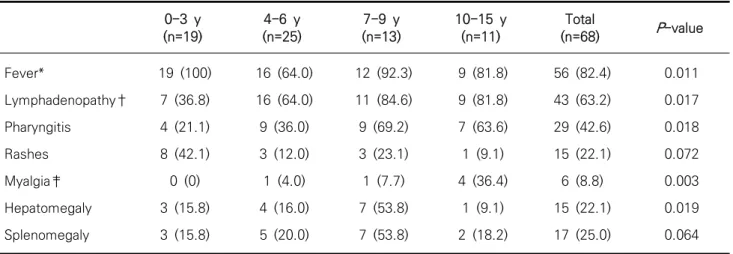
Fever was the most common presentation (82.2%), followed by cervical lymph node enlarge- ment (63.2%), pharyngitis (42.6%), rash (22.1%), myalgia (8.8%), and headache (2.9%). The duration of fever before admission was mean 4.7 days (range: 1–14 days). The mean durations of fever by age group were 4.5 days (0–3 y), 3.3 days (4–6 y), 6.2 days (7–9 y), and 6.1 days (10–15 y; P = 0.094). The duration of hospital admission was mean 5.0 days (range: 1–11 days). The mean dura- tion of admission decreased from 7.1 days in 2010 to 4.3 d ays in 2014 (P = 0.049). Use of antibiotics before admission was common (63.2%; n = 43).
Sixteen cases reported no history of antibiotics use (23.5%), and 9 cases did not respond.
The presentation of clinical features varied ac-
cording to age group (Table 2). Fever was more common in children 0–3 group than the 4–6 group (P = 0.018). Cervical lymphadenopathy was more common in children 7–9 group than the 0–3 group (P = 0.048). Myalgia was more common in 10–15 group than the 0–3 group (P = 0.007).
In the 0–3 group, all patients had fever. The number of patients with pharyngitis increased with age (r = 0.360, P = 0.003), and this was accom- panied by cervical lymphadenopathy (r = 0.411, P = 0.002), protracted fever (r = 0.259, P = 0.033), and rash (r = 0.244, P = 0.045). Myalgia was only observed in one patient each in the 4–6 (4.0%) and 7–9 groups (7.7%), and four patients in the 10–15 group (34.6%). It increased with age (r = 0.353, P = 0.003). Fatigue was only described by Fig. 3. Monthly distribution of EBV-associated IM cases in a hospital in Korea.
one patient in the 10–15 group (9.1%). We did not find a correlation between the occurrence of rash and a history of antibiotic use. The mean size of palpable cervical lymph nodes at physical exami- nation was 1.7 cm (range: 0.5–4 cm). The mean size of enlarged cervical lymphadenopathy measured by Computed Tomography(CT) or Ultrasonography was 2.2 cm (range: 1.5–4 cm).
Splenomegaly and hepatomegaly at physical examination or investigated by ultrasonography were observed in 22.1% and 25.0% of patients, respectively. No difference in the frequency of hepatomegaly (P = 0.071) or splenomegaly (P = 0.203) was observed among the four age groups.
Splenomegaly and hepatomegaly frequently co-occurred (n = 17, r = 0.758, P < 0.001).
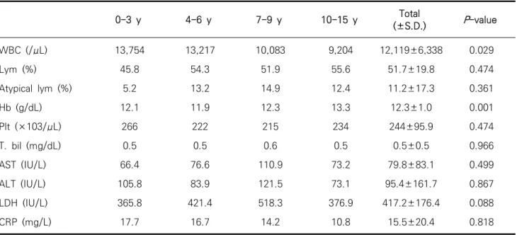
Laboratory findings also differed among age groups (Table 3). Hb levels were higher in the 10–
15 group than the 0–3 group (Scheffe post hoc analysis, P = 0.001) and WBC counts decreased with age (P = 0.029), which may reflect normal
changes in Hb and WBC counts with age. There were no differences in the proportions of lympho- cytes and atypical lymphocytes among groups, al- though the proportion of atypical lymphocytes appeared lower in the 0–3 group (P = 0.474). The proportion of atypical lymphocytes correlated with the presence of splenomegaly (r = 0.407, P
= 0.001) and hepatomegaly (r = 0.417, P < 0.001).
There were no differences in the levels of total bilirubin, AST, ALT, LDH, or CRP among the four age groups (P > 0.05).
The frequencies of leucopenia, leukocytosis, thrombocytopenia, elevated AST or ALT, elevated total bilirubin, or elevated LDH did not differ among age groups (Table 4). Thrombocytopenia was observed in 14 patients: 2 (10.5%) 0–3, 3 (12.0%) 4–6, 6 (23.1%) 7–9 , and 3 (27.3%) 10–15 group (P = 0.543). Abnormal liver function tests (AST > 45 U/L or ALT > 45 U/L) were observed in 29 patients (range: 46–380 U/L). Elevated AST or ALT was correlated with hepatomegaly (14 of 0-3 y
(n=19)
4-6 y (n=25)
7-9 y (n=13)
10-15 y (n=11)
Total
(n=68) P-value
Fever*
Lymphadenopathy†
Pharyngitis Rashes Myalgia‡
Hepatomegaly Splenomegaly
19 (100) 7 (36.8) 4 (21.1) 8 (42.1) 0 (0) 3 (15.8) 3 (15.8)
16 (64.0) 16 (64.0) 9 (36.0) 3 (12.0) 1 (4.0) 4 (16.0) 5 (20.0)
12 (92.3) 11 (84.6) 9 (69.2) 3 (23.1) 1 (7.7) 7 (53.8) 7 (53.8)
9 (81.8) 9 (81.8) 7 (63.6) 1 (9.1) 4 (36.4)
1 (9.1) 2 (18.2)
56 (82.4) 43 (63.2) 29 (42.6) 15 (22.1) 6 (8.8) 15 (22.1) 17 (25.0)
0.011 0.017 0.018 0.072 0.003 0.019 0.064 Values are presented as number(%).
Table 2. Frequency of signs and symptoms associated with infectious mononucleosis according to age group
19 patients with hepatomegaly; r = 0.470, P <
0.001). Elevated total bilirubin (range: 1.66–3.72 mg/dL) was observed in one patient in each of the age groups. One of these reasons exhibited increasing total and direct bilirubin that those pa- tients were transferred to another health center under suspicion of autoimmune hepatitis caused by EBV infection.
Abdominal ultrasonography (USG) was per- formed in 30 patients because of abnormal liver function test results, hepatomegaly, and/or sple- nomegaly: 6 (31.6%) 0–3, 12 (48.0%) 4–6, 9 (69.2%) 7–9, and 4 (36.4%) 10–15 group. Twenty-nine per- cent of patients (n = 20) was checked for pharynx CT scan because of enlarged cervical lymph nodes (n = 18) or airway obstruction (n = 2). The steroids
0-3 y 4-6 y 7-9 y 10-15 y Total
(±S.D.) P-value WBC (/μL)
Lym (%)
Atypical lym (%) Hb (g/dL) Plt (×103/μL) T. bil (mg/dL) AST (IU/L) ALT (IU/L) LDH (IU/L) CRP (mg/L)
13,754 45.8
5.2 12.1
266 0.5 66.4 105.8 365.8 17.7
13,217 54.3 13.2 11.9 222 0.5 76.6 83.9 421.4
16.7
10,083 51.9 14.9 12.3 215 0.6 110.9 121.5 518.3 14.2
9,204 55.6 12.4 13.3 234 0.5 73.2 73.1 376.9
10.8
12,119±6,338 51.7±19.8 11.2±17.3 12.3±1.0 244±95.9
0.5±0.5 79.8±83.1 95.4±161.7 417.2±176.4 15.5±20.4
0.029 0.474 0.361 0.001 0.474 0.966 0.499 0.867 0.088 0.818 Table 3. Laboratory findings by patient age group
0-3 y 4-6 y 7-9 y 10-15 y Total
Leukopenia*
Leukocytosis†
Thrombocytopenia Elevated AST or ALT Elevated T.bil Elevated LDH
0 3 (15.8) 2 (10.5) 8 (42.1%)
1 (5.2) 4 (21.1)
1 (4.0) 1 (4.0) 3 (12.0) 13 (52.0)
1 (4.0) 8 (32.0)
1 (7.6) 0 (0) 3 (23.1) 8 (61.5) 1 (7.6) 6 (46.2)
0 (0) 0 (0) 3 (27.3) 5 (45.5) 1 (9.1) 1 (9.1)
2 (2.9) 4 (5.9) 11 (16.2) 34 (50.0) 4 (5.9) 19 (27.9) Values are presented as number(%).
*Leukopenia is defined as less than 3,000/mm3 of WBC counts
†Leukocytosis is defined as more than of 20,000/mm3 WBC counts
Table 4. Frequency of abnormal laboratory findings among 68 pediatric patients with Epstein-Barr virus-associated infectious mononucleosis
were used to treat patients with prolonged fever, more than 10 days (n = 3), airway obstruction (n
=2), thrombocytopenia (n = 1), and angioedema (n = 1). One patient with fever for more than 15 days was diagnosed with infection-associated he- mophagocytic syndrome (IAHS) and transferred to another health center for chemotherapy.
DISCUSSION
Incidence of EBV-associated IM according to patient age and date
In the present study, we found that the number of EBV-associated cases of IM increased from 8 to 26 between 2011 and 2014. The number of cas- es we observed in 2013 and 2014 is greater than that reported in previous studies based in Korea (17 cases in Kwangju between 1990 and 1996,6 and 18 cases in Seoul between 1989 and 1992).5 The increase in IM cases in 2013 and 2014 was associated with an increase in the number of young children under 6 years old. Our results in- dicate that most of the IM appears among pre- school children as like the results of Mexican children.9 The most common ages in our study were 2 and 5 years, similar to what was seen in another Korean study (3 and 5 years),8 but slightly different from the children in the Mexico study (5 to 7.5 years).9
IM in preadolescents is not rare, but it is more commonly recognized among adolescents and young adults. This is partly because of a lack of
recognition of the syndrome in preadolescents.17 EBV is transmitted in saliva and deep kissing is considered one of the most important risk factors for infection.18 Close contact between young chil- dren in daycare centers or schools may be con- ducive to EBV transmission. We found that 27.9%
of the EBV-associated IM cases in our hospital were under age of 3 years, similar to what was observed among Danish children (27%).10 In China, seroprevalence of anti-EBV antibodies was found to exceed 50% before age 3, which likely derives from the practices of mouth feeding ba- bies or premasticating food.12 However, this does not explain the high rate of IM observed in very young children here or in Denmark.10
The present study describes the clinical features of EBV-associated IM among children in the Jinju region. Fever was the most common clinical pre- sentation regardless of age, as was the case in studies in Denmark,10 in China,14 and elsewhere in Korea.8 We observed fever most often in chil- dren under 3 years old. Moreover, the mean dura- tion of fever was 4.7 days, which is shorter than that in the study of Son et al.’(7.7 days).8 Thus, young children with a persistent fever and more than 3 d of pharyngitis with palpable neck lymph nodes should be suspected to have IM caused by EBV.
In Canada and Mexico, the most frequent clin- ical feature of IM was lymphadenopathy (72% in Canada and 89.5% in Mexico).9,15 In the present study, lymphadenopathy was the second most common presentation and was more commonly
seen in children in the 7–9 age group.
Skin rashes tended to present more often among children under 3 years old, but this did not have statistical support. In China, rash was more fre- quent in children under 6 years.14
Myalgia did not present in children aged 0–3 years. The absence in our study could have arisen because very young children cannot describe their symptoms fully or because the medical pro- fessional did not report myalgia.
In China, hepatomegaly and splenomegaly were more common in patients under 6 years old and tended to become less frequent with older age, which may be relevant to the mature degree of immune system.14 In other countries, the in- cidence of hepatomegaly and splenomegaly ap- pear not to vary by age.8,10,15 We observed more hepatomegaly and splenomegaly in children aged 7–9 years as compared with our other age groups.
Because physical examinations are inaccurate for detecting hepatosplenomegaly, 30 patients (44.1%) underwent abdominal USG. Measured in this way, a greater proportion of patients were positive for hepatosplenomegaly.
Elevated WBC counts and atypical lymphocy- tosis are important indicators of IM.10,15 Greater than 10% atypical lymphocytes is a typical labo- ratory finding of EBV-associated IM.16 We ob- served the mean percentage atypical lymphocytes in children under 3 years old to be lower than in older children, even though total WBC counts were highest in the 0–3 age group.
The mean levels of AST, ALT, LDH, and CRP
were higher than the normal range, consistent with existing literature. Transaminasemia was ob- served in 42.1% to 61.5% of children in the four age groups, similar to results from Canada (47.0%) and Demark (53.7%) but lower than in Bucheon (26.6–76.9%).8 The number of children with ab- normal liver function was greater than that of children with hepatomegaly. Thrombocytopenia was seen in 16.2% of patients; only one was treated with steroid. Slightly elevated levels of CRP (15 mg/L) were observed here as in Bucheon (26 mg/L). One patient in the 4–6 y group was diag- nosed with IAHS.
We observed atypical complications in two pa- tients: autoimmune hepatitis and IAHS. In Seoul, 1 of 18 IM patients was diagnosed with IAHS.5 Hemophagocytic syndrome (HLH) is a life-threat- ening disease and half of all HLH cases are asso- ciated with EBV infection.3 High levels of ferritin could facilitate the diagnosis of HLH in patients with progressive hepatosplenomegaly, lympha- denopathy, and pancytopenia.
In the present study, about one-third of patients underwent pharyngeal CT scan because of cer- vical lymphadenopathy. A lack of a response to antibiotics and cervical lymph nodes > 3 cm in size should differentiate IM from tuberculosis, lymphoma, and Kikuchi disease.
There were several limitations to this study.
First, this was a retrospective evaluation of chil- dren positive for EBV-VCA IgM antibodies.
Second, the patients underwent examinations by multiple medical residents. Third, a small number
of patient were followed up, so we could not assess the long-term impact of EBV infection. And, last, this study was performed in one hospital located in a suburban area.
In conclusion, our results show that EBV-asso- ciated IM is not uncommon in children under 3 years old. We suggest that febrile children who are 0–3 years of age and who present with cervical lymphadenopathy and rash should be suspected to be infected with EBV, even if the percentage of atypical lymphocytes is low. Further study of variation in symptoms among children is needed, as are long-term follow-up studies to investigate the incidence of EBV-associated cancer in Korea.
Acknowledgement
The clinical data used in this study were pro- vided by the Gyeongsang National University Hospital, which is a member of the National Biobank of Korea, which is supported by the Ministry of Health, Welfare and Family Affairs. All samples derived from the National Biobank of Korea were obtained with informed consent under institutional review board approved protocols.
Conflict of interest: none
Financial support: This work supported by Development Fund Foundation, Gyeongsang National University, 2015.
REFERENCES
1. Higgins CD, Swerdlow AJ, Macsween KF, Harrison N, Williams H, McAulay K, et al. A study of risk factors for acquisition of Epstein-Barr virus and its subtypes. J Infect Dis. 2007;195:474-82.
2. Luzuriaga K, Sullivan JL. Infectious mononucleosis.
N Engl J Med. 2010;362:1993-2000.
3. Bolis V, Karadedos C, Chiotis I, Chaliasos N, Tsabouri S. Atypical manifestations of Epstein- Barr virus in children: a diagnostic challenge.
J Pediatr (Rio J). 2016;92:113-21.
4. Murata T, Sato Y, Kimura H. Modes of infection and oncogenesis by the Epstein-Barr virus. Rev Med Virol. 2014;24:242-53.
5. Moon WY, Oh SH, Ko TS, Park YS, Moon HN, Hong CY, et al. Infectious mononucleosis in children. J Korean Pediatr Soc. 1994;37:822-31.
6. Choi JS, Kim TH, Park HY, Lim SC. Clinical analysis of infectious mononucleosis. Korean J Otolaryngol- Head Neck Surg. 1997;40:914-21.
7. Oh SH, Lee YA, Moon WY, Ko TS, Park YS, Moon HN, et al. Prevalence of Epstein-Barr virus (EBV) antibody in Korean children. J Korean Pediatr Soc. 1994;37:804-11.
8. Son KH, Shin MY. Clinical features of Epstein-Barr virus-associated infectious mononucleosis in hospitalized Korean children. Korean J Pediatr.
2011;54:409-13.
9. González Saldaña N, Monroy Colín VA, Piña Ruiz G, Juárez Olguín H. Clinical and laboratory char- acteristics of infectious mononucleosis by Epstein-Barr virus in Mexican children. BMC Res
Notes. 2012;5:361.
10. Topp SK, Rosenfeldt V, Vestergaard H, Christiansen CB, Von Linstow ML. Clinical char- acteristics and laboratory findings in Danish chil- dren hospitalized with primary Epstein-Barr virus infection. Infect Dis (Lond). 2015;47:908-14.
11. Wang Y, Li J, Ren YY, Zhao H. The levels of liver enzymes and atypical lymphocytes are higher in youth patients with infectious mononucleosis than in preschool children. Clin Mol Heptol.
2013;19:382-8.
12. Xiong G, Zhang B, Huang MY, Zhou H, Chen LZ, Feng QS, et al. Epstein-Barr virus (EBV) in- fection in Chinese children: a retrospective study of age-specific prevalence. PLoS One. 2014;9:
e99857.
13. Dowd JB, Palermo T, Brite J, McDade TW, Aiello A. Seroprevalence of Epstein-Barr virus infection in U.S. children ages 6-19, 2003-2010. PLoS One.
2013;8:e64921.
14. Gao LW, Xie ZD, Liu YY, Wang Y, Shen KL.
Epidemiologic and clinical characteristics of in- fectious mononucleosis associated with Epstein- Barr virus infection in children in Beijing, China.
World J Pediatr. 2011;7:45-9.
15. Odame J, Robinson J, Khodai-Booran N, Yeung S, Mazzulli T, Stephens D, et al. Correlates of illness severity in infectious mononucleosis. Can J Infect Dis Med Microbiol. 2014;25:277-80.
16. Ventura KC, Hudnall SD. Hematologic differences in heterophile-positive and heterophile- neg- ative infectious mononucleosis. Am J Hematol.
2004;76:315-8.
17. Balfour HH Jr, Dunmire SK, Hogquist KA. Infectious mononucleosis. Clin Transl Immunology. 2015;4:
e33.
18. Balfour HH Jr, Odumade OA, Schmeling DO, Mullan BD, Ed JA, Knight JA, et al. Behavioral, virologic, and immunologic factors associated with acquisition and severity of primary Epstein-Barr virus infection in university students. J Infect Dis. 2013;207:80-8.