Acute limb ischemia can have 6P symptoms like pain, paralysis, paresthesia, pulselessness, pallor and poikilothermia, and if the imminent situation of tissue loss such as gangrene in the lower ex- tremity occurs it may threaten limb viability.1-3 The incidence of acute limb ischemia is about 14 people among 100,000 people per year, and the mortality rate within 30 days is approximately 15% and the amputation rate within 30 days is 10 to 30%.4 The most common causes of acute artery occlusion are thrombosis and embolism occurring in the underlying atherosclerotic
lesion.5,6 Risk factors of arterial thrombosis in- clude smoking, hypertension, diabetes, dyslipi- demia, obesity, age, hyperhomocysteinemia and specific hypercoagulable state.7,8
The treatment of acute limb ischemia includes drug therapy using anticoagulation therapy, a percutaneous endovascular procedure and a sur- gical reperfusion procedure, while additional therapies during the percutaneous endovascular procedure include catheter directed thrombol- ysis, aspiration thrombectomy and mechanical thrombectomy.1,5,9 In the current aging society
Case Report
Endovascular treatment of acute limb ischemia due to thrombotic occlusion of the suprainguinal artery
Byung Woo Kang, Jun Ho Bae, Deuk Young Nah, Jin Wook Chung, Byeong Joo Jo, Jun Gi Park
Division of Cardiology, Department of Internal Medicine, Dongguk University College of Medicine, Gyeongju-si, Gyeongsangbuk-do, Korea
Acute limb ischemia (ALI) is a serious condition requiring prompt intervention due to a sudden decrease in limb perfusion threatening limb viability. Treatment of ALI depends on the clinical status of the affected limb and patient comorbidities. Surgical therapy has been the historical standard of care for restoring limb perfusion;
however, percutaneous endovascular intervention has been shown to be a promising treatment option in selected patients of ALI at high surgical risk. We report on a case of a 75-year-old man with ALI caused by thrombotic occlusion of the suprainguinal artery, successfully treated with endovascular therapy including stent insertion and thrombus aspiration and catheter-directed urokinase infusion in view of the clinical findings and imaging studies.
Key Words: Endovascular procedures, Ischemia, Lower extremity, Thrombolytic therapy
Corresponding Author: Jun Ho Bae, Division of Cardiology, Department of Internal Medicine, Dongguk University Gyeongju Hospital, Dongguk University College of Medicine, 87, Dongdae-ro, Gyeongju-si, Gyeongsangbuk-do, 38067, Korea
Tel: +82-54-770-8587 Fax: +82-54-770-8378 E-mail: [email protected]
Received:
Revised:
Accepted:
Mar. 25, 2015 Aug. 24, 2015 Sep. 16, 2015
Acute limb ischemia and endovascular treatment
with increasing numbers of elderly patients ex- hibiting vascular and comorbid diseases who are difficult to operate on, advanced mechanical technology has allowed the percutaneous endo- vascular procedure to emerge as an initial treat- ment of these patients. We report use of the percu- taneous endovascular procedure including thrombus and catheter directed thrombolysis in the case of an elderly male with acute limb ische- mia due to the thrombotic occlusion of the com- mon iliac artery, for which surgery is the standard treatment.
CASE
A 75-year old male came to the hospital with the main complaint of pain in his left leg, which suddenly started two days ago while resting. At the time of hospitalization, he complained the paresthesia in his toes but he didn’t have muscle asthenia. The patient had a history of stent in- sertion due to acute myocardial infarction 12 years ago in this hospital and he had intermittent claudication in the left lower limb six months before coming to the hospital. He had no history of diabetes and hypertension, and he had a history of 50 pack per year smoking and chronic ob- structive pulmonary disease.
At the time of hospitalization, the vital signs
were 130/80 mmHg for blood pressure, 36.5℃
for body temperature, 65 times/min for pulse rate and 20 times/min for respiration rate. The left limb was pale and cold, and the pulse of femoral, popliteal arteries and anterior and pos- terior tibial arteries were not palpable. The ankle brachial index was 0.57 on the right and the left was not measurable. Other peripheral blood tests including hemoglobin, white blood cells, plate- lets, chemical tests, coagulation and lipid status were in the normal range. ECG showed normal sinus rhythm and the left ventricular ejection frac- tion on the transthoracic echocardiography was reduced to 37%.
Based on the above symptoms, physical exami- nation and test results, the patient was prelimi- narily diagnosed with acute limb ischemia. Due to his clinical category IIa (only minor paresthesia on toes and no problem with motor ability on the lower limb),10 reduced avacularization in the left ventricular due to aging, and accompanied chronic obstructive pulmonary disease, he was in a high risk group for surgery. Since the severity of ischemia was IIa he was expected to have a good prognosis following an endovascular proce- dure so this was planned as an initial treatment.
First, injection of unfractionated heparin was started and an emergency angiogram was per- formed to accurately locate the occlusive vessel and obtain a definite diagnosis. The vessel access
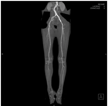
site was prepared against hemorrhagic complica- tion due to the use of thrombolytic agent, and since the left iliac artery was suspected to be the site of the lesion, the right common femoral artery apposite to the lesion was selected as the access site. For puncturing the right common femoral artery an 6-Fr sheath (St. Jude, Minnesota, U.S.A) was inserted and a 5-Fr Judkins right coro- nary artery 3.5 ㎝ curved catheter (Terumo, Tokyo, Japan) was advanced to the left common femoral artery, and the angiogram was performed. As a result, the complete occlusion was observed at the lower part of the ostium of left common iliac artery (Fig. 1A).
After taking the diagnostic angiogram, we switched to an 8-Fr flexor contra guiding sheath (Cordis, California, U.S.A). The 0.014 inch guide
wire with 5-Fr Glide catheter (Terumo, Tokyo, Japan) went through the lesion without any sub- stantial resistance and advanced to the left super- ficial femoral artery. The angiogram using an 5-Fr Glide catheter confirmed that the guide wire was positioned in the vessel and an additional angiogram was performed to check the vessel lesion located in the popliteal arteries and the lower part. As a result of the angiogram, the sus- pected lesion to have the complete occlusion by an embolus could be observed at the terminal region of the popliteal artery (Fig. 1B). Based on the sudden occurrence of symptoms only two days ago and relative ease with which the guide wire passed through the lesion, we determined that a considerable organized thrombus was not present at the lesion and thus we decided to per- Fig. 1. (A) Initial angiography shows total occlusion of the left common iliac artery.
(B) After guide wire and glide catheter are passed the left common iliac artery lesion, angiography shows total occlusion of the popliteal artery.
Acute limb ischemia and endovascular treatment
form the catheter aspiration thrombectomy first.
The aspiration thrombectomy was performed several times at the common femoral artery lesion using 5-Fr Heartrail guide catheter (Terumo, Tokyo, Japan). After performing thrombectomy, the blood flow of TIMI (thrombolysis in my- ocardial infarction) 1 was obtained and, addition- ally, angioplasty was performed using a Powerflex 6.0×120 mm balloon (Cordis, California, U.S.A).
After balloon angioplasty, blood flow was re-opened to the popliteal artery (Fig. 2) but the significant underlying stenosis was left in the common femoral artery, so the SMART 8.0×100 mm stent (Cordis, California, U.S.A) was inserted.
At the angiogram performed after stent insertion,
the successful stent expansion was confirmed and TIMI 2 to 3 of prospective blood flow to the pop- liteal artery was confirmed (Fig. 3A). In the pop- liteal artery lesion, to recover blood flow in at least one vessel in anterior and posterior tibial arteries to the ankle, an aspiration thrombectomy using 5-Fr Heartrail directed catheter, and bal- loon angioplasty using Sleek 2.5×220 ㎜ balloon (Cordis, California, U.S.A) were performed in an- terior and posterior tibial arteries, and 100,000 units of urokinase was injected directly into the vessel using a 5-Fr Glide catheter (Terumo, Tokyo, Japan) to each anterior and posterior tibial ar- teries, repeatedly. After the procedure, the blood flow of the posterior tibial artery and peroneal artery did not recover, but the blood flow of the anterior tibial artery was successfully recovered to the dorsalis pedis artery (Fig. 3B). To melt the residue thrombus, a 5-Fr multi-sideport infusion catheter (Cook, Bloomington, U.S.A) was posi- tioned from the popliteal artery to the posterior tibial artery and urokinase was continuously in- jected by 50,000 units per hour. 12 hours after the infusion, the angiogram was performed again.
The results showed that a small amount of throm- bus remained but the blood flow of the anterior and posterior tibial arteries and fibular artery were confirmed to be well maintained up to tip- toes (Fig. 4). The patient didn't complain about the pain anymore and the pulses of the dorsalis Fig. 2. After several aspiration thrombectomy with a
5-Fr Heartrail guiding catheter and balloon angioplasty, angiography shows significant luminal stenosis of the left common iliac artery and intraluminal filling defects in the left common and external iliac artery.
Fig. 3. (A) After aspiration thrombectomy, balloon angioplasty and the stent insertion at the left common iliac artery, angiography shows good flow restoration to the distal part popliteal artery. (B) After aspiration thrombectomy, balloon angioplasty and intrathrombus urokinase injection at anterior and posterior tibial artery, angiography shows good flow restoration to the dorsalis pedis artery (white arrow head).
Acute limb ischemia and endovascular treatment
Fig. 4. Follow-up angiography after continuous urokinase infusion (50,000 IU/hr) with a multi-sideport infusioncatheter for 12 hours shows good patent anterior tibial artery, posterior tibial artery and peroneal artery and good antegrade flow to foot.
pedis artery and posterior tibial artery could be palpated and the angle brachial index had in- creased to 0.98. Continuous oral anticoagulation therapy was maintained by changing heparin to warfarin. After two months, computer tomog- raphy angiography showed the stent in the com- mon femoral artery. There were no signs of steno- sis and thrombus in the popliteal artery, the ante- rior and posterior tibial arteries or the peroneal artery and the blood flow was well maintained (Fig. 5).
DISCUSSION
Acute arterial occlusion is caused by thrombo- sis in a blood vessel that underwent bypass or in an original blood vessel with atherosclerosis or by thromboembolism occurring in the heart or aneurysm. The causes of acute limb ischemia in patients who do not have atherosclerosis in- clude artery damage due to the accident, aortic dissection, bakers cyst, popliteal entrapment and vasospasm.6,9 The induction incidence of throm- bosis and embolism, which are the cause of acute limb ischemia, accounts for 85% and 15% of cases respectively.5 Relatively low levels of fibrin and erythrocytes exemplify the characteristics of ar- terial thrombosis, and instead it is mainly white thrombi composed of platelets, and occurs at
the site where an atherosclerotic plaque rupture occurred due to high shear stress.7,8 In most cases, it is difficult to distinguish the thrombosis and embolism clinically and it is impossible to dis- tinguish in 10 to 15% of cases. In the case of the above patient, the left occlusion of the com- mon femoral artery was presumed to be thrombo- sis, considering his old age, 50 pack per year smoking, long-term myocardial infarction, clau- dication only in the left lower limb from 6 months ago and sinus rhythm in ECG. Also, the occlusion of the popliteal artery was determined to have occurred due to the occlusion of thromboembo- lism in common femoral artery.11
In patients with acute limb ischemia, in order to save the limb, a medical examination by inter- view that includes leg symptoms and their causes, atherosclerosis risk factors and comorbid dis- eases should be performed.2,9,12 Since pulse pal- pation should not be trusted, measurement of the peripheral pulse using Doppler should be considered in patients with suspected ischemia1,5 and the interview and physical examination are important in determining the severity and reversibility. Medical imaging methods such as computer tomography angiography, magnetic resonance angiography or duplex sonography can be used to plan the strategy for diagnosis and interventional procedures. Angiography has the disadvantage of being invasive and using a
Acute limb ischemia and endovascular treatment
contrast agent, but it has the advantage of de- termining the precise lesion position and showing the terminal branches of the artery in detail, so when selecting therapeutic methods it helps a lot, especially when considering the endovascular treatment.
The selection of reperfusion procedure is de- termined by clinical considerations including the urgent extent of the ischemic event, severity, pe- riod, anatomical location of the lesion, type of vessel (artery or implanted vessel), comorbid dis- ease, risk of procedure and contraindication of therapy.1,2,5,9 When there is a complete occlusion of the common femoral artery like in this patient, the surgical reperfusion procedure was consid- ered as a standard therapy for a while. However, there may be several limitations in the early stages and it shows high pre- and post-operative mortality. In several recent studies the endovas- cular procedure, including thrombolysis, showed equivalent results to the surgery, so it can be used as an initial treatment in selective patients.
In endovascular thrombolysis using drugs, the injection of thrombolytic agent directly to the thrombosis by continuous infusion using the catheter is commonly used to reduce the hemor- rhagic complications due to drugs and to melt the thrombus effectively. To accelerate the dis- solution and to shorten the treatment time, a high dose can be injected within the first few
hours. In one study, a high dose (250,000 IU/hr for 4 hours followed by 125,000 IU/hr) and low dose (50,000 IU/hr) of urokinase injection were all reported to be similarly effective on a periph- eral thrombus13 so it is thought that the dosage can be determined depending on the clinical sta- tus of the patient and severity of the lesion.
Using aspiration thrombectomy with pharma- cologic thrombolysis can reduce the time for re- perfusion and reduce the bleeding complications, consequently making it possible to have endovas- cular therapy in acute limb ischemia.5,9 However, aspiration thrombectomy can induce distal em- bolism so the procedure should be performed carefully. In this case, the patient had acute limb ischemia caused by complete occlusion in the
Fig. 5. Computed tomography angiography 2 months later shows a well expanded stent at the left common iliac artery, good patency of the popliteal
left common femoral artery. Surgical intervention might be considered as an initial treatment ac- cording to the traditional standard treatment manner and recommendation based on the occlu- sion location, however, due to the accompanied comorbid disease and clinical severity of IIa, en- dovascular therapy was considered appropriate.
Relatively all endovascular procedures such as catheter aspiration thrombectomy, catheter di- rected urokinase injection and procedures using balloon and stent were used. The endovascular procedure was performed in the popliteal artery, trying to recover the blood flow of at least one vessel in the anterior and posterior tibial arteries and successful results were obtained. Even though there is a stenosis at the distal end of the right popliteal artery, clinical symptoms like claudica- tion were not evident so the conservative treat- ment was performed. For the occlusion of the distal end of left anterior tibial artery, there are possibilities of chronic occlusion and residual thrombus, so antithrombotic drug was in use.
According to recent meta-analysis comparing the thrombolysis and surgical reperfusion ther- apy in patients with acute limb ischemia, there were no difference in the two methods with re- spect to their effects, but in the case of performing thrombolysis, it was reported that the incidence of bleeding and hemorrhagic stroke had increased. Therefore, occurrence of complica-
tions should be observed closely after using the thrombolytic agent. The percutaneous endovas- cular procedure in acute limb ischemia has com- parable effect to surgery, but the procedure may be a better initial treatment since it has less of a burden on the patients. However, in order to use this method as an initial treatment, patient screening is very important and the occurrence of complications should be observed closely after the treatment.
REFFRENCES
1. Norgren L, Hiatt WR, Dormandy JA, Nehler MR, Harris KA, Fowkes FG, et al. Inter-society con- sensus for the management of peripheral arterial disease (TASC II). J Vasc Surg 2007;45:S5-67.
2. Hirsch AT, Haskal ZJ, Hertzer NR, Bakal CW, Creager MA, Halperin JL, et al. ACC/AHA 2005 practice guidelines for the management of pa- tients with peripheral arterial disease (lower ex- tremity, renal, mesenteric, and abdominal aort- ic): a collaborative report from the American Association for Vascular Surgery/Society for Vascular Surgery, Society for Cardiovascular Angiography and Interventions, Society for Vascular Medicine and Biology, Society of Interventional Radiology. Circulation 2006
;113:e463-654.
Acute limb ischemia and endovascular treatment
3. Creager MA, Kaufman JA, Conte MS. Acute limb ischemia. N Engl J Med 2012;366:2198-206.
4. Dormandy J, Heeck L, Vig S. Acute limb ischemia.
Semin Vasc Surg 1999;12:148-53.
5. Walker TG. Acute Limb Ischemia. Tech Vasc Interv Radiol 2009;12:117-29.
6. Costantini V, Lenti M. Treatment of acute occlu- sion of peripheral arteries. Thromb Res 2002;106:V285-94.
7. Lowe GD. Common risk factors for both arterial and venous thrombosis. Br J Haematol 2008
;140:488-95.
8. Franchini M, Mannucci PM. Association between venous and arterial thrombosis: clinical implications. Eur J Intern Med 2012;23:333-7.
9. Patel NH, Krishnamurthy VN, Kim S, Saad WE, Ganguli S, Gregory Walker T, et al. Quality im- provement guidelines for percutaneous manage- ment of acute lower-extremity ischemia. J Vasc Interv Radiol 2013;24:3-15.
10. Rutherford RB, Baker JD, Ernst C, Johnston KW, Porter JM, Ahn S, et al. Recommended standards for reports dealing with lower extremity ischemia:
revised version. J Vasc Surg 1997;26:517-38.
11. Ouriel K, Veith FJ, Sasahara AA. A comparison of recombinant urokinase with vascular surgery as initial treatment for acute arterial occlusion of the legs. N Engl J Med 1998;338:1105-11.
12. Karnabatidis D, Spiliopoulos S, Tsetis D, Siablis D. Quality improvement guidelines for percuta- neous catheter-directed intra-arterial thrombol- ysis and mechanical thrombectomy for acute low- er-limb ischemia. Cardiovasc Intervent Radiol 2011;34:1123-36.
13. Cragg AH, Smith TP, Corson JD, Nakagawa N, Castaneda F, Kresowik TF, et al. Two urokinase dose regimens in native arterial and graft occlu- sions: Initial results of a prospective, randomized clinical trial. Radiology 1991;178:681-6.