Introduction
External apical root resorption (EARR) is a complication of orthodontic treatment. Loss of apical root material is unpredictable and irreversible when extending into dentine.
Extensive orthodontic root resorption can compromise the benefits of a successful orthodontic outcome.
There is no single explanation why teeth resorb to vari- ous degrees; however a number of factors taken together may affect external apical root resorption. Brezniak and Wasserstein1-4reviewed and explained the factors that might affect root resorption including biological factors such as previous trauma and systemic disease, and mech- anical factors such as using Class II elastics and long treat- ment duration. Knowing the causal factors would allow clinicians to predict the incidence, location, and severity of root resorption before the commencement of orthodon- tic treatment.
The objectives of this study were to evaluate the incid-
External apical root resorption in maxillary incisors in orthodontic patients:
associated factors and radiographic evaluation
Kamonporn Nanekrungsan, Virush Patanaporn*, Apirum Janhom**, Narumanus Korwanich***
Department of Dentistry, Overbrook Hospital, Chiang Rai, Thailand
*Division of Orthodontics, Faculty of Dentistry, Chiang Mai University, Chiang Mai, Thailand
**Division of Oral and Maxillofacial Radiology, Faculty of Dentistry, Chiang Mai University, Chiang Mai, Thailand
***Division of Community Dentistry, Faculty of Dentistry, Chiang Mai University, Chiang Mai, Thailand ABSTRACT
Purpose: This study was performed to evaluate the incidence and degree of external apical root resorption of maxil- lary incisors after orthodontic treatment and to evaluate particular associated factors related to external apical root resorption.
Materials and Methods: The records and maxillary incisor periapical radiographs of 181 patients were investigated.
Crown and root lengths were measured and compared on the pre- and post-treatment periapical radiographs. Crown length was measured from the center of the incisal edge to the midpoint of the cemento-enamel junction (CEJ). Root length was measured from the CEJ midpoint to the root apex. A correction factor for the enlargement difference was used to calculate root resorption.
Results: The periapical radiographs of 564 teeth showed that the average root resorption was 1.39±1.27 (8.24±
7.22%) and 1.69±1.14 mm (10.16±6.78%) for the maxillary central and lateral incisors, respectively. The results showed that the dilacerated or pointed roots, maxillary premolar extraction cases, and treatment duration were highly significant factors for root resorption (p⁄0.001). Allergic condition was a significant factor at p⁄0.01. Age at the start of treatment, large overjet, and history of facial trauma were also factors significantly associated with root resorp- tion (p⁄0.05). There was no statistically significant difference in root resorption among the factors of gender, overbite, tongue-thrusting habit, types of malocclusion, and types of bracket.
Conclusion: These results suggested that orthodontic treatment should be carefully performed in pre-treatment extrac- tion patients who have pointed or dilacerated roots and need long treatment duration. (Imaging Sci Dent 2012; 42 : 147-54)
KEY WORDS: Root Resorption; Radiography, Dental; Orthodontics
Received April 11, 2012; Revised July 12, 2012; Accepted August 3, 2012 Correspondence to : Dr. Kamonporn Nanekrungsan
Department of Dentistry, Overbrook Hospital, 17 Singhaklai Road, Chiangrai 57000, Thailand
Tel) 66-53-711366, Fax) 66-53-711367, E-mail) [email protected]
Copyright ⓒ 2012 by Korean Academy of Oral and Maxillofacial Radiology
This is an Open Access article distributed under the terms of the Creative Commons Attribution Non-Commercial License (http://creativecommons.org/licenses/by-nc/3.0) which permits unrestricted non-commercial use, distribution, and reproduction in any medium, provided the original work is properly cited.
Imaging Science in Dentistry∙pISSN 2233-7822 eISSN 2233-7830
ence and degree of EARR of the maxillary incisors after orthodontic treatment and to evaluate some particular fac- tors related to EARR of the maxillary incisors.
Materials and Methods
Sample selection
Three hundred fifty patients who completed orthodontic treatment during 1995-2005 in the post-graduate clinic of the Department of Orthodontics, Chiang Mai University were examined. This study was based on 181 of those patients who satisfied the study criteria: complete maxil- lary and mandibular arches, fixed appliance treatment, and availability of pre-treatment and treatment records and of pre- and post-treatment intra-oral periapical radiographs.
Additionally, subjects were excluded from the study if crown dimensions were altered during the treatment period due to tooth fracture or abrasion or if their radiographs fell within the criteria of exclusion: the projection was poor, the crown or apex was not fully visible, or the cemento- enamel junction (CEJ) was blurred.
Examination of records
Pre-treatment data and treatment variables were recorded including gender, age at start of treatment, overjet, overbite (in mm), root shape, history of facial trauma, tongue-thrust- ing habit (yes or no), allergic condition (yes or no), types of malocclusion, history of maxillary premolar extraction, types of bracket, and treatment duration (Table 1).
Examination of periapical radiographs
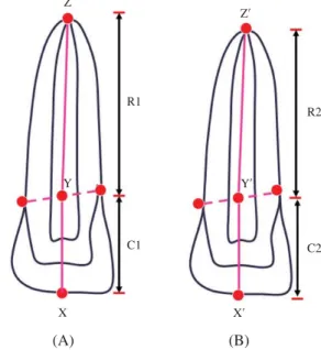
The pre- and post-treatment radiographs of the maxil- lary incisors were measured by a digital vernier caliper (KEIBA®, Sanjo, Japan) with a fine tip measuring 0.01 mm. The crown length was measured from the median CEJ point, the midpoint between the mesial and distal CEJ, to the mid-incisal point of the crown. Root length was measured from the median CEJ point to the most apical point of the root (Fig. 1).
Any image enlargement between pre- and post-treatment radiographs was corrected by using the crown length reg- istrations, assuming crown lengths to be unchanged over the observation period. Therefore, the ratio between the initial crown length (C1) and post-treatment crown length (C2) determined the correction factor (CF). For this reason, teeth with obviously fractured or abraded crowns were excluded from the study. A correction factor was calculated to relate the pre- and post-treatment root length of each particular tooth as shown in the following equations. Then the EARR per tooth in millimeters was calculated:
Correction factor (CF)==C1/C2 EARR==R1-(R2×CF)
EARR was also expressed as the percentage shortening
Table 1.The examination factors and their units of measurement for external apical root resorption per (Mann-Whitney U and Kru- skal-Wallis H test)
Examination factor Unit of measurement P value
Gender Male/Female 0.378
Age at start of treatment Years (‹16/¤16) 0.025*
Overjet mm (⁄1, 1-4, ¤4-6, ¤6) 0.038*
Overbite mm (‹0, ¤0-3, ¤3-5, ¤5) 0.092
Root shape Normal/Abnormal ⁄0.001‡
(dilacerated or pointed)
History of facial trauma Yes/no 0.014*
Tongue-thrusting habit Yes/no 0.411
Allergic condition Yes/no 0.003†
Types of malocclusion Cl I, II, III 0.946 Upper premolar extraction Yes/no ⁄0.001‡ Types of bracket Standard/Preadjusted 0.748
Treatment duration Months ⁄0.001‡
(1-12, 13-24, 25-36, ›37)
*P⁄0.05, †P⁄0.01, ‡P⁄0.001
Z
Y Y′
X
R1
C1 C2
R2 Z′
X′
(A) (B)
Fig. 1. The points and distance measured on the examination of periapical radiographs. (A) Pre-treatment: X==mid-incisal point of crown, Y==midpoint of CEJ, Z==most apical point of root, C1== crown length before treatment, R1==root length before treatment.
(B) post-treatment: X′==mid-incisal point of crown, Y′==midpoint of CEJ, Z′==most apical point of root, C2==crown length after treat- ment, R2==root length after treatment.
per tooth. Since the differences in root lengths of various teeth make comparisons of root resorption values in mil- limeters less meaningful, the percentage value is a better comparative value for EARR.
Percentage of EARR per tooth==(EARR×100)/R1
Statistical analysis
Descriptive statistics were calculated for analysis of EARR for all variables for all maxillary incisors. Mann- Whitney U and Kruskal-Wallis H test were used to deter- mine the association between EARR and associated factors.
Reproducibility of the measurements was established by having the same examiner take measurements twice on 20 randomly-selected radiographs and using Pearson’s pro- duct-moment correlation. The test showed high correlation (r==0.81) between the first and second measurements. The correlation was significant at p⁄0.05 level.
Results
Those 564 teeth from 181 cases fulfilled the criteria for sample selection. The results showed that the mean amount
of EARR in the maxillary incisors was 1.55±1.30 mm (9.32±7.78%) of the root length. The average EARR for each maxillary incisor was 1.41±1.23 mm for the right central incisor, 1.62±1.26 mm for the right lateral incisor, 1.37±1.43 mm for the left central incisor, and 1.76±1.28 mm for the left lateral incisor, respectively. The percentage of EARR per tooth was 8.24±7.10, 9.63±7.80, 8.24±
8.54, and 10.93±7.57, respectively (Table 2).
The maxillary lateral incisor showed more EARR than the maxillary central incisor. The means and percentages of EARR in the maxillary central and lateral incisors were
Table 2.Mean external apical root resorption (EARR) (mm) and mean percentage of external apical root resorption (EARR) per tooth for each maxillary incisor
Tooth n mean EARR (mm) mean EARR (%)
11 150 1.41±1.23 8.24±7.10
12 131 1.62±1.26 9.63±7.80
21 149 1.37±1.43 8.24±8.54
22 134 1.76±1.28 10.93±7.57
11 and 21 299 1.39±1.27 8.24±7.22
12 and 22 265 1.69±1.14 10.16±6.78
11: maxillary right central incisor, 12: maxillary right lateral incisor, 21:
maxillary left central incisor, 22: maxillary left lateral incisor
Table 3.Numbers, percentage of external apical root resorption (EARR) per tooth, and interquartile ranges in maxillary incisors in each group of variable factors and their significance levels
Associated factor Number of teeth Percentage EARR per tooth Interquartile range p Age at start of treatment
≤16 years old 338 7.79 10.12 0.025*
¤16 years old 226 9 9
Overjet group (mm)
⁄1 77 7.68 7.71 0.038*
1-4 250 8.09 11.03
¤4-6 146 7.7 10.36
¤6 91 9.56 10.94
Root shape
Normal 508 7.8 9.76 ⁄0.001‡
Pointed or dilacerated 56 13.28 10.89
Allergic condition
Without allergic condition 472 7.89 9.96 0.003†
With allergic condition 88 10.06 11.05
History of trauma
Without history of trauma 539 8.07 9.84 0.014*
With history of trauma 25 14.75 17.31
Treatment plan
Non-extraction 159 5.57 9.85 ⁄0.001‡
Upper premolar extraction 405 8.94 9.57
Treatment duration (Months)
1-12 21 8.57 6.61 ⁄0.001‡
13-24 154 6.21 9.18
25-36 273 8.42 10.59
≥37 116 10.49 11.13
1.39±1.27 mm (8.24±7.22%) and 1.69±1.14 mm (10.16
±6.78%), respectively (Table 2). There was a statistically significant difference in EARR between the central and lateral incisors at p⁄0.05.
The degree of EARR was classified into 3 categories according to the percentage of resorption: mild (‹10%), moderate (10-20%), and severe (¤20%). It was found that 59.6% of the investigated teeth expressed a mild degree of EARR, 31.9% had moderate EARR, and 8.5% had severe EARR.
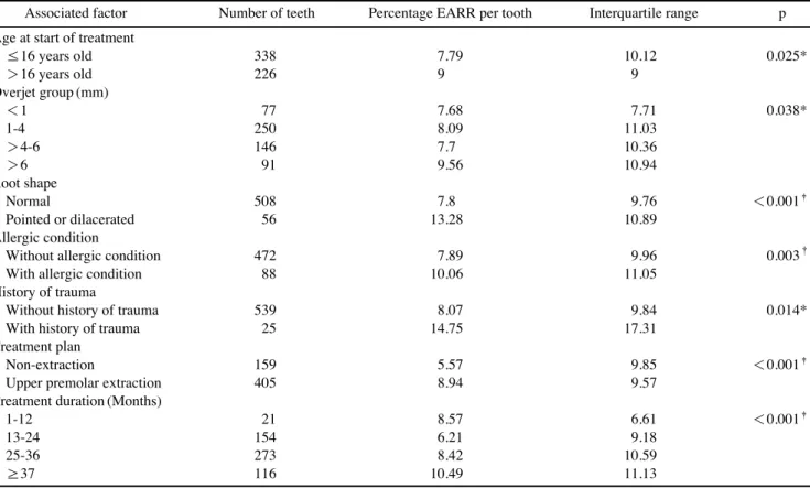
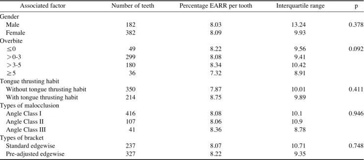
Of all the factors examined in this study, statistically sig- nificant differences in EARR were found among the pati- ents according to the following variables: age at start of treatment, overjet, root shape, history of trauma, incidence of allergy, cases of maxillary premolar extraction, and treat- ment duration (Table 3). Gender, overbite, tongue-thrust- ing habit, types of malocclusion, and types of bracket were not associated with EARR (Table 4).
Cases of EARR in dilacerated maxillary lateral incisors and in maxillary incisors with a history of trauma are shown in Figures 2 and 3, respectively.
Discussion
There are many diagnostic aids for detecting EARR.
However, this study used periapical radiographs because they provide less distortion and fewer superimposition errors than do panoramic or lateral cephalomatric radio- graphs.5,6 Although they could not assess the amount of
Table 4.Numbers, percentage of external apical root resorption (EARR) per tooth, and interquartile ranges in maxillary incisors in each group of variable factors
Associated factor Number of teeth Percentage EARR per tooth Interquartile range p Gender
Male 182 8.03 13.24 0.378
Female 382 8.09 9.93
Overbite
≤0 49 8.22 9.56 0.092
¤0-3 299 8.08 9.41
¤3-5 180 8.34 10.42
≥5 36 7.32 8.91
Tongue thrusting habit
Without tongue thrusting habit 350 7.87 10.01 0.411
With tongue thrusting habit 214 8.75 9.89
Types of malocclusion
Angle Class I 416 8.08 10.1 0.946
Angle Class II 107 8.06 10.9
Angle Class III 41 8.36 8.78
Types of bracket
Standard edgewise 237 8.07 10.71 0.748
Pre-adjusted edgewise 327 8.22 9.35
A B
A B
Fig. 2.A. Dilacerated maxillary lateral incisor before orthodontic treatment. B. Moderate external apical root resorption of dilacerated maxillary lateral incisor after orthodontic treatment.
Fig. 3. A. Traumatized maxillary teeth before orthodontic treat- ment. B. Severe external apical root resorption of the traumatized maxillary incisors after orthodontic treatment.
root loss in buccal and lingual surfaces, they provided the most appropriate information with the least irradiation to patients. Periapical radiographs and correction factors have been used to measure the amount of root shortening.7-9 Brezniak et al10,11found this combination to be the best method for the assessment of root resorption. However, periapical radiographs using the paralleling technique should be used. Cone beam computed tomography (dental CT) might be suggested to evaluate three-dimensional EARR for further studies.
It was difficult to compare the frequency and severity of EARR in this study with that reported by other studies due to the differences in techniques and methods employed.
However, as in most previous studies, this study found that EARR occurred in the maxillary lateral incisors more fre- quently and with greater severity than it did in the maxil- lary central incisors.12,13
There are many possible explanations for why maxillary lateral incisors were more severely affected. Among all of the teeth, the maxillary lateral incisors demonstrated the highest percentages of abnormal root shapes or narrow roots.14,15Our data confirmed that 7.1% of maxillary lateral incisors had pointed or dilacerated root shapes but only 2.84% were found in maxillary central incisors. It is hypo- thesized that the more narrowed or shortened the root, the more force might be distributed to the root surface. More- over, dilacerated roots would be more likely to be resorbed since more force would be orthodontically applied to move or torque the root than would be the case with normal root shapes.
Several studies described EARR after orthodontic treat- ment and its associated risk factors. The comparison of associated factors in previous studies and the present study is shown in Table 5. This result indicated that EARR after orthodontic treatment might be a consequence of a com- plex combination of individual biology and the effects of mechanical factors.
Age at start of treatment
A common question is whether older patients experience more EARR than do younger patients. Thilander et al16 described the physiological changes of tissues in adults that might be involved in the root resorption process. In adults, the periodontal ligament becomes less vascular, aplastic, and narrow; the bone more dense, avascular, and aplastic; and the cementum wider. These changes are refl- ected in a higher susceptibility to EARR in adults. This explanation supports the results of this study, which found
Table 5.Comparison of factors associated with external apical root resorption(EARR) between some previous studies and the present study Age at startHistory ofTongue Allergic Types of Treatment planTypes of bracket Treatment AuthorsGenderOverjetOverbiteRoot shapethrusting (Standard edgewise of treatmenttrauma habitconditionmalocclusion(Ext./Non-Ext.) vs Straight wire)duration Linge and Linge 1991NoNoYesNo-YesYes----- Mirabella and --NoNoYes; long, narrow,---No-NoNo Årtun 1995abnormal root Baumrind et al 1996MaleNoNoNo----NoNo-Yes Levander et al 1998NoNo--Yes; apically bent------Yes Mavragani et al 2000NoYesNoNoNoYesNo---Standard¤No Straight wire Sameshima and SinclairNoadult¤YesNoYes; pointed, ------- 2001childrendilacerated Brin et al 2003--Yes--No--Class II div 1¤--Yes ¤Class I Nishioka et al 2005----Yes; RootNoNoYes-NoNo- abnormality Mohandesan et al 2007Female--------YesNoYes Present studyNoolder¤YesNoYes; pointed,YesNoYesNoYesNoYes youngerdilacerated
that a higher age at the beginning of treatment (age group
¤16 years) resulted in significantly more EARR than did a lower age (age group ‹16 years) at p⁄0.05. However, some previous reports17-19 found no statistical difference in EARR with age.
Overjet
Overjet was significantly associated with EARR in maxil- lary incisors. Greater overjet was a powerful predictor of EARR. This finding is in agreement with the findings of some authors13,17,20 who believed that overjet might be regarded as a risk factor for EARR. The explanation was that the correction of large overjet often required fixed appliance treatment, in which anterior teeth were moved long distances to reduce maxillary anterior protrusion and active torque with rectangular wire was also required. Both active torque and movement of teeth through long distances have been shown to result in EARR.
Root shape
In this study, abnormal root shape was categorized into dilacerated and pointed root shapes. We found that the dilacerated or pointed roots were highly significant factors for EARR. This finding is in agreement with the findings of previous studies.13,14,21
There are possible explanations for why dilacerated and pointed roots had more EARR. Dilacerated roots are more likely to be resorbed than normal roots since more force is orthodontically applied to move or torque dilacerated roots. Pointed roots have more EARR because higher stress is distributed over a smaller root apex than over the larger apex of a normal root shape when tipping or torque move- ment is used.
History of trauma
According to many studies,17,22,23trauma is an important risk factor for EARR. This might be explained by the fact that at the moment of impact, a significant amount of energy is expended in driving the tooth into its socket.
Traumatic forces compress the periodontal ligament and crush the alveolar socket wall. EARR following dental trauma along a root surface appears to be a sequela of wound healing events, where a periodontal ligament has been lost due to acute trauma.24 There were not many traumatic cases in this study and there was very little infor- mation about intensity, location, and type of trauma. This dearth of information minimizes the importance of this
finding. Consequently, more intensive investigation of such factors as type of traumatic dental injury associated with EARR should be included in further studies.
Allergic condition
Allergic condition and asthma were risk factors for EARR in this investigation. These findings confirmed those of earlier studies.25-27Inflammation is an integral part of tis- sue response to orthodontic force in the early stage of tooth movement. In this process, immune cells migrate out of the capillaries into the periodontal ligament and interact with locally residing cells by elaborating a large array of signal molecules. The presence of primed leucocytes in the peripheral blood which originate in diseased organs supports the possible association between root resorption and pathologic conditions, including allergic conditions and asthma, which involve the immune system.27
Treatment plan (Extraction/Non-Extraction)
Extraction of premolar teeth for orthodontic treatment was also found to be a significant factor for EARR in this study. Patients who underwent maxillary premolar tooth extraction had more severe EARR than did those patients who were treated with non-extraction. This finding is in agreement with the report of Mohandesan et al.28However, Baumrind et al18and McFadden et al29found no difference in EARR between the non-extraction and extraction groups.
Basically, in cases of premolar extraction, the remaining teeth are usually moved relatively long distances, particu- larly when maxillary incisors are retracted to reduce the protrusion. On the other hand, if the extraction space is used to relieve crowding, the maxillary incisors are not moved such great distances. Therefore, EARR in maxil- lary incisors may depend on the distance that the anterior teeth are moved in cases of maxillary premolar extraction.
Treatment duration
In this study, there were highly significant differences in EARR between treatment duration groups (within 1, 2, 3, and over 3 years of treatment). These findings agree with some previous studies.18,19Baumrind et al18found that the increased length of treatment time was positively associated with increased EARR. Their results suggested that there was 0.38 mm of resorption during each year of orthodontic treatment. Levander and Malmgren19 divided total treat- ment time into treatment groups: within 1 year, 2 years, and more than 2 years. They found statistically significant
differences among the groups and concluded that total treatment duration was significantly related to EARR.
In contrast, Linge and Linge7and Mirabella and Artun14ex- plained that appliances might be present for longer periods without creating force on the teeth. Therefore, treatment duration was not used as a predictor for root resorption.
In conclusion, the mean EARR in this study was 1.55±
1.30 mm (9.32±7.78%) of the initial root length. Maxil- lary lateral incisors showed more EARR than maxillary central incisors. The age at start of treatment, dilacerated or pointed roots, excessive overjet, allergy, history of trauma, upper premolar extractions, and treatment duration were found to be the associated factors for EARR during the course of orthodontic treatment (p⁄0.05).
Acknowledgments
Our sincere appreciation is expressed to Dr. Janya Apis- ariyakul for her help and valuable assistance throughout this study. We would like to thank all of the orthodontists who had collected the patient’s records and finished the courses of orthodontic treatment. We also thank all of the patients who permitted access to their treatment records for this project. Finally, we are grateful to Dr. M. Kevin O Carroll, Professor Emeritus of the University of Mis- sissippi School of Dentistry, and Faculty Consultant at Chiang Mai University Faculty of Dentistry, for his assis- tance in the preparation of the manuscript.
References
1. Brezniak N, Wasserstein A. Root resorption after orthodontic treatment: Part 1. Literature review. Am J Orthod Dentofacial Orthop 1993; 103 : 62-6.
2. Brezniak N, Wasserstein A. Root resorption after orthodontic treatment: Part 2. Literature review. Am J Orthod Dentofacial Orthop 1993; 103 : 138-46.
3. Brezniak N, Wasserstein A. Orthodontically induced inflam- matory root resorption. Part I: The basic science aspects. Angle Orthod 2002; 72 : 175-9.
4. Brezniak N, Wasserstein A. Orthodontically Induced Inflam- matory root resorption. Part II: The clinical aspects. Angle Orthod 2002; 72 : 180-4.
5. Leach HA, Ireland AJ, Whaites EJ. Radiographic diagnosis of root resorption in relation to orthodontics. Br Dent J 2001; 190 : 16-22.
6. Sameshima GT, Asgarifar KO. Assessment of root resorption and root shape: periapical vs panoramic films. Angle Orthod 2001; 71 : 185-9.
7. Linge BO, Linge L. Apical root resorption in upper anterior teeth. Eur J Orthod 1983; 5 : 173-83.
8. Blake M, Woodside DG, Pharoah MJ. A radiographic compar-
ison of apical root resorption after orthodontic treatment with the edgewise and Speed appliances. Am J Orthod Dentofacial Orthop 1995; 108 : 76-84.
9. Mavragani M, Vergari A, Selliseth NJ, Boe OE, Wisth PL. A radiographic comparison of apical root resorption after ortho- dontic treatment with a standard edgewise and a straight-wire edgewise technique. Eur J Orthod 2000; 22 : 665-74.
10. Brezniak N, Goren S, Zoizner R, Shochat T, Dinbar A, Was- serstein A, et al. The accuracy of the cementoenamel junction identification on periapical films. Angle Orthod 2004; 74 : 496- 500.
11. Brezniak N, Goren S, Zoizner R, Dinbar A, Arad A, Wassers- tein A. A comparison of three methods to accurately measure root length. Angle Orthod 2004; 74 : 786-91.
12. Mirabella AD, Artun J. Prevalence and severity of apical root resorption of maxillary anterior teeth in adult orthodontic pati- ents. Eur J Orthod 1995; 17 : 93-9.
13. Sameshima GT, Sinclair PM. Predicting and preventing root resorption: Part I. Diagnostic factors. Am J Orthod Dentofacial Orthop 2001; 119 : 505-10.
14. Mirabella AD, Artun J. Risk factors for apical root resorption of maxillary anterior teeth in adult orthodontic patients. Am J Orthod Dentofacial Orthop 1995; 108 : 48-55.
15. Artun J, Smale I, Behbehani F, Doppel D, Van’t Hof M, Kuij- pers-Jagtman AM. Apical root resorption six and 12 months after initiation of fixed orthodontic appliance therapy. Angle Orthod 2005; 75 : 919-26.
16. Thilander B, Rygh P, Reitan K. Tissue reactions in orthodontics.
In: Graber TM, Vanarsdall RL Jr. Orthodontics: current prin- ciples and techniques. 3rd ed. St. Louis: Mosby; 2000. p. 117- 125.
17. Linge L, Linge BO. Patient characteristics and treatment vari- ables associated with apical root resorption during orthodontic treatment. Am J Orthod Dentofacial Orthop 1991; 99 : 35-43.
18. Baumrind S, Korn EL, Boyd RL. Apical root resorption in orthodontically treated adults. Am J Orthod Dentofacial Orthop 1996; 110 : 311-20.
19. Levander E, Malmgren O. Evaluation of the risk of root resorp- tion during orthodontic treatment: a study of upper incisors.
Eur J Orthod 1988; 10 : 30-8.
20. Brin I, Tulloch JF, Koroluk L, Philips C. External apical root resorption in Class II malocclusion: a retrospective review of 1- versus 2-phase treatment. Am J Orthod Dentofacial Orthop 2003; 124 : 151-6.
21. Smale I, Artun J, Behbehani F, Doppel D, van’t Hof M, Kuij- pers-Jagtman AM. Apical root resorption 6 months after initia- tion of fixed orthodontic appliance therapy. Am J Orthod Dento- facial Orthop 2005; 128 : 57-67.
22. Malmgren O, Goldson L, Hill C, Orwin A, Petrini L, Lundberg M. Root resorption after orthodontic treatment of traumatized teeth. Am J Orthod 1982; 82 : 487-91.
23. Hamilton RS, Gutmann JL. Endodontic-orthodontic relation- ships: a review of integrated treatment planning challenges.
Int Endod J 1999; 32 : 343-60.
24. Andreasen JO, Andreasen FM. Root resorption following trau- matic dental injuries. Proc Finn Dent Soc 1992; 88 : 95-114.
25. Nishioka M, Ioi H, Nakata S, Nakasima A, Counts A. Root resorption and immune system factors in the Japanese. Angle
Orthod 2006; 76 : 103-8.
26. Owman-Moll P, Kurol J. Root resorption after orthodontic treatment in high- and low-risk patients: analysis of allergy as a possible predisposing factor. Eur J Orthod 2000; 22 : 657-63.
27. Krishnan V, Davidovitch Z. Cellular, molecular, and tissue- level reactions to orthodontic force. Am J Orthod Dentofacial Orthop 2006; 129 : 469.e1-32.
28. Mohandesan H, Ravanmehr H, Valaei N. A radiographic analy- sis of external apical root resorption of maxillary incisors dur- ing active orthodontic treatment. Eur J Orthod 2007; 29 : 134-9.
29. McFadden WM, Engstrom C, Engstrom H, Anholm JM. A study of the relationship between incisor intrusion and root shortening. Am J Orthod Dentofacial Orthop 1989; 96 : 390-6.