INTRODUCTION
Pituitary adenoma accounts for 10–20% of all intracranial tumors, and is a highly angiogenic tumor that secretes vascu- lar endothelial growth factor (VEGF) [1,2]. Angiogenesis or neovascularization plays an important role in tumor growth, which is determined by angiogenesis stimulatory factor and its inhibitors, VEGF and endostatin. VEGF is secreted by the normal pituitary glands and all types of adenomas may be in- volved in pituitary tissue growth [3-5]. Endostatin is one of a
Analysis of Circulating Endostatin and Vascular Endothelial Growth Factor in Patients with Pituitary Adenoma Treated by Stereotactic Radiosurgery: A Preliminary Study
Kyung-Min Lee, Seong-Hyun Park, Ki-Su Park, Jeong-Hyun Hwang, Sung-Kyoo Hwang
Department of Neurosurgery, Kyungpook National University Hospital, Daegu, Korea
Received May 4, 2015 Revised June 2, 2015 Accepted July 16, 2015 Correspondence Seong-Hyun Park
Department of Neurosurgery, Kyungpook National University Hospital, 130 Dongdeok-ro, Jung-gu, Daegu 41944, Korea
Tel: +82-53-200-5652 Fax: +82-53-423-0504 E-mail: [email protected]
Background The purpose of this study was to investigate plasma levels of endostatin and vascular endothelial growth factor (VEGF) in normal subjects and in patients with pituitary adenoma and to eval- uate change in these levels following stereotactic radiosurgery (SRS) for pituitary adenoma.
Methods Peripheral venous blood was collected from five patients with pituitary adenoma be- fore SRS using Gamma Knife and at the 1 week and 1 month follow-up visits. Plasma endostatin and VEGF levels were measured using commercially available enzyme-linked immunosorbent assay kits.
Peripheral blood samples were obtained from 10 healthy volunteers as controls.
Results Mean baseline plasma endostatin level (105.3 ng/mL, range, 97.0–120.2 ng/mL) in patients with pituitary adenoma was higher than that of the healthy controls (86.6 ng/mL, range, 71.3–
98.2 ng/mL) (p=0.001). Mean plasma VEGF level was 89.5 pg/mL (range, 24.1–171.8 pg/mL) in pa- tients with pituitary adenoma at baseline and 29.3 pg/mL (range, 9.2–64.3 pg/mL) in the control group (p=0.050). Plasma endostatin level changed to 106.6 ng/mL 1 week after SRS and decreased to 95.9 ng/mL after 1 month. Plasma VEGF level following SRS decreased to 74.1 pg/mL after 1 week and 79.0 pg/mL after 1 month. There was a trend toward decreased plasma endostatin and VEGF concentra- tions 1 month after SRS compared to baseline levels (p=0.195, p=0.812, respectively).
Conclusion Plasma endostatin and VEGF levels in patients with pituitary adenoma were signifi- cantly elevated over controls at baseline, which decreased from baseline to 1 month after SRS for pi- tuitary adenomas.
Key Words Endostatins; Gamma Knife radiosurgery; Pituitary adenoma;
Stereotactic radiosurgery; Vascular endothelial growth factor.
number of endogenously generated anti-angiogenic protein fragments that have anti-tumoral activity in a murine model [6].
Surgical resection is the initial treatment for pituitary ade- noma; however, delayed recurrence after complete resection can occur in 24–80% of cases [7]. Stereotactic radiosurgery (SRS) has been used for pituitary adenoma in cases when the tumor has recurred or was not completely removed. Although pituitary adenoma control rates after SRS are high, analysis of angiogenic factors after Gamma Knife radiosurgery (GKS) may be needed to understand the anti-tumoral and anti-an- giogenic effect of GKS [2].
Angiogenesis plays an important role in the progression of pituitary adenomas; however, no study has evaluated plasma endostatin and VEGF levels in patients with pituitary adeno-
This is an Open Access article distributed under the terms of the Creative Commons Attribution Non-Commercial License (http://creativecommons.org/licenses/by-nc/3.0) which permits unrestricted non-commercial use, distribution, and reproduction in any medium, provided the original work is properly cited.
Copyright © 2015 The Korean Brain Tumor Society, The Korean Society for Neuro- Oncology, and The Korean Society for Pediatric Neuro-Oncology
ma before and after radiosurgery. In this study, we evaluated plasma endostatin and VEGF levels in patients with pituitary adenoma before and after SRS.
MATERIALS AND METHODS Patient population
Five patients with pituitary adenoma, all of whom provid- ed written informed consent, were enrolled in this prospective protocol that was reviewed and approved by the Institutional Review Board of our hospital (No. KNUH 2012-07-021-001).
All patients underwent SRS following surgical resection. SRS was considered for patients with recurrent or residual pitu- itary adenoma after an initial transsphenoidal resection. Three patients experienced three operations and two patients un- derwent one surgery before SRS. All pituitary adenomas were pathologically proven to be non-functioning pituitary adeno- mas. Peripheral blood samples donated from 10 volunteers wi- thout known malignancy or pregnancy were used as controls.
Sample collection and enzyme-linked immunosorbent assays for endostatin and VEGF
Peripheral venous whole blood was obtained before radio- surgery (baseline sample) and 1 week and 1 month after SRS.
Blood samples from study participants were collected in eth- ylenediaminetetraacetic acid tubes and centrifuged at 3,200 rpm for 10 minutes at 4°C. The supernatant including the plasma fraction was transferred to a microtube and frozen im- mediately at -80°C until analysis. The analysis was performed with commercially available enzyme-linked immunosorbent assays kits (Quantikine Human VEGF Immunoassay, R&D Systems, Minneapolis, MN, USA), according to the manu- facturer’s instructions. All patient samples collected were as- sayed simultaneously.
Radiosurgical technique and volume measurement
SRS was carried out using a Gamma Knife Model C (Elekta AB, Stockholm, Sweden). T1-, T2-, and enhanced T1-weight- ed magnetic resonance imaging (MRI) with a slice thickness of 2 mm was used for three-dimensional reconstructions and treatment planning. The MRIs were transferred to a worksta- tion for post-processing and analysis. The Gamma Plan sys- tem was used to determine the GKS for all patients. Multiple small isocenters were used to deliver a highly conformal dose to the tumor. Tumor volume ranged from 1.1 to 4.9 mL (mean, 2.3 mL). Mean marginal dose was 13.6 Gy (range, 12–15 Gy).The 50% isodose line was used as the margin in all patients.
Maximum radiation dose was 24–30 Gy (mean, 27.2 Gy). The size of each tumor before and 6 months and 1 and 2 years af- ter GKS was measured retrospectively using the Gamma Plan
workstation and a picture archiving communication system.
Tumor progression was defined as an increase in tumor vol- ume of at least 10%. Tumor regression was defined as at least a 10% decrease in tumor volume. Tumors that were ±10% of their original volume were defined as stable [7].
Statistical analysis
All analyses were performed using SPSS 18.0 for Windows (SPSS Inc., Chicago, IL, USA). Because the data were not dis- tributed normally, they were analyzed using the Wilcoxon’s rank-sum test, the Mann-Whitney U-test. Correlations were assessed using the nonparametric Spearman’s rank correlation analysis. p-values<0.05 were considered significant.
RESULTS
Of the five consecutive patients enrolled, three were men and two were women (mean age, 46.6 years; range, 40–55 years). Six men and four women with a mean age of 47.6 years (range 30–55 years) were included as controls. Blood samples were drawn from five pituitary adenoma patients before SRS and 1 week after SRS, and 1 month after SRS. The patient and tumor characteristics as well as the radiosurgical parameters, are summarized in Table 1. Four tumors had decreased in size and one tumor had increased in size at the 2 year MR follow- up (Table 2). No radiosurgical complications were noted.
Plasma endostatin and VEGF concentrations in controls
Mean plasma endostatin and VEGF concentrations in con- trols were 86.6 ng/mL and 29.3 pg/mL, respectively. There was no significant differences in mean plasma endostatin as well as VEGF levels between men and women. There was no cor- relation between mean plasma concentrations of endostatin and VEGF levels and age.
Plasma endostatin and VEGF concentrations in patients
Mean baseline plasma endostatin level was 105.3 ng/mL Table 1. The patient and tumor characteristics and radiosurgical parameters
Variable Mean (range)
Age (yr) 46.6 (40–55)
Gender (male:female) 3:2
Tumor volume (mL) 2.3 (1.1–4.9)
Tumor size 2 year after radiosurgery (%)
Decreased 4 (80)
Increased 1 (20)
Margin dose (Gy) 13.6 (12–15)
Maximal dose (Gy) 27.2 (24–30)
(range, 97.0–120.2 ng/mL) in patients with pituitary adenoma, which was significantly higher than that in the control group (mean, 86.6 ng/mL; range, 71.3–98.2 ng/mL) (p=0.001) (Fig.
1). There was no significant difference in plasma endostatin levels between men and women (p=0.638). No correlation was found between endostatin level and age (p=0.873) or tumor size (p=0.391). Mean plasma VEGF level was 89.5 pg/mL (range, 24.1–171.8 pg/mL) at baseline in patients with pitu-
itary adenoma and 29.3 pg/mL (range, 9.2–64.3 pg/mL) in the control group (p=0.050) (Fig. 2). Plasma VEGF levels in pa- tients with pituitary adenoma were higher than those in the control group; however, no significant difference was ob- Table 2. Demographic and clinical characteristics of 5 pituitary adenoma patients
Patient
No. Age/
sex
Tumor volume (mL) Margin dose (Gy)
Endostatin (ng/mL) VEGF (pg/mL)
Before SRS
2 yr after SRS
Before SRS
1 wk after SRS
1 mo after SRS
Before SRS
1 wk after SRS
1 mo after SRS
1 55/ F 4.9 1.8 12 103.6 95.7 91.6 171.8 212.2 157.9
2 46/M 2.5 4.8 14 120.2 123.9 115.0 61.4 85.1 82.1
3 41/M 1.3 0.8 14 104.7 99.1 91.5 159.6 38.3 122.9
4 51/M 1.1 0.7 13 97.0 110.1 83.3 30.8 13.4 12.5
5 40/F 1.7 1.4 14 101.0 104.4 98.2 24.1 21.4 19.8
SRS, stereotactic radiosurgery; VEGF, vascular endothelial growth factor
Control
*12
Pituitary adenoma
Endostatin (ng/mL)
130 120 110 100 90 80 70
Fig. 1. Plasma concentrations of endostatin in controls and pa- tients with pituitary adenoma (p=0.001).
Control Pituitary adenoma
VEGF (pg/mL)
200
150
100
50
0
Fig. 2. Plasma concentrations of vascular endothelial growth factor (VEGF) in controls and patients with pituitary adenoma (p=0.050).
70 75 80 85 90 95 100 r=-0.188, p=0.603
VEGF (pg/mL)
Endostatin (ng/mL)
60
40
20
0
Fig. 3. Correlation between serum concentrations of endostatin and vascular endothelial growth factor (VEGF) in the control group (r=-0.188, p=0.603).
0 50 100 150 200 r=0.500, p=0.391
VEGF (pg/mL)
Endostatin (ng/mL)
125 120 115 110 105 100 95
Fig. 4. Correlation between serum concentrations of endostatin and vascular endothelial growth factor (VEGF) in patients with pi- tuitary adenoma (r=0.500, p=0.391).
served between the two groups. No correlation was found in plasma VEGF levels between men and women (p=0.863).
No correlation was observed between VEGF level and age (p=
0.285) or tumor size (p=0.391).
No correlation was found between plasma endostatin and VEGF levels in the control group (r=-0.188, p=0.603) (Fig. 3).
No correlation was observed between plasma endostatin and VEGF levels in patients with pituitary adenoma (r=0.500, p=
0.391) (Fig. 4).
Plasma endostatin and VEGF concentrations after radiosurgery
The effect of SRS for pituitary adenomas was evaluated by sequential measurement of plasma endostatin and VEGF con- centrations before SRS and 1 week and 1 month after SRS.
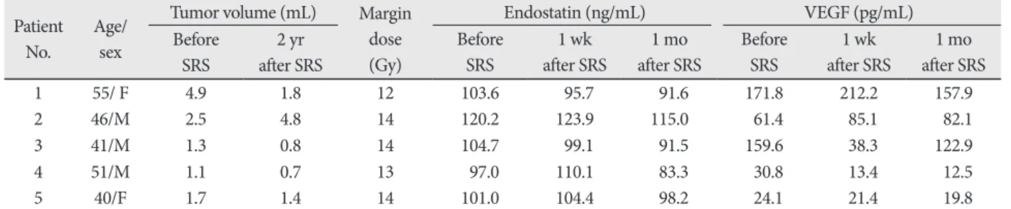
Mean plasma endostatin level changed to 106.6 ng/mL 1 week
after SRS and then dropped to 95.9 ng/mL after 1 month (Fig.
5). There was a trend toward decreased plasma endostatin concentrations 1 month after SRS compared to baseline lev- els (p=0.195). Plasma endostain concentration 1 month after SRS was still elevated over control levels, even though it was not significantly different from control levels (p=0.140).
Mean plasma VEGF level following SRS decreased to 74.1 pg/mL at 1 week and 79.0 pg/mL at 1 month (Fig. 6). Mean plasma VEGF level 1 month after SRS decreased compared to pretreatment level; however, there was no significant differ- ence from baseline levels (p=0.812). Plasma VEGF level 1 month after SRS was still significantly increased over controls (p=0.033).
Four patients with a decrease in tumor size experienced a constant decrease in endostatin and VEGF concentrations.
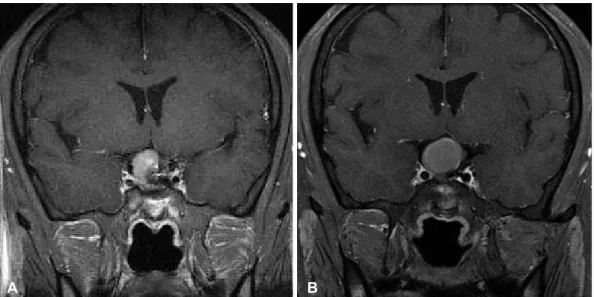
One patient with an enlarged pituitary adenoma after SRS had an elevated VEGF level and a slightly decreased end- ostatin level (Fig. 7). This patient underwent surgical resection for a recurrent tumor 2 years after SRS. No patient had radia- tion necrosis following SRS.
DISCUSSION
Angiogenesis is essential for the development and progres- sion of brain tumors. Angiogenesis is thought to be induced by an imbalance between pro- and anti-angiogenic factors. VEGF is the most potent angiogenic factor known so far and end- ostatin is a potent endogenous angiogenesis inhibitor [3,8,9].
Endostatin and VEGF have recently been implicated in the pathophysiology of pituitary adenoma. VEGF is a tyrosine ki- nase that plays an important role in angiogenesis and modu- lation of vascular permeability. VEGF-A binds with high specificity to VEGF receptor-1 and VEGF receptor-2 on vas- cular endothelial cells. These receptors modulate downstream signaling pathways affecting various cellular processes [10].
Increased plasma levels of VEGF are observed in patients with pituitary adenomas, compared to those in controls were de- tected [4]. Endostatin is a cleaved fragment of collagen XVIII, and inhibits migration of endothelial cells and induces endo- thelial cell apoptosis [6]. Plasma endostatin concentrations are significantly higher in patients with pituitary adenoma com- pared to those in controls [3].
Patients with pituitary adenoma usually undergo surgical resection; however, complete resection is often difficult. SRS is an effective and well-tolerated management option for recur- rent or residual pituitary adenoma [2]. Recurrent or residual tumors are related to significant morbidity over the lifetime of a patient. Monitoring tumor response with biomarkers may be useful to improve long-term treatment outcomes of patients who undergo SRS for pituitary adenoma.
Before 2
* 12
1 week SRS
1 month
Endostatin (ng/mL)
130 120 110 100 90 80
Fig. 5. Plasma concentrations of endostatin before and after ste- reotactic radiosurgery (SRS).
Before
6
1 week SRS
1 month
VEGF (pg/mL)
250 200 150 100 50 0
Fig. 6. Plasma concentrations of vascular endothelial growth fac- tor (VEGF) before and after stereotactic radiosurgery (SRS).
We evaluated plasma endostatin and VEGF levels in pa- tients with pituitary adenoma before/after SRS and in a con- trol group. We sought to understand angiogenic mechanisms regulating pituitary adenomas and the anti-angiogenic effect of SRS by studying changes in plasma endostain and VEGF levels in patients with pituitary adenoma undergoing SRS. A better understanding of the role of abnormal angiogenesis in patients with pituitary adenoma may lead to improved patient management and novel therapeutic options.
Plasma endostain and VEGF levels in patients with pituitary adenoma
Plasma endostain and VEGF levels were significantly higher in patients with pituitary adenoma at baseline in comparison to the controls in this study, which is consistent with those of previous studies [3,4]. Although several studies have found that abnormal local expression of endostain and VEGF plays a role in angiogenesis in pituitary adenomas, the cause of in- creased endostain and VEGF still remains unclear [1,3-6].
Feldman et al. [11,12] found that circulating endostatin and VEGF levels increase in patients with clear cell renal cancer and colorectal cancer with liver metastasis. They suggested that the correlation between circulating endostatin and VEGF may be associated with secretion of proteases that cleave endostatin from collagen XVII, both from tumor and endo- thelial cells. VEGF upregulates the release of proteases. Ele- vated plasma endostain levels may be a defense mechanism to protect the host from angiogenesis by VEGF [13]. Angio- genesis inhibitors are effectively suppress growth of experi- mental pituitary adenoma [14].
Plasma VEGF is detectable in the peripheral blood of nor-
mal healthy volunteers [9]. The origin and biologic role of VEGF in healthy subjects are unknown; however, this finding suggests a role for endothelial mitogens in the maintenance of physiologic endothelial integrity [8]. In healthy controls, a significant negative correlation between circulating endostatin and VEGF levels was found [3]. It results from balance mecha- nism between angiogenic and anti-angiogenic factors. Our results showed that VEGF and endostatin levels in the periph- eral blood in patients with pituitary adenoma increased at the same time. Gruszka et al. [3] suggested that simultaneous elevation of endostatin and VEGF may attenuate the pro-an- giogenic action of VEGF and could be responsible for the ra- ther weak neovascularization of pituitary adenomas. Pituitary adenomas are mostly benign and have lower vascular density than that of non-tumorous glands. Low microvascular density or inhibited angiogenesis may be a partial explanation for the relatively low growth potential observed in pituitary tumors [3,5,15].
Change in plasma endostatin and VEGF levels
The plasma endostatin and VEGF levels in the patients with pituitary adenoma dropped 1 month after SRS but were still elevated over control levels, confirming abnormal angiogene- sis in pituitary pathophysiology. This finding suggests that the natural history of a pituitary adenoma is related to disruption of endostatin and VEGF expression. Our result suggests that Gamma Knife–irradiated pituitary adenoma tissue has sig- nificantly less angiogenic activity than that of a previously un- treated pituitary adenoma. Our results support the anti-an- giogenic effects of SRS, which may be important for treating pituitary adenomas.Fig. 7. Enhanced T1-weighted (A) magnetic resonance image (MRI) obtained in a 46-year-old man with a pituitary adenoma. Stereotactic radiosurgery (SRS) was performed with a margin dose of 14 Gy to the 50% isodose line. The MRI obtained 2 years after SRS shows an in- crease of tumor volume from 2.5 mm3 to 4.8 mm3 (B). Plasma endostatin concentrations slightly decreased and vascular endothelial growth factor increased 1 month after SRS.
A B
Unlike four patients, one patient experienced increased endostain and VEGF levels after SRS. That patient experienced a tumor recurrence 2 years after SRS. Systemic expression of endostatin and VEGF may be related to the development, pro- gression in pituitary adenoma patients. O’Sullivan et al. [16]
reported that the 5- and 10-year actuarial rates of recurrence or growth of a residual adenoma after resection are 24.4% and 51.5%, respectively. Monitoring peripheral blood biomarkers in patients with pituitary adenoma may be valuable for those with highly angiogenic tumors, which tend to recur. Serial plasma endostatin and VEGF measurements may be a useful to monitor the treatment efficacy of SRS for angiogenic tumor cells in the future.
The main limitations of our study were the small number of patients and the short follow-up period. Sufficient plasma endostatin and VEGF data in a control group and abnormal levels in patients with pituitary adenoma are needed to use them as biomarkers and a diagnostic tool to monitor the tumor response after SRS. A controlled systematic prospective study with a longer follow-up is necessary to investigate the anti-an- giogenic effect of SRS in patients with pituitary adenoma.
In conclusions, plasma endostatin and VEGF levels in pa- tients with pituitary adenoma are elevated over controls at baseline and decrease 1 month after SRS. This was a prelimi- nary study on the potential association between plasma end- ostatin and VEGF levels and pituitary adenoma. Our results provide evidence for further work related to determining whether plasma endostatin and VEGF levels could be useful to monitor the tumor response after SRS.
Conflicts of Interest
The authors have no financial conflicts of interest.
REFERENCES
1. Sánchez-Ortiga R, Sánchez-Tejada L, Moreno-Perez O, Riesgo P, Niveiro M, Picó Alfonso AM. Over-expression of vascular endothelial growth factor in pituitary adenomas is associated with extrasellar growth and recurrence. Pituitary 2013;16:370-7.
2. Sheehan JP, Starke RM, Mathieu D, et al. Gamma Knife radiosurgery for the management of nonfunctioning pituitary adenomas: a multi- center study. J Neurosurg 2013;119:446-56.
3. Gruszka A, Kunert-Radek J, Pawlikowski M, Stepien H. Serum end- ostatin levels are elevated and correlate with serum vascular endothelial growth factor levels in patients with pituitary adenomas. Pituitary 2005;
8:163-8.
4. Komorowski J, Jankewicz J, Stepień H. Vascular endothelial growth fac- tor (VEGF), basic fibroblast growth factor (bFGF) and soluble interleu- kin-2 receptor (sIL-2R) concentrations in peripheral blood as markers of pituitary tumours. Cytobios 2000;101:151-9.
5. Turner HE, Harris AL, Melmed S, Wass JA. Angiogenesis in endocrine tumors. Endocr Rev 2003;24:600-32.
6. O’Reilly MS, Boehm T, Shing Y, et al. Endostatin: an endogenous in- hibitor of angiogenesis and tumor growth. Cell 1997;88:277-85.
7. Lee CC, Kano H, Yang HC, et al. Initial Gamma Knife radiosurgery for nonfunctioning pituitary adenomas. J Neurosurg 2014;120:647-54.
8. Kraft A, Weindel K, Ochs A, et al. Vascular endothelial growth factor in the sera and effusions of patients with malignant and nonmalignant dis- ease. Cancer 1999;85:178-87.
9. Vermeulen S, Young R, Li F, et al. A comparison of single fraction radio- surgery tumor control and toxicity in the treatment of basal and non- basal meningiomas. Stereotact Funct Neurosurg 1999;72 Suppl 1:60-6.
10. Nonoguchi N, Miyatake S, Fukumoto M, et al. The distribution of vas- cular endothelial growth factor-producing cells in clinical radiation necrosis of the brain: pathological consideration of their potential roles.
J Neurooncol 2011;105:423-31.
11. Feldman AL, Alexander HR Jr, Bartlett DL, et al. A prospective analysis of plasma endostatin levels in colorectal cancer patients with liver me- tastases. Ann Surg Oncol 2001;8:741-5.
12. Feldman AL, Tamarkin L, Paciotti GF, et al. Serum endostatin levels are elevated and correlate with serum vascular endothelial growth factor levels in patients with stage IV clear cell renal cancer. Clin Cancer Res 2000;6:4628-34.
13. Zucker S, Mirza H, Conner CE, et al. Vascular endothelial growth factor induces tissue factor and matrix metalloproteinase production in endo- thelial cells: conversion of prothrombin to thrombin results in progelati- nase A activation and cell proliferation. Int J Cancer 1998;75:780-6.
14. Páez Pereda M, Ledda MF, Goldberg V, et al. High levels of matrix me- talloproteinases regulate proliferation and hormone secretion in pitu- itary cells. J Clin Endocrinol Metab 2000;85:263-9.
15. Stepien HM, Kołomecki K, Pasieka Z, Komorowski J, Stepień T, Kuzdak K. Angiogenesis of endocrine gland tumours--new molecular targets in diagnostics and therapy. Eur J Endocrinol 2002;146:143-51.
16. O’Sullivan EP, Woods C, Glynn N, et al. The natural history of surgically treated but radiotherapy-naïve nonfunctioning pituitary adenomas.
Clin Endocrinol (Oxf) 2009;71:709-14.