Acute pancreatitis is a common disease with a wide variety of complications. Most cases of acute pancre- atitis are mild with rapid recovery and good prognosis.
However, in 20-30% of cases, the clinical course can be severe with life-threatening complications such as extensive pancreatic necrosis, pancreatic abscess formation, and vascular complications (1).
Pancreatic pseudocyst is a well-known complication of acute or chronic pancreatitis. And vascular compli- cations related to pancreatic pseudocyst include pseudoaneurysm formation, arterial hemorrhage, and venous thrombosis. Pseudocyst rupture into the portal vein is a very rare complication and only a few cases
are reported in literature (2-6). We report a case of fistula formation between the pseudocyst and the portal vein, diagnosed noninvasively by ultrasonogra- phy (US), computed tomography (CT), and magnetic resonance imaging (MRI).
A 50-year-old man presented with a one-week history of abdominal pain. The patient was a heavy alcoholic and had been previously diagnosed with alcoholic liver cirrhosis and chronic pancreatitis. At admission, hemodynamic status was stable and physical examination showed no substantial abnormal- ities. His admission laboratory findings were signifi- cant for elevated serum amylase 2,685 U/L (normal;
25-125 U/L) and lipase 1,269 U/L (normal; 7-25 U/L). Liver function tests were unremarkable.
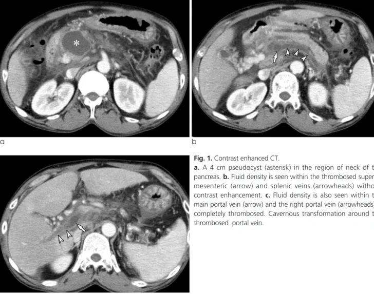
Abdominal CT showed mild peripancreatic inflam- matory change with mild dilatation of pancreatic duct and a 4 cm pseudocyst in the region of neck of the pancreas (Fig. 1a). Fluid density was seen within the main and left portal, superior mesenteric, and splenic veins without contrast enhancement, and the pseudo-
CASE REPORT INTRODUCTION
�Received; April 16, 2014�Revised; June 16, 2014
�Accepted; June 17, 2014
Corresponding author : Young Hwan Lee, M.D.
Department of Radiology, School of Medicine, Catholic University of Daegu, 3056-6, Daemyung-4 dong, Nam-gu, Daegu 705-718, Korea.
Tel. 82-53-650-4329, Fax. 82-53-650-4339, E-mail : [email protected] This is an Open Access article distributed under the terms of the Creative Commons Attribution Non-Commercial License (http://creativecommons.org/licenses/by- nc/3.0/) which permits unrestricted non-commercial use, distribution, and reproduction in any medium, provided the original work is properly cited.
Non-invasive MR Demonstration of the Fistula between Pancreatic Pseudocyst and Portal Vein:
A Case Report
Sung Min Kim, Young Hwan Lee, Ung Rae Kang
Department of Radiology, School of Medicine, Catholic University of Daegu, Daegu, Korea
Pancreatic pseudocyst rupture into the portal vein is a very rare complication and only three reported cases were con- firmed using MRI. We report the case of a 50-year-old man with fistula formation between the pseudocyst and the portal vein, confirmed noninvasively by MRI. T2-weighted MR images and magnetic resonance cholangiopancreatography showed fluid signal intensity within the portal, superior mesenteric, and splenic veins, and a direct communication between the pseudocyst and the portal vein.
Index words : Pancreatic pseudocyst∙Portal vein thrombosis∙Pancreatitis∙Magnetic resonance imaging∙MRCP Case Report
cyst appeared to be connected to the splenoportal confluence (Fig. 1b, c). The right portal vein was completely thrombosed and there was cavernous transformation around the portal vein (Fig. 1b). On ultrasonography, the main and left portal, superior mesenteric, and splenic veins were filled with the mixture of residual thrombus and fluid, without color blood flow. And communication between the pseudo- cyst and the splenoportal confluence appeared to be present.
MRI was performed to confirm a communication of the pseudocyst with the portal vein, and consisted of breath-hold axial T1-weighted spoiled gradient recalled echo (SPGR) images (TR/TE, 175/4; flip angle, 60 ), axial T2-weighted single-shot fast spin- echo (FSE) images (TR/TE, 1,058/151), respiratory- triggered heavily T2-weighted FSE 3-dimensional
magnetic resonance cholangiopancreatography (MRCP) (TR/TE, 3,750/676; slice thickness, 1.4 mm), and breath-hold fat-saturated contrast-enhanced axial T1-weighted 3-dimensional SPGR images (TR/TE, 4.5/2.2; flip angle, 12 ; slice thickness, 4 mm).
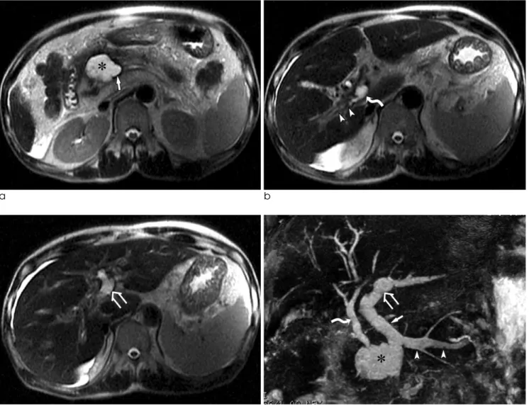
T2-weighted MR images showed fluid signal intensity within the main and left portal, superior mesenteric, and splenic veins, similar signal intensity to that of the pseudocyst (Fig. 2a-c). Coronal maximum-intensity projection (MIP) of MRCP confirmed a direct communication between the pseudocyst and the portal vein (Fig. 2d). On the other hand, the right portal vein thrombosis showed intermediate signal on T1-weighted imaging (Fig. 2e), intermediate to low signal intensity on T2-weighted imaging (Fig. 2b), and no contrast enhancement (Fig.
2f), consistent with the finding of complete thrombo-
a b
c
Fig. 1. Contrast enhanced CT.
a. A 4 cm pseudocyst (asterisk) in the region of neck of the pancreas. b. Fluid density is seen within the thrombosed superior mesenteric (arrow) and splenic veins (arrowheads) without contrast enhancement. c. Fluid density is also seen within the main portal vein (arrow) and the right portal vein (arrowheads) is completely thrombosed. Cavernous transformation around the thrombosed portal vein.
sis. The diagnosis of rupture of pseudocyst into the portal vein was made with the findings of MRI, and no further invasive diagnostic modality was required.
Since the patient’s symptom was mild, he received conservative treatment. After 6 months, a follow-up abdominal CT showed decrease in size of the pseudo- cyst and obliteration of the portal vein.
Pancreatic pseudocyst is a common complication of acute or chronic pancreatitis occurring in 20% to 40%
of cases, and may be associated with a spectrum of venous and arterial vascular complications including thrombosis of the portosplenic venous system.
Rupture of pancreatic pseudocyst into the portal vein is a very rare complication with only a few cases described in the literature (2-6). Most reported cased were diagnosed by invasive methods, such as endoscopic retrograde cholangiopancreatography (ERCP) or surgery. Only three reported cases were confirmed using MRI (3, 4, 6).
MRCP is a very useful imaging modality for evalua- tion of fluid-filled structures through the use of heavily T2-weighted sequences and has an essential role in the DISCUSSION
Fig. 2. a-c. Single-shot FSE T2-weighted images show a communication of the pseudocyst (asterisk) with the splenoportal confluence (arrow). Fluid signal intensity is seen within the main portal (curved arrow) and left portal (open arrow) veins. Right portal vein thrombosis (arrowheads) shows intermediate to low signal intensity. d. Coronal MIP MRCP image clearly demonstrates a direct communication of the pseudocyst (asterisk) with the splenoportal confluence. Fluid signal intensity is seen within the main portal (arrow), left portal (open arrow), and splenic (arrowheads) veins. Bile duct (curved arrow).
c d
a b
noninvasive evaluation of pancreaticobiliary pathol- ogy. In our case, the presence of static fluid within the portal vein was clearly defined on T2-weighted imaging and MRCP, which showed quite different signal intensity from that of thrombosis. MRI also demonstrated a communication of the pseudocyst with the portal vein. CT and US are useful in diagnosis of portal vein thrombosis, and may provide a presump- tive diagnosis of communication between the pseudo- cyst and portal vein but are not confirmatory.
The precise pathophysiologic mechanism of fistula formation between the pseudocyst and the portal vein remains unclear. It has been postulated that portal vein thrombosis is the initial stage of disease (3, 5).
Thrombotic complications are thought to be related to stasis, spasm, and mass effect from surrounding inflamed pancreas, and direct intimal damage of the venous wall by the liberated enzymes (1, 7). The next stage is erosion of the wall of the vein and lysis of the fresh thrombus by pancreatic enzyme within the pseudocyst. Finally the thrombus is substituted by fluid-like material from the pseudocyst. In our case, the main and left portal veins were filled with the mixture of residual thrombus and fluid and right portal vein was remained completely thrombosed.
These findings may support the previous hypothesis of mechanism of fistula formation.
The management guideline has not been clearly established because of a few reported cases. The
treatment depends on the severity of patient’s symptom. In terms of mild symptoms, such as in our case, conservative treatment can be helpful. Patients with severe symptom have been treated surgically, but their postoperative mortality rate was high. Recently, improvement of the pancreatic ductal flow through endoscopic pancreatic duct stent insertion has been suggested as a possible treatment alternative (8).
We have made a diagnosis of a fistula formation between the pseudocyst and the portal vein using MRI noninvasively. MRI is thought be a powerful diagnostic modality for confirmation of fluid within the portal vein and detection of a fistula in this rare complica- tion. Additionally, there was concomitant existence of complete thrombosis in the right portal vein and the mixture of residual thrombus and fluid in the left and main portal veins in our case. It is worthwhile to explain the pathomechanism of fistula formation between the pseudocyst and the portal vein.
References
1. Mortele KJ, Mergo PJ, Taylor HM, et al. Peripancreatic vascular abnormalities complicating acute pancreatitis: contrast-enhanced helical CT findings. Eur J Radiol 2004;52:67-72
2. Procacci C, Mansueto GC, Graziani R, et al. Spontaneous rupture of a pancreatic pseudocyst into the portal vein.
Cardiovasc Intervent Radiol 1995;18:399-402
3. Riddell A, Jhaveri K, Haider M. Pseudocyst rupture into the portal vein diagnosed with MRI. Br J Radiol 2005;78:265-268 4. Yoon SE, Lee YH, Yoon KH, Choi CS, Kim HC, Chae KM.
f e
Fig. 2. e. T1-weighted SPGR image. Right portal vein thrombosis (arrowheads) shows intermediate signal intensity. Low signal intensity is seen within the main portal vein (curved arrow). f. Fat-saturated contrast-enhanced axial T1-weighted SPGR image. No contrast enhancement is seen in the right (arrowheads) and main portal vein (curved arrow).
Spontaneous pancreatic pseudocyst-portal vein fistula present- ing with pancreatic ascites: strength of MR cholangiopancreatog- raphy. Br J Radiol 2008;81:e13-16
5. Dawson BC, Kasa D, Mazer MA. Pancreatic pseudocyst rupture into the portal vein. South Med J 2009;102:728-732
6. Dayala M, Sharmaa R, Madhusudhana KS, Gargb PK. MRI diagnosis of rupture of pancreatic pseudocyst into portal vein:
case report and review of literature. Ann Gastroenterol
2014;27:173-176
7. Belli M, Jennings CM, Nakielny RA. Splenic and portal venous thrombosis: a vascular complication of pancreatic disease demonstrated on computed tomography. Clin Radiol 1990;41:13-16
8. Noh R, Kim HJ. A pancreatic pseudocyst-portal vein fistula closed by endoscopic pancreatic stent insertion. Gastrointest Endosc 2010;72:1103-1105
통신저자 : 이영환, (705-120) 대구시 남구 대명4동 3056-6, 대구가톨릭대학교 의과대학 영상의학교실 Tel. (053) 650-4329 Fax. (053) 650-4339 E-mail: [email protected]
자기공명영상을 이용하여 비침습적으로 진단된 췌장 가성낭종과 간문맥 사이의 누공: 증례 보고
대구가톨릭대학교 의과대학 영상의학교실 김성민∙이영환∙강웅래
간문맥으로 파열된 췌장 가성낭종은 매우 드문 합병증이며, 자기공명영상으로 확진된 경우는 3예만 보고되어 있다.
저자들은 50세 남자 환자에서 가성낭종과 간문맥 사이에 형성된 누공을 자기공명영상을 이용하여 비침습적으로 확진 한 증례를 보고한다. T2강조영상 및 자기공명 담체관조영술에서 간문맥, 상장간막정맥, 그리고 비장정맥 내에 액체신 호가 보였으며, 가성낭종과 간문맥이 직접 연결된 소견을 보였다.
대한자기공명의과학회지 18:171-175(2014)