http://dx.doi.org/10.5624/isd.2015.45.4.233
Introduction
Maxillary third molars are generally less difficult to ex- tract than mandibular third molars.1 The surgical removal of maxillary third molars is usually associated with low rates of complications and low morbidity.2 However, sur- gical extraction of the upper third molar can cause serious complications, such as displacement into adjacent ana- tomic spaces.3,4
The most frequent complications are fracture of the
maxillary tuberosity, root fracture, and perforation of the maxillary sinus.5,6 The level of the occlusal plane, contact with the second molar, and the relationship of the molar to the maxillary sinus have been found to be significant predictors of surgical difficulty.1 An occlusal surface of the third molar deeper than the cementoenamel junction of the second molar can make maxillary third molar ex- traction more difficult.7,8 Contact of the third molar with the second molar root has been associated with difficulty in the surgical removal of impacted third molars.1 The possibility of accidental displacement was found to in- crease when the third molar was deeply impacted and when it was close to or in the maxillary sinus.9
Displacement of maxillary third molars has been as- sociated with an inadequate clinical and radiographic examination, improper surgical technique, insufficient
Assessment of maxillary third molars with panoramic radiography and cone-beam computed tomography
Yun-Hoa Jung1, Bong-Hae Cho1,*
1Department of Oral and Maxillofacial Radiology, School of Dentistry, Pusan National University, Yangsan, Korea
AbstrAct
Purpose: This study investigated maxillary third molars and their relation to the maxillary sinus using panoramic radiography and cone-beam computed tomography(CBCT)
Materials and Methods: A total of 395 maxillary third molars in 234 patients were examined using panoramic radio- graphs and CBCT images. We examined the eruption level of the maxillary third molars, the available retromolar space, the angulation, the relationship to the second molars, the number of roots, and the relationship between the roots and the sinus.
results: Females had a higher frequency of maxillary third molars with occlusal planes apical to the cervical line of the second molar(Level C) than males. All third molars with insufficient retromolar space were Level C. The most common angulation was vertical, followed by buccoangular. Almost all of the Level C molars were in contact with the roots of the second molar. Erupted teeth most commonly had three roots, and completely impacted teeth most commonly had one root. The superimposition of one third of the root and the sinus floor was most commonly associated with the sinus floor being located on the buccal side of the root.
conclusion: Eruption levels were differently distributed according to gender. A statistically significant association was found between the eruption level and the available retromolar space. When panoramic radiographs showed a superimposition of the roots and the sinus floor, expansion of the sinus to the buccal side of the root was generally observed in CBCT images.(Imaging Sci Dent 2015; 45: 233-40)
Key words: Maxilla; Molar, Third; Maxillary sinus; Cone-Beam Computed Tomography; Radiography, Panoramic
Copyright ⓒ 2015 by Korean Academy of Oral and Maxillofacial Radiology
This is an Open Access article distributed under the terms of the Creative Commons Attribution Non-Commercial License(http://creativecommons.org/licenses/by-nc/3.0) which permits unrestricted non-commercial use, distribution, and reproduction in any medium, provided the original work is properly cited.
Imaging Science in Dentistry·pISSN 2233-7822 eISSN 2233-7830
*This work was supported by a two-year research grant from Pusan National Univer- sity.
Received July 30, 2015; Revised August 21, 2015; Accepted September 3, 2015
*Correspondence to : Prof. Bong-Hae Cho
Department of Oral and Maxillofacial Radiology, Pusan National University Dental Hospital, Beomeo-ri, Mulgeum-eup, Yangsan-si, Gyeongsangnam-do 50612, Korea Tel) 82-55-360-5261, Fax) 82-55-360-5029, E-mail) [email protected]
visibility, and excessive force during extraction.2,3,10 Al- though iatrogenic tooth displacement was found to be rare during the extraction of maxillary third molars, max- illary third molars have been accidentally displaced into adjacent anatomic spaces.2 Depending on the direction of force application, the maxillary third molar can be dis- placed superiorly to the maxillary sinus,11 posteriorly to the infratemporal fossa,3,4,10 posterolaterally to the buccal space,12 posterosuperiorly to the pterygopalatine space,13 or posteromedially into the lateral pharyngeal space.14 Inadequate bone height buccal and distal to the molar has been associated with a higher risk of displacement into the buccal space.12
Insufficient clinical and radiographic examination is an important factor that could lead to accidental tooth dis- placement.2,7 Accurate radiographic localization of the tooth is a prerequisite for both an initial extraction and the extraction of a displaced tooth.15 The preoperative assess- ment should include a detailed morphologic analysis of the third molar and its relationship to adjacent structures and surrounding tissues.16 The identification of predictive variables associated with adverse events may be helpful in reducing complications.17
Panoramic radiography is the standard preoperative imaging modality.8,18 However, it is difficult to precisely assess the angulation of the third molar and the position of the root relative to the maxillary sinus on panoramic radiographs. Computed tomography or cone-beam com- puted tomography(CBCT) is capable of providing more exact information regarding the position of the maxillary third molars.
This study was performed to investigate maxillary third molars and their relation to the maxillary sinus using pan- oramic radiography and CBCT.
Materials and Methods
The subjects of this retrospective study were randomly selected from patients who visited Pusan National Uni- versity Dental Hospital for extraction of the upper third molars and who underwent panoramic radiography and CBCT imaging in 2013. The study sample consisted of 395 maxillary third molars from 234 patients aged 20 years and older, of which 196 were on the right and 199 were on the left. The patients comprised 120 males and 114 fe- males with a mean age of 28.5 years(range, 20-67 years).
Patients with pathology in the maxillary posterior teeth or who were missing the second molar were excluded from the study.
All panoramic radiographs were taken with a Proline XC machine(Planmeca Co., Helsinki, Finland). CBCT scans were acquired with a PaX-Zenith 3D scanner(Vatech Co., Hwasung, Korea). The scanning parameters were set at 110kVp, 24 seconds, 5.7mA, a voxel size of 0.2 mm, and field of view of 16 cm×14cm. The CBCT volumet- ric data were reconstructed using the Ez3D Plus Profes- sional CBCT software(Vatech Co., Hwasung, Korea).
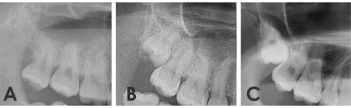
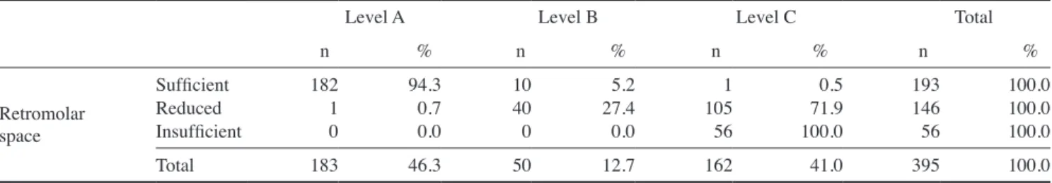
We examined the eruption level of the maxillary third molars, the available retromolar space, their relationship to the adjacent second molar, and their relationship to the maxillary sinus on panoramic radiographs. The eruption level of the maxillary third molars was assessed according to their relationship to the occlusal plane of the adjacent second molar using the Pell and Gregory classification system:19 level A; the occlusal plane of the third molar is at the same level as the adjacent tooth; level B, the occlu- sal plane of the third molar is between the occlusal plane and the cervical line of the adjacent tooth; and level C, the occlusal plane of the third molar is apical to the cer- vical line of the adjacent tooth(Fig. 1). The available ret- romolar space was measured as the distance between the distal surface of the second molar crown and the cortex of the maxillary tuberosity, and the space was categorized as sufficient(space greater than or equal to the mesiodistal length of the third molar), reduced(space greater than half and less than the entire mesiodistal length of the third mo- lar), and insufficient(space less than half the mesiodistal length of the third molar)(Fig. 2). The relationship to the adjacent second molar was classified as no contact, con- tact with the crown, and contact with the root. The rela- tionship of the maxillary third molar to the sinus on pan- oramic radiographs was classified into five types: class 1, the sinus floor is above the roots; class 2, the sinus floor touches the root tips; class 3, the sinus floor is superim-
A B C
Fig. 1. Eruption levels of maxillary third molars on panoramic radiographs. A. Level A, the occlusal plane of the third molar is at the same level as the adjacent second molar. B. Level B, the occlusal plane of the third molar is between the occlusal plane and the cervical line of the adjacent tooth; C. Level C, the occlusal plane of the third molar is apical to the cervical line of the adjacent tooth.
posed on up to one third of the root; class 4, the sinus floor is superimposed on up to two thirds of the root; and class 5, the sinus floor extends up to the tooth cervix(Fig.
3).On CBCT images, the angulation of the maxillary third molars, the number of roots, and the horizontal relation- ship between the roots of the third molars and the sinus were investigated. The angulation of the maxillary third molar with respect to the long axis of the second molar was classified as vertical, buccoangular, linguoangular,
buccolingual, mesioangular, distoangular, horizontal, and inverted using a modified version of Winter’s classifica- tion(Fig. 4). The number of roots was classified into one fused root, two roots, three roots, and four roots. Class 3, class 4, and class 5 relationships on panoramic radio- graphs were subclassified into five types according to the horizontal relationship between the roots of the third mo- lars and the sinus on CBCT images: type B, the lowest point of the maxillary sinus floor is located on the buccal side of the root; type C, the root projects into the sinus;
type P, the lowest point of the sinus floor is located on the palatal side of the root; type M, the lowest point of the maxillary sinus floor is located on the mesial side of the maxillary third molar; and type D, the lowest point of the maxillary sinus floor is located on the distal side of the maxillary third molar(Fig. 5).
The statistical analysis consisted of descriptive cross- tabulations showing frequency distributions among the selected categorical variables. Data were analyzed using the chi-square test and Fisher’s exact test. P-values less than 0.05 were considered to indicate statistically signifi- cant differences. The statistical analyses were performed using SPSS version 21.0(IBM Corp., Armonk, NY, USA).
A B C
Fig. 2. Classification of the available retromolar space on panora
mic radiographs. A. Sufficient, a space greater than or equal to the mesiodistal length of the third molar; B. Reduced, a space greater than half and less than the mesiodistal length of the third molar;
C. Insufficient, a space less than half the mesiodistal length of the third molar.
Fig. 3. The relationship between the root of the maxillary third molars and the sinus on panoramic radiographs. A. Class 1, the sinus floor is above the roots. B. Class 2, the sinus floor touches the root tips. C. Class 3, the sinus floor is superimposed on up to one third the root. D.
Class 4, the sinus floor is superimposed on up to two thirds of the root. E. Class 5, the sinus floor extends up to the tooth cervix.
A B C D E
Fig. 4. Classification of the angulation of the maxillary third molars on conebeam computed tomography images. V, vertical; B, buccoan- gular; L, linguoangular; BL, buccolingual; M, mesioangular; D, distoangular; H, horizontal; I, inverted.
V B L BL
M D H I
results
The eruption levels of the 395 maxillary third molars included in this study were classified as follows: level A for 183(46.3%), level B for 50(12.7%), and level C for 162(41.0%). Level C was more common in females
(49.0%) than in males(33.5%). The distribution of the eruption levels was significantly different between gen- ders(P<0.01)(Table 1).
A total of 182 maxillary third molars with sufficient retromolar space were level A(94.3%), 105 third molars with reduced space were level C(71.9%), and all third
Fig. 5. The horizontal relationship between the root of the maxillary third molars and the sinus on cone-beam computed tomography im- ages. A. Type B, the lowest point of the sinus floor is located on the buccal side of the root. B. Type C, the root is projects into the sinus.
C. Type P, the lowest point of the sinus is located on the palatal side of the root. D. Type M, the lowest point of the sinus is located on the mesial side of the third molar. E. Type D, the lowest point of the sinus is located on the distal side.
A B C D E
Table 1. Comparison of the eruption levels of the maxillary third molars between genders
Male Female Total
n % n % n %
Eruption level
Level A Level B Level C
110 25 68
54.212.3 33.5
73 25 94
38.013.0 49.0
183 50 162
46.312.7 41.0
Total 203 100.0 192 100.0 395 100.0
Level A: the occlusal plane of the third molar is at the same level as the adjacent second molar, Level B: the occlusal plane of the third molar is between the occlusal plane and the cervical line of the adjacent tooth, Level C: the occlusal plane of the third molar is apical to the cervical line of the adjacent tooth.
P<0.01, chi-square test.
Table 2. Eruption levels of the maxillary third molars according to the extent of retromolar space
Level A Level B Level C Total
n % n % n % n %
Retromolar space
Sufficient Reduced Insufficient
1821 0
94.30.7 0.0
1040 0
27.45.2 0.0
1051 56
71.90.5 100.0
193146 56
100.0 100.0 100.0
Total 183 46.3 50 12.7 162 41.0 395 100.0
P<0.01, chi-square test.
Table 3. Angulation of the maxillary third molars according to the extent of retromolar space Angulation of the
maxillary third molar
V B L BL M D H I Total
n % n % n % n % n % n % n % n % n %
Retromolar space
Sufficient Reduced Insufficient
16459 10
85.040.4 17.9
2543 4
13.029.5 7.1
06 6
0.04.1 10.7
03 9
0.02.1 16.1
273 22
18.51.6 39.3
05 3
0.03.4 5.4
12 1
0.5 1.4 1.8
01 1
0.0 0.7 1.8
193146 56
48.937.0 14.2
Total 233 59.0 72 18.2 12 3.0 12 3.0 52 13.2 8 2.0 4 1.0 2 0.5 395 100
V: vertical, B: buccoangular, L: linguoangular, BL: buccolingual, M: mesioangular, D: distoangular, H: horizontal, I: inverted.
molars with insufficient space were level C. The chi- square test showed statistically significant associations between the eruption level and the available retromolar
space(P<0.01)(Table 2).
The most common angulation of the maxillary third molars was vertical(59.0%), followed by buccoangular
Table 4. Eruption levels of the maxillary third molars according to their relationship to the second molar
Level A Level B Level C Total
n % n % n % n %
Relation to second molar
No contact Contact with crown Contact with root
1830 0
100.00.0 0.0
350 15
70.00.0 30.0
10 161
0.60.0 99.4
2181 176
55.20.3 44.6
Total 183 100.0 50 100.0 162 100.0 395 100.0
P<0.01, Fisher’s exact test.
Table 5. Relationship between the number of roots and the eruption level of the maxillary third molars
Level A Level B Level C Total
n % n % n % n %
Number of roots
One root Two roots Three roots Four roots
7723 821
42.112.6 44.80.5
2210 171
44.020.0 34.02.0
8336 421
51.222.2 25.90.6
18269 1413
46.117.5 35.70.8
Total 183 100.0 50 100.0 162 100.0 395 100.0
P<0.01, Fisher’s exact test.
Table 6. Relation of the maxillary third molars to the maxillary sinus according to eruption level
Level A Level B Level C Total
n % n % n % n %
Relation of the third molar to the maxillary sinus
Class 1 Class 2 Class 3 Class 4 Class 5
3263 7314 1
17.534.4 39.97.7 0.5
44 2218 2
8.08.0 44.036.0 4.0
174 3583 23
10.52.5 21.651.2 14.2
4084 130115 26
10.121.3 32.929.1 6.6
Total 183 100.0 50 100.0 162 100.0 395 100.0
Class 1: sinus floor is above the roots, Class 2: sinus floor touches the root tips, Class 3: sinus floor is superimposed on up to one third of the root, Class 4:
sinus floor is superimposed on up to two thirds of the root, Class 5: sinus floor extends up to the tooth cervix. P<0.01, chi-square test.
Table 7. The horizontal relationship between the maxillary third molars and the maxillary sinus according to the extension of the maxil- lary sinus on panoramic radiographs
Class 3 Class 4 Class 5 Total
n % n % n % n %
Horizontal relationship between the third molar and sinus
Type B Type C Type P Type M Type D
6357 45 1
48.543.8 3.13.8 0.8
4937 235 1
42.632.2 20.04.3 0.9
74 141 0
26.915.4 53.83.8 0.0
11998 1042 2
43.936.2 15.53.7 0.7
Total 130 100.0 115 100.0 26 100.0 271 100.0
Type B: the lowest point of the sinus floor is located on the buccal side of the root, Type C: the root projects into the sinus, Type P: the lowest point of the sinus is located on the palatal side of the root, Type M: the lowest point of the sinus is located on the mesial side of the third molar, Type D: the lowest point of the sinus is located on the distal side.
(18.2%) and mesioangular(13.2%). Vertical angulation (85.0%) was most frequent in molars with sufficient ret- romolar space, as well as in molars with reduced space (40.4%). The buccoangular orientation was the second most common(29.5%) in molars with reduced space, and mesioangular angulation(39.3%) was the most common in molars with insufficient space(Table 3).
All level A maxillary third molars were in contact with the crown of the second molars, while 30% of level B molars and 99.4% of level C molars were in contact with the root of the second molar(Table 4). Maxillary third molars with three roots were most frequent in level A mo- lars(44.8%), and the presence of one fused root was most frequent in level C molars(51.2%)(Table 5).
Class 3 was most common in level A and level B mo- lars, and class 4 was most common in level C molars (Table 6). When the sinus floor was superimposed on the root of the maxillary third molars, type B was most fre- quent in class 3 and class 4 molars, and type M was most frequent in class 5 molars(Table 7).
discussion
We investigated maxillary third molars and assessed their relationship to the maxillary sinus on panoramic radiographs and CBCT images. Careful radiographic lo- calization of the third molar is a prerequisite for surgical extraction in order to prevent complications.15
Some studies have found impaction of the third molar to be common in females.20-23 Others have reported no gender predilection for third molar impaction.24,25 The higher frequency reported in females has been explained by gender-based differences in growth patterns. Females usually stop growing when the third molars just begin to erupt, whereas in males, the jaws continue to grow while the third molars erupt, creating more space for third molar eruption.26 In this study, impacted third molars were more common in females than in males.
The etiology of third molar impaction has been investi- gated in many international studies. Several factors have been reported as possible causes of third molar impaction, including a lack of space distal to the permanent second molar, delayed third molar mineralization, and early physical maturation.26 Factors commonly associated with third molar impaction include a shortage of space avail- able for eruption, and third molar impaction was found to be more likely to occur when the retromolar space was in- adequate.27 These studies found that the retromolar space was closely related to the eruption level of maxillary third
molars.
Increased third molar angulation has also been found to be significantly linked to third molar impaction.27 Hash- emipour et al.20 evaluated the angulation of maxillary third molars using panoramic radiography, and the most common angulation was vertical, followed by distal. In our results, the most common angulation was vertical, followed by buccoangular. This could have been because we assessed the angulation of the maxillary third molars using CBCT images and added the categories of buccoan- gular, linguoangular, and buccolingual, which could not be assessed on panoramic radiographs. Buccoangular or buccolingual third molars could have been classified as distoangular on panoramic radiographs.
Greater proximity between the third and second molars has been associated with an additional risk of surgical difficulty.1,17 A close relationship between these teeth re- duces the space between the distal surface of the second molar and the mesial surface of the third molar, impeding access to the tip of the elevator.17 Our results showed that most level C molars were in contact with the root of the adjacent second molar.
The root number and morphology have been found to be even more important, because these factors were asso- ciated with more issues in surgical management, although this trend did not reach statistical significance.1 In our re- sults, one fused root was most frequent, followed by three roots, and four roots were rarely observed.
It is important to know the anatomical relationship bet- ween the maxillary sinus and the third molar in preoper- ative treatment planning for maxillary third molars.1 The more the sinus floor was projected onto the root on pan- oramic radiographs, the greater the relative probability of oroantral communication.18 If no superimposition of the root and sinus floor was visible on the panoramic ra- diographs, no oroantral perforation was to be expected.18 However, the relative probability of oroantral perforation increased in class 3 molars, and was significantly higher in class 4 and class 5 molars.18 In our results, the super- imposition of the sinus and the root of the maxillary third molars was observed in more than two thirds of panoram- ic radiographs. The depth of the impaction of the maxil- lary third molars was associated with a greater likelihood of oroantral perforation.28,29 In our results, eruption level C showed more superimposition between the sinus and root than levels A or B.
When the sinus floor is superimposed on the root on panoramic radiographs, it is necessary to obtain more in- formation about the relationship between the sinus and
the third molars in order to prevent sinus perforation.30 We used CBCT images to identify the horizontal relation- ship between the sinus and the third molars, and the sinus floor was most commonly found to be located on the buc- cal side of the root.
In order to minimize the risk of complications, surgical difficulty should not be underestimated. We should iden- tify variables predictive of a higher risk of complications during removal of the maxillary third molars and take action accordingly. Further research is needed to evaluate the correlation of predictive radiographic factors with the occurrence and types of complications.
In conclusion, the eruption level showed a different dis- tribution between males and females. A statistically sig- nificant association was found between the eruption level and the available retromolar space. Expansion of the si- nus to the buccal side of the root was the most frequently seen pattern on CBCT when the panoramic radiographs showed a superimposition of the roots and the sinus floor.
references
1. de Carvalho RW, de Araújo Filho RC, do Egito Vasconcelos BC. Assessment of factors associated with surgical difficulty during removal of impacted maxillary third molars. J Oral Maxillofac Surg 2013; 71: 839-45.
2. Patel M, Down K. Accidental displacement of impacted max- illary third molars. Br Dent J 1994; 177: 57-9.
3. Dimitrakopoulos I, Papadaki M. Displacement of a maxillary third molar into the infratemporal fossa: case report. Quintes- sence Int 2007; 38: 607-10.
4. Gómez-Oliveira G, Arribas-García I, Alvarez-Flores M, Gre- goire-Ferriol J, Martínez-Gimeno C. Delayed removal of a maxillary third molar from the infratemporal fossa. Med Oral Patol Oral Cir Bucal 2010; 15: e509-11.
5. Oberman M, Horowitz I, Ramon Y. Accidental displacement of impacted maxillary third molars. Int J Oral Maxillofac Surg 1986; 15: 756-8.
6. Sverzut CE, Trivellato AE, Lopes LM, Ferraz EP, Sverzut AT.
Accidental displacement of impacted maxillary third molar: a case report. Braz Dent J 2005; 16: 167-70.
7. Koerner KR. The removal of impacted third molars. Princi- ples and procedures. Dent Clin North Am 1994; 38: 255-78.
8. Bouquet A, Coudert JL, Bourgeois D, Mazoyer JF, Bossard D.
Contributions of reformatted computed tomography and pan- oramic radiography in the localization of third molars relative to the maxillary sinus. Oral Surg Oral Med Oral Pathol Oral Radiol Endod 2004; 98: 342-7.
9. Iwai T, Chikumaru H, Shibasaki M, Tohnai I. Safe method of extraction to prevent a deeply-impacted maxillary third molar being displaced into the maxillary sinus. Br J Oral Maxillofac Surg 2013; 51: e75-6.
10. Selvi F, Cakarer S, Keskin C, Ozyuvaci H. Delayed removal of a maxillary third molar accidentally displaced into the in-
fratemporal fossa. J Craniofac Surg 2011; 22: 1391-3.
11. Durmus E, Dolanmaz D, Kucukkolbsi H, Mutlu N. Accidental displacement of impacted maxillary and mandibular third mo- lars. Quintessence Int 2004; 35: 375-7.
12. Kocaelli H, Balcioglu HA, Erdem TL. Displacement of a maxillary third molar into the buccal space: anatomical im- plications apropos of a case. Int J Oral Maxillofac Surg 2011;
40: 650-3.
13. Ozer N, Uçem F, Saruhanoğlu A, Yilmaz S, Tanyeri H. Re- moval of a maxillary third molar displaced into pterygopala- tine fossa via intraoral approach. Case Rep Dent 2013; 2013:
392148.
14. Bobo M, Werther JR. Self-induced displacement of a maxil- lary molar into the lateral pharyngeal space. Int J Oral Maxil- lofac Surg 1998; 27: 38-9.
15. Lee D, Ishii S, Yakushiji N. Displacement of maxillary third molar into the lateral pharyngeal space. J Oral Maxillofac Surg 2013; 71: 1653-7.
16. Nakamori K, Tomihara K, Noguchi M. Clinical significance of computed tomography assessment for third molar surgery.
World J Radiol 2014; 6: 417-23.
17. Carvalho RW, Araújo-Filho RC, Vasconcelos BC. Adverse events during the removal of impacted maxillary third molars.
Int J Oral Maxillofac Surg 2014; 43: 1142-7.
18. Pourmand PP, Sigron GR, Mache B, Stadlinger B, Locher MC.
The most common complications after wisdom-tooth remov- al: part 2: a retrospective study of 1,562 cases in the maxilla.
Swiss Dent J 2014; 124: 1047-51.
19. Pell GJ, Gregory BT. Impacted mandibular third molars:
classification and modified techniques for removal. Dent Dig 1933; 9: 330-8.
20. Hashemipour MA, Tahmasbi-Arashlow M, Fahimi-Hanzaei F.
Incidence of impacted mandibular and maxillary third molars:
a radiographic study in a Southeast Iran population. Med Oral Patol Oral Cir Bucal 2013; 18: e140-5.
21. Quek SL, Tay CK, Tay KH, Toh SL, Lim KC. Pattern of third molar impaction in a Singapore Chinese population: a retrospective radiographic survey. Int J Oral Maxillofac Surg 2003; 32: 548-52.
22. Hugoson A, Kugelberg CF. The prevalence of third molars in a Swedish population. An epidemiological study. Community Dent Health 1988; 5: 121-38.
23. Murtomaa H, Turtola L, Ylipaavalniemi P, Rytömaa I. Status of the third molars in the 20- to 21-year-old Finnish universi- ty population. J Am Coll Health 1985; 34: 127-9.
24. Hattab FN, Rawashdeh MA, Fahmy MS. Impaction status of third molars in Jordanian students. Oral Surg Oral Med Oral Pathol Radiol Endod 1995; 79: 24-9
25. Haidar Z, Shalhoub SY. The incidence of impacted wisdom teeth in a Saudi community. Int J Oral Maxillofac Surg 1986;
15: 569-71.
26. Bishara SE. Impacted maxillary canines: a review. Am J Orth- od Dentofacial Orthop 1992; 101: 159-71.
27. Almpani K, Kolokitha OE. Role of third molars in orthodon- tics. World J Clin Cases 2015; 3: 132-40.
28. Lim AA, Wong CW, Allen JC Jr. Maxillary third molar: pat- terns of impaction and their relation to oroantral perforation. J Oral Maxillofac Surg 2012; 70: 1035-9.
29. Rothamel D, Wahl G, d’Hoedt B, Nentwig GH, Schwarz F, Becker J. Incidence and predictive factors for perforation of the maxillary antrum in operations to remove upper wisdom teeth: prospective multicentre study. Br J Oral Maxillofac
Surg 2007; 45: 387-91.
30. Jung YH, Cho BH. Assessment of the relationship between the maxillary molars and adjacent structures using cone beam computed tomography. Imaging Sci Dent 2012; 42: 219-24.