4)서 론
골섬유이형성 (Fibrous dysplasia of bone)은 Lichtenstein 과 Jaffe에 의해 최초로 명명되었으며 골이 섬유성 조직으로 대치되어 기형 및 병적 골절을 유발하는 질환이다[1,2]. 만 성적인 임상 경과를 보이며 어떤 부위의 골에도 발병될 수
접수일자: 2004년 11월 17일 통과일자: 2005년 3월 21일
책임저자: 우정택, 경희대학교 의과대학 내분비대사내과
있으나 일반적으로 두개골 및 장골 (long bone)에 호발하는 것으로 알려져 있다.
골섬유이형성은 그 임상상에 따라 단골성 (monostotic), 다골성 (polyostotic), 그리고 McCune-Albright 증후군의 3 가지 아형으로 분류할 수 있다[3]. 특히 McCune-Albright 증 후군(MAS)은 다골섬유이형성 (polyostotic fibrous dyspla- sia)과 함께 피부 색소침착 (cutaneous pigmentation), 사춘기 조발증(sexual precosity), 성장호르몬 분비과다, 갑상선 및 부신 기능항진 등의 다양한 내분비 기능장애를 그 특징으로
골섬유이형성 환자에서 Gsα단백 유전자 돌연변이에 관한 연구
경희대학교 의과대학 내분비대사내과1, 내분비연구소2, 경희대학교 의과대학 부속병원 병리과3
이상열1․우정택1,2․고관표1,2․오승준1,2․김성운1․김진우1,2․김영설1․박용구3
Mutational Analysis of Gsα Protein in Fibrous dysplasia of the Bone
Sang Youl Rhee1, Jeong-taek Woo1,2, Gwanpyo Koh1,2, Seungjoon Oh1,2, Sung Woon Kim1, Jin-Woo Kim1,2, Young Seol Kim1 and Yong-Koo Park3
Department of Endocrinology and Metabolism1, Research Institute of Endocrinology2, Department of Pathology3, School of Medicine, Kyung Hee University, Seoul, Korea
ABSTRACT
Background: Fibrous dysplasia of the bone (FD) is a benign fibrous bone lesion which usually involves the long bones of the extremities. FD may be asymptomatic, but often leads to bone deformity and pathological fracture. The disease is caused by a somatic mutation in the Gsα protein, which is responsible for intracellular signal transduction.
Methods: Mutations in the GNAS1 gene, which codes for Gsα protein, was investigated in 34 patients with monostotic and polyostotic FD and McCune-Albright syndrome. DNA was extracted from formalin-fixed, paraffin embedded bone tissues, and exons 8 and 9 of the GNAS1 gene amplified using a polymerase chain reaction (PCR). Subsequently, plasmid cloning and DNA sequencing analysis were performed.
Results: The PCR was successfully performed in 5 patients with monostotic FD. However, the sequencing analysis failed to identify any significant point mutations in exons 8 or 9 of GNAS1. Nevertheless, 3 point mutations were observed in the intron of the GNAS1 gene in 2 samples.
Conclusion: In addition to the previously known somatic mutations of the GNAS1 gene, this study suggests that fibrous dysplasia of the bone might be associated with another point mutations of the GNAS1 gene (J Kor Soc Endocrinol 20:142~147, 2005).
ꠏꠏꠏꠏꠏꠏꠏꠏꠏꠏꠏꠏꠏꠏꠏꠏꠏꠏꠏꠏꠏꠏꠏꠏꠏꠏꠏꠏꠏꠏꠏꠏꠏꠏꠏꠏꠏꠏꠏꠏꠏꠏꠏꠏꠏꠏꠏꠏꠏꠏꠏꠏꠏꠏꠏꠏꠏꠏꠏꠏꠏꠏꠏꠏꠏꠏꠏꠏꠏꠏꠏꠏꠏꠏꠏꠏꠏꠏꠏꠏꠏꠏꠏꠏꠏꠏꠏꠏꠏ
Key Words: Fibrous dysplasia, McCune-Albright syndrome, GNAS1, GTP-Binding Protein alpha Subunits, Gs
한다[4].
골섬유이형성의 원인은 Gsα단백을 부호화 (coding)하는 GNAS1 유전자에서 exon 8의 201번째 위치의 Arginine (Arg201)이 Cysteine (Cys201) 또는 Histidine (His201)으로 치 환되는 변이에 의한다고 알려져 있다[5, 6]. Gsα단백은 세 포의 신호 전달에 관여하는 heterotrimeric G단백의 일종으 로 세포내의 adenylyl cyclase의 활성을 증가시켜 세포내 신 호전달(signal trnasduction)에 작용하는 2차전령(second me- ssenger)인 cyclic adenosine monophosphate (cAMP)의 생 성을 증가시키는 역할을 한다. Gsα단백에서의 돌연변이는 G단백의 불활성에 관여하는 GTP가수분해효소 (GTPase)의 기능을 저하시켜 adenylyl cyclase의 활성을 지속화하여 cAMP에 반응하여 증식하는 세포들의 과기능을 유발하는 것으로 알려져 있다. Arg201이외에 exon 9의 227번째 Glycine(Gln227)의 돌연변이 역시 Gsα단백의 GTPase 활성 저 하를 유발하는 것으로 알려져 있으며[7,8] 이와 같은 Gsα단 백의 돌연변이는 골 외에도 뇌하수체, 갑상선 및 부신 등의 여러 다양한 내분비 질환을 유발하는 원인이 된다.
골섬유이형성에 있어 이러한 Gsα단백의 돌연변이는 cAMP의 증가에 의한 조골전구세포 (Osteoblastic precursor cell)의 분화과정에 장애가 발생하여 세포의 퇴축 (cellular retraction)및 비정상적인 골세포의 침착(cellular deposition) 을 유발하는 것으로 알려져 있다[9,10]. 특히 MAS에서 골 섬유이형성 및 내분비 증상은 개체의 조직 분화시 일어난 Gsα단백 유전자의 체성 돌연변이가 섞임증 (mosaicism)과 관련되어 다양한 임상증상을 보이는 것으로 설명되고 있다 [11,12]. 하지만 단골 및 다골섬유이형성 환자들의 경우 Gs α단백 유전자의 돌연변이를 확인한 최근의 여러 연구[9,13, 14]에도 불구하고 병변이 국소적이고 골 이외 다른 장기의 임상증상을 보이는 경우가 드물며, 비교적 소수의 환자들을 대상으로 연구가 진행되었기 때문에 아직 논란의 여지가 있 다[15].
본 연구는 우리나라 환자들에 있어서 골섬유이형성의 병 태생리와 그 특징에 대한 유전학적 의의를 확인하기 위해 진행되었다. 정형외과적 수술을 받은 환자들 중 조직생검을 통해서 골섬유이형성으로 진단된 환자들을 대상으로 시행 하였으며 다골성 및 단골성, 그리고 MAS환자를 망라하였 다. 이들의 조직생검 표본에서 DNA를 추출하여 GNAS1 유전자에서의 체성 돌연변이 유무를 확인하고자 하였다.
대상 및 방법 1. 대상
1991년부터 2001년까지 경희대학교 의과대학 부속병원 에서 수술을 받은 환자들 중 조직생검을 통해서 골섬유이형 성증으로 진단된 환자를 대상으로 하였다. 대상 환자들의
나이와 성, 그리고 이완된 골의 종류에 대해서는 기록을 참 조하여 후향적으로 조사하였다.
2. 표본에서의 파라핀의 제거
수술 후 골의 생검표본은 포르말린으로 고정 후 파라핀 처리된 상태로 기존의 연구들에서 적용된 실험 방법을 참조 하여 파라핀을 제거하였다[14,16]. 2 mL 용량의 실험튜브에 10 μm 두께의 절편으로 나뉘어진 생검표본 10개를 넣고 크 실렌 1 mL을 넣은 뒤 55℃에서 15분간 유지하였다. 이후 Centrifuge 5402 (Brinkmann Co., NY, USA)를 이용하여 10000 g에서 15분간 원심분리 후 상청액을 제거하였다. 상 청액을 제거한 표본에 크실렌과 동량의 100% 에탄올을 가 하여 2회 세척 후 55℃에서 10분간 건조하였다. 위와 같은 과정을 각 표본당 3회 반복하여 조직 내에서 파라핀을 제거 하였다.
3. 조직에서의 DNA의 추출
파라핀을 제거한 조직에서의 DNA의 추출은 QIAmpⓇ DNA mini kit (QIAGEN Inc., CA, USA)을 사용하였다.
각 조직샘플이 들어있는 실험튜브에 360 μL의 ATL lysis buffer와 40 μL의 proteinase K를 가한 뒤 보텍스 교반기를 사용하여 혼합하였다. 혼합한 튜브를 56℃에서 매 1시간마 다 교반기로 혼합하며 조직이 완전히 융해될 때까지 유지하 였다. 이를 원심분리 및 상청액 제거를 통해 불순물을 제거 한 뒤 QiagenⓇ DNA extraction column을 이용해 DNA를 추 출하였다.
4. 중합효소연쇄반응 (Polymerase chain react- ion, PCR)
추출한 DNA에서 GNAS1유전자의 정방향 및 역방향으 로 exon 8의 Arg201과 exon 9의 Gln227을 포함하는 각각의 시동체 (Primer)를 사용하여 중합효소 연쇄반응 (PCR)을 시 행하였다 (Table 1). 중합효소연쇄반응은 thermal cycler (Gene and PCR system 9600, Perkin-Elmer, MA, USA)를 사용하였고 시행조건은 각 1회의 주기마다 94℃에서 30초 간 변성 (Denaturation), 50℃에서 1분간 불림 (Annealing), 72℃에서 1분간 확대 (Extension)를 반복하였으며 최종 주 기에서 72℃, 15분의 조건으로 최종확대 (Final extension) 를 시행하였다.
5. 플라스미드 TOPOⓇ cloning
PCR을 시행하여 얻은 DNA 표본을 TOPO TA cloningⓇ Kit (Invitrogen corporation, CA, USA)를 이용하여 유전자 복제 (cloning)를 시행하였다. PCR을 완료한 DNA 표본에 PCR-TOPOⓇvector, 증류수, 염(Salt solution)을 가하여 TOPOⓇ cloning reaction을 유발하였고 이를 냉온에서
E.coli와 혼합하였다. 이 혼합물을 42℃에서 30초간 열충격 (heat shock)을 유발하여 PCR로 증폭된 DNA가 포함된 플 라스미드를 E.coli 내에 삽입하였다. 이를 ampicillin이 포함 된 배지에서 배양하여 플라스미드가 삽입된 E.coli만을 선 택적으로 증식시켰다.
6. DNA 염기서열의 분석
선택한 E.coli에서 플라스미드 DNA를 분리하여 3100 genetic analyzer (Applied Biosystems, CA, USA)를 이용 하여 복제한 DNA의 염기서열 분석을 시행하였으며 이를 Vector NTI suite 7 (Invitrogen corporation, CA, USA)을 이용하여 정상 염기서열과 비교하였다.
결 과 1. 환자들의 특성
1991년부터 2001년까지 경희대학교 의과대학 부속병원 에서 수술을 받은 환자들 중 조직생검을 통해서 단골 혹은 다골섬유이형성으로 진단된 환자는 총 40명이었다. 이 환자 들 중 조직표본을 확보할 수 있었던 34명의 환자들을 대상 으로 하였다. 34명의 환자 중 26명의 환자가 단골섬유이형 성으로 진단되었고 7명은 다골섬유이형성, 그리고 1명은 MAS로 진단되었다. 이들의 평균연령은 68.53 ± 2.0세였으 며 남자는 18명, 여자는 16명이었다. 이환된 골은 대부분 장 골(long bone)로 대퇴골 및 경골이 각기 19예와 8예로 다수
를 차지하였고 그 외 상완골, 요골의 순이었다 (Table 2).
2. DNA 돌연변이의 확인
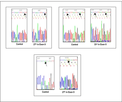
DNA의 추출을 시도한 34개의 생검 표본중 단지 5개의 표본에게서만 중합효소 연쇄반응이 가능한 정도의 적절한 DNA를 얻을 수 있었으며 이는 모두 단골섬유이형성 환자 들의 표본이었다. 이 표본들의 DNA를 유전자 복제를 이용 하여 GNAS1 유전자의 exon 8의 Arg201과 exon 9의 Gln227 부위의 염기서열을 확인하였다. 염기서열의 확인결과 5개의 표본 모두에서 exon의 염기서열 변이는 관찰되지 않았다.
하지만 2명의 환자 표본에서 intron부위의 염기서열 변화를 확인하였다 (Fig. 1).
고 찰
저자들은 10여 년에 걸친 표본 수집을 통해 다골 및 단 골섬유이형성 그리고 MAS을 망라하는 다수의 표본을 확보 하였다. 이를 통해 우리나라 환자들에게 있어서 골섬유이형 성에 이환된 골의 빈도 및 종류 등의 기초적 정보에서부터 현재 알려진 GNAS1 유전자에서 Arg201의 체성 돌연변이와 동시에 다른 내분비 질환들에 있어서 Gsα단백의 GTPase 활성 저하에 관여하는 것으로 알려진 바 있는 exon 9의 Gln227의 변이에 대해서도 살펴보았다. 그러나 다수의 표본 을 확보하였음에도 불구하고 표본에서 파라핀을 제거하고 DNA를 추출하는데 있어 단지 5개의 표본에서만 DNA를 확 보할 수 있었다. 낮은 DNA의 수율과 더불어 5개의 표본이 모두 단골섬유이형성 환자의 표본으로, 이는 본 연구에서 의도했던 여러 골섬유이형성 아형간의 비교를 불가능하게 하는 큰 제한점이 되었다.
Xylene과 QIAmpⓇ DNA mini kit을 이용한 DNA 추출 은 골수내 림프양세포 (lymphoid cell)를 비롯한 각종 파라 핀 처리 표본에서의 DNA 추출시 일반적으로 이용되어온 방법이다[16,17]. 하지만 파라핀 처리된 조직에서 사용되는 기존의 방법은 비교적 낮은 DNA 수율을 보여 충분한 DNA 의 획득이 곤란한 문제점이 있었다[18]. 금번 실험에서 DNA 수율이 크게 낮은 이유는 충분한 DNA를 얻기 어려 운 파라핀 처리된 조직의 특성에도 그 원인이 있겠지만 골 수에 비해 상대적으로 표본 내 단위면적당 세포의 양이 적 Table 1. Oligonucleotide Primers Used for PCR Amplification of Exon 8 and Exon
9 in the GNAS1 Gene
Exon in GNAS1 gene Strand Oligonucleotide sequence Exon 8
Exon 9
Sense Antisense
Sense Antisense
5-CTCTGAGCCCTCTTTCCCAAACTAC-3 5-GGTTATTCCAGAGGGACTGGGGTG-3 5-GGTTTCTTGACATTCACCCC-3 5-CGAAGATGATGGCAGTCACA-3
Table 2. Sites of Involved Bone in Patients with Fibrous Dysplasia
Involved Bone Cases Percentage Femur
Tibia Humerus
Radius Ulnar Metacarpal bone
Ilium Rib Talus
19 8 3 2 1 1 1 1 1
51.4%
21.6%
8.1%
5.4%
2.7%
2.7%
2.7%
2.7%
2.7%
Total 37 100%
은 골 조직의 특성과도 관련이 있을 것으로 판단된다. 따라 서 일차적으로 파라핀 처리된 조직에 있어 좀 더 많은 양의 절편을 사용하여 DNA 수율을 높일 수 있을 것으로 기대한 다. 또한 DNA 추출 방법 자체에 대한 개선의 여지가 있어 Sato 등[19], Shi 등[20]을 비롯한 다수의 연구자들이 파라 핀 처리된 표본에 열 (heat)처리를 통하여 높은 DNA 수율 을 보고하고 있는 실정으로 향후 유사한 실험에 있어 개선 된 실험방법의 도입이 필요할 것으로 보인다.
본 연구에서 단골섬유이형성 환자들의 Gsα단백 유전자 염기서열 분석결과 이들의 DNA에서는 기존에 알려진 exon 8의 Arg201, exon 9의 Gln227을 포함한 exon에서의 염기서 열의 변화를 관찰할 수 없었다. 오히려 기존에 알려지지 않 은 intron의 염기서열 변화를 환자 2명의 표본에서 확인할 수 있었는데 intron의 염기서열 변화는 단백질 구조의 변화 와는 관계가 없기 때문에 관련성이 없을 가능성이 높다. 다 만 기존의 연구 역시 10명 이내의 비교적 작은 수의 표본을 대상으로 하고 있으며 Candeliere 등[21]은 exon 8의 Arg201 위치에 Cys201 혹은 His201이 아닌 Serine으로의 변이를 보 고한 바 있어 기존에 알려진 exon 8의 Arg201 외에 다양한 돌연변이와 관련되어 있을 가능성을 배제하지 못한다. 또한 본 연구에서는 GNAS1 유전자의 다른 exon의 염기서열 분
석을 시행하지 않았기 때문에 다른 exon에서의 돌연변이의 가능성을 완전히 배제할 수 없다.
최근 골섬유이형성 환자에서 섬유모세포 (fibroblast)의 증 식에 관계된 Platelet-derived growth factor (PDGF-B)의 β -chain이 증가되어 있음이 발견된 바 있다[22]. PDGF-B의 발현은 사춘기, 임신 그리고 경구피임약 등의 체내 성호르 몬의 증가와도 관련성이 있으며 이러한 성장인자의 증가가 골섬유이형성의 발생에 중요한 역할을 하는 것으로 추정된 다[23]. 또한 Fraser 등[24]은 3명의 골섬유이형성 환자에서 병변에서 PTHrP (PTH-related protein)가 과발현됨을 보고 하여 골섬유이형성의 발생에 있어 Gsα단백의 돌연변이 이 외의 다른 원인과의 관련성을 시사하였다.
여러 제한점에도 불구하고 본 연구는 골섬유이형성 환자 의 병변에서 유전학적 검사를 통해 우리나라 환자에서 골섬 유이형성의 정확한 병태생리에 대해 최초로 연구하는 데 그 의의가 있다. 또한 본 연구는 앞으로 조직학적인 진단 뿐 아 니라 유전학적 진단을 통해 골섬유이형성을 정확하게 감별 진단 할 수 있는 기초적인 자료로 활용될 수 있으며 비록 표본수가 적지만 기존의 결과에 대한 추가적인 검토 및 연 구가 필요함을 시사하고 있다.
Fig. 1. Analyses of gene mutations in patients with fibrous dysplasia. The figures show the intron sequences of the GNAS1 gene. Point mutations (arrowed) were observed in the introns of the 27th and 33rd samples, and these were amplified by using a primer for exon 8. Another point mutation was observed in an intron of the 27th sample, and this was amplified by using a primer for exon 9.
Control 27thin Exon 9
Control 27thin Exon 8 Control 33rdin Exon 8
요 약
연구배경: 골섬유이형성은 골이 섬유성 조직으로 대치되 어 기형 및 병적 골절을 유발하는 질병으로 단골성 (mo- nostotic), 다골성 (polyostotic), 그리고 McCune-Albright 증 후군의 3가지 아형으로 분류할 수 있다. 그 원인은 세포 내 신호전달을 담당하는 Gsα단백의 GNAS1 유전자 체성 돌연 변이이며, 섞임증 (mosaicism)에 의한 다양한 임상상을 보 인다.
방법: 저자들은 생검에서 골섬유이형성으로 진단된 34명 의 환자들을 대상으로 이들의 GNAS1 유전자 돌연변이를 살펴보았다. 파라핀 처리된 골 생검 표본에서 DNA를 추출 하고 GNAS1 유전자 부위를 중합효소 연쇄반응으로 증폭 시켰다. 이 DNA를 플라스미드 내에 삽입 및 증폭한 후 DNA의 염기서열 분석을 시행하였으며 이를 정상 염기서열 과 비교하였다.
결과: 34명의 환자중 단지 5명의 환자에게서만 중합효소 연쇄반응이 가능한 양의 DNA를 얻을 수 있었으며 이들은 모두 단골섬유이형성 환자들이었다. 5명의 환자 모두에게서 이미 알려진 GNAS1유전자의 exon에서의 염기서열의 변이 는 관찰할 수 없었으나 2명의 환자에서 intron부위의 염기 서열 변화를 확인하였다.
결론: 골섬유이형성은 기존에 알려진 Gsα단백의 체성 돌 연변이 이외의 다른 기전과도 관련성이 있을 것으로 추정되 며 향후 좀더 많은 환자를 대상으로 한 연구가 필요할 것으 로 판단된다.
참 고 문 헌
1. Lichtenstein L: Polyostotic fibrous dysplasia. Arch Surg 36:874-898, 1938
2. Lichtenstein L, Jaffe H: Fibrous dysplasia of a bone:
Condition affecting one, several or many bones, graver cases of which may present abnormal pigme- ntation of skin, premature sexual development, hyper- thyroidism or still other extraskeletal abnormalities.
Arch Pathol 33:777, 1942
3. Fletcher C, Unni K, Mertens F: Pathology and Genetics of Tumours of Soft Tissue and Bone: World Health Organization Classification of Tumours.
pp341-342, Lyon, IARC Press, 2002
4. Tinschert S, Gerl H, Gewies A, Jung HP, Nurnberg P:
McCune-Albright syndrome: Clinical and molecular evidence of mosaicism in an unusual giant patient.
Am J Med Genet 83:100-108, 1999
5. Weinstein LS, Shenker A, Gejman PV, Merino MJ,
Friedman E, Spiegel AM: Activating mutations of the stimulatory G protein in the McCuneAlbright syn- drome. N Engl J Med 325:1688- 1695, 1991 6. Weinstein LS, Yu S, Warner DR, Liu J: Endocrine
manifestations of stimulatory G protein alpha-subunit mutations and the role of genomic imprinting. Endocr Rev 22:675-705, 2001
7. Coleman DE, Berghuis AM, Lee E, Linder ME, Gil- man AG, Sprang SR: Structures of active conform- ations of Gi alpha 1 and the mechanism of GTP hydrolysis. Science 265:1405-1412, 1994
8. Sondek J, Lambright DG, Noel JP, Hamm HE, Sigler PB: GTPase mechanism of Gproteins from the 1.7-A crystal structure of transducin alpha-GDP-AIF-4.
Nature 372:276-279, 1994
9. Marie PJ, de Pollak C, Chanson P, Lomri A: Inc- reased proliferation of osteoblastic cells expressing the activating Gs alpha mutation in monostotic and polyostotic fibrous dysplasia. Am J Pathol 150:1059- 1069, 1997
10. Riminucci M, Fisher LW, Shenker A, Spiegel AM, Bianco P, Gehron Robey P: Fibrous dysplasia of bone in the McCune-Albright syndrome: Abnormalities in bone formation. Am J Pathol 151:1587-1600, 1997 11. Ringel MD, Schwindinger WF, Levine MA: Clinical
implications of genetic defects in G proteins. The mole- cular basis of McCune-Albright syndrome and Albright hereditary osteodystrophy. Medicine (Baltimore) 75:
171-184, 1996
12. Spiegel AM: Inborn errors of signal transduction: Mu- tations in G proteins and G protein-coupled receptors as a cause of disease. J Inherit Metab Dis 20:
113-121, 1997
13. Alman BA, Greel DA, Wolfe HJ: Activating mutations of Gs protein in monostotic fibrous lesions of bone. J Orthop Res 14:311-315, 1996
14. Sakamoto A, Oda Y, Iwamoto Y, Tsuneyoshi M: A comparative study of fibrous dysplasia and osteo- fibrous dysplasia with regard to Gsα mutation at the Arg201 codon: Polymerase chain reaction-restriction fragment length polymorphism analysis of paraffin- embedded tissues. J Mol Diagn 2:67-72, 2000 15. Bianco P, Riminucci M, Majolagbe A, Kuznetsov SA,
Collins MT, Mankani MH, Corsi A, Bone HG, Wientroub S, Spiegel AM, Fisher LW, Robey PG:
Mutations of the GNAS1 gene, stromal cell dysfunction,
and osteomalacic changes in non-McCune-Albright fibrous dysplasia of bone. J Bone Miner Res 15:120- 128, 2000
16. Wickham CL, Boyce M, Joyner MV, Sarsfield P, Wi- lkins BS, Jones DB, Ellard S: Amplification of PCR products in excess of 600 base pairs using DNA extracted from decalcified, paraffin wax embedded bone marrow trephine biopsies. Mol Pathol 53:19-23, 2000
17. Provan AB, Hodges E, Smith AG, Smith JL: Use of paraffin wax embedded bone marrow trephine biopsy specimens as a source of archival DNA. J Clin Pathol 45:763-765, 1992
18. Wu L, Patten N, Yamashiro CT, Chui B: Extraction and amplification of DNA from formalinfixed, par- affin-embedded tissues. Appl Immunohis tochem Mol Morphol 10:269-274, 2002
19. Sato Y, Sugie R, Tsuchiya B, Kameya T, Natori M, Mukai K: Comparison of the DNA extraction methods for polymerase chain reaction amplification from formalin-fixed and paraffin-embedded tissues. Diagn Mol Pathol 10:265-271, 2001
20. Shi SR, Datar R, Liu C, Wu L, Zhang Z, Cote RJ,
Taylor CR: DNA extraction from archival formalin- fixed, paraffin-embedded tissues: Heatinduced retri- eval in alkaline solution. Histochem Cell Biol 122:
211-218, 2004
21. Candeliere GA, Roughley PJ, Glorieux FH: Polymerase chain reaction-based technique for the selective enri- chment and analysis of mosaic Arg201 mutations in G alpha s from patients with fibrous dysplasia of bone.
Bone 21:201-206, 1997
22. Alman BA, Naber SP, Terek RM, Jiranek WA, Gol- dberg MJ, Wolfe HJ: Platelet-derived growth factor in fibrous musculoskeletal disorders: A study of path- ologic tissue sections and in vitro primary cell cultures.
J Orthop Res 13:67-77, 1995
23. Stevens-Simon C, Stewart J, Nakashima, II, White M:
Exacerbation of fibrous dysplasia associated with an adolescent pregnancy. J Adolesc Health 12:403-405, 1991
24. Fraser WD, Walsh CA, Birch MA, Durham B, Dillon JP, McCreavy D, Gallagher JA: Parathyroid horm- one-related protein in the aetiology of fibrous dys- plasia of bone in the McCune Albright syndrome. Clin Endocrinol (Oxf) 53:621-628, 2000