www.jpis.org
pISSN 2093-2278 eISSN 2093-2286 Copyright © 2012 Korean Academy of PeriodontologyThis is an Open Access article distributed under the terms of the Creative Commons Attribution Non-Commercial License (http://creativecommons.org/licenses/by-nc/3.0/).
Improvement of osteogenic potential of biphasic calcium phosphate bone substitute coated with two concentrations of expressed recombinant human bone morphogenetic protein 2
Hyunmin Choi1, No-Je Park1, Otgonbold Jamiyandorj1, Kyung-Hee Choi2, Min-Ho Hong3, Seunghan Oh4, Young-Bum Park1, Sungtae Kim5,*
1Department of Prosthodontics, Yonsei University College of Dentistry, Seoul, Korea
2Cowell R&D Institute, Cowell Medi, Busan, Korea
3Department of Dental Biomaterials and Bioengineering, Research Institute of Dental Biomaterials and Bioengineering, Yonsei University College of Dentistry, Seoul, Korea
4Department of Dental Biomaterials, Institute of Biomaterials-Implant, Wonkwang University School of Dentistry, Iksan, Korea
5Department of Periodontology, Dental Research Institute, Seoul National University School of Dentistry, Seoul, Korea
Purpose: The aim of this study was to determine whether biphasic calcium phosphate (BCP) bone substitute with two differ- ent concentrations of Escherichia coli-expressed recombinant human bone morphogenetic protein 2 (ErhBMP-2) enhances new bone formation in a standardized rabbit sinus model and to evaluate the concentration-dependent effect of ErhBMP-2.
Methods: Standardized, 6-mm diameter defects were made bilaterally on the maxillary sinus of 20 male New Zealand white rabbits. Following removal of the circular bony windows and reflection of the sinus membrane, BCP bone substitute without coating (control group) was applied into one defect and BCP bone substitute coated with ErhBMP-2 (experimental group) was applied into the other defect for each rabbit. The experimental group was divided into 2 subgroups according to the concen- tration of ErhBMP-2 (0.05 and 0.5 mg/mL). The animals were allowed to heal for either 4 or 8 weeks and sections of the aug- mented sinus and surrounding bone were analyzed by microcomputed tomography and histologically.
Results: Histologic analysis revealed signs of new bone formation in both the control and experimental groups with a statis- tically significant increase in bone formation in experimental group 1 (0.05 mg/mL ErhBMP-2 coating) after a 4-week healing period. However, no statistically significant difference was found between experimental group 1 and experimental group 2 (0.5 mg/mL ErhBMP-2 coating) in osteoinductive potential (P<0.05).
Conclusions: ErhBMP-2 administered using a BCP matrix significantly enhanced osteoinductive potential in a standardized rabbit sinus model. A concentration-dependent response was not found in the present study.
Keywords: Bone morphogenetic protein 2, Bone regeneration, Bone substitute, Maxillary sinus, Rabbits.
INTRODUCTION
Recombinant human bone morphogenetic protein-2 (rh-
BMP-2) derived from BMP gene transfected Escherichia coli (ErhBMP-2) has been developed and considered to be a pos- sible alternative to rhBMP-2 derived from the BMP gene
Received: Jun. 10, 2012; Accepted: Aug. 3, 2012
*Correspondence: Sungtae Kim
Department of Periodontology, Dental Research Institute, Seoul National University School of Dentistry, 101 Daehak-ro, Jongno-gu, Seoul 110-744, Korea E-mail: [email protected], Tel: +82-2-2072-4712, Fax: +82-2-744-0051
Recently ErhBMP-2, which showed biological activity through the post-translational refolding technique, has been reported to induce ectopic bone formation and noticeably enhanced new bone formation in a dose-dependent manner [2,5]. A supraphysiologic dose of BMP is usually applied in clinical situations to induce osteogenesis effectively [6].
However, there exist several adverse effects such as ectopic bone formation, cyst-like bone void formation, and soft tis- sue swelling [7-9]. Therefore, if possible, a lower dose of BMP should be applied to prevent these possible adverse effects.
Biphasic calcium phosphate (BCP) bone substitute has been widely used as a defect-filling material for periodontal and dental implant surgery and has shown favorable clinical out- comes [10]. The BCP bone substitute used in the present study is hydroxyapatite (HA) coated with β-tricalcium phosphate (β-TCP) in a 7:3 ratio that showed an ideal BCP degradation rate in previous studies [10,11]. This BCP is usually used as an osteoconductive matrix that maintains space for new bone formation. It also has osteoinductive potential which is limit- ed but higher than than pure HA or β-TCP [11,12]. The com- bination of BCP as osteoconductive matrix with osteogenic cells and osteoinductive growth factors such as BMP may be even more effective in bone formation than autograft, the latter of which has, until now, been considered the gold stan- dard of bone substitutes [13]. There exist micro- and macro- pores in the BCP bone substitute surface. Micropores are known to be sites of nucleation for biological apatite precipi- tation [14]. Macropores are potential spaces for new bone maturation, and possible sites for entrapment of BMP-2 [15].
There have been studies in which BMP-2 was applied to surgical defects in animal models. Collagen membrane, col- lagen block, and particulated or block type BCP bone substi- tute have been applied as a matrix for BMP-2 [16-18]. Colla- gen is the most often applied carrier. However, considering its poor structural integrity and minimal osteoconductivity, collagen cannot be an ideal matrix for BMP-2 [16,17]. BCP might be a better osteoconductive matrix for BMP-2. In ad- dition, BCP coated with BMP-2 could be better than BCP moistened with diluted BMP-2 because of the inaccurate dosing and uncontrolled flow with the moistening method [18,19]. The purpose of this study was to evaluate the differ- ence in bone forming ability of BCP coated with two different concentrations (0.5 mg/mL and 0.05 mg/mL) of ErhBMP-2 in rabbit sinus defects.
MATERIALS AND METHODS
Animals
Twenty male New Zealand white rabbits (40 maxillary sinus
defects) weighing 2.5 to 3.0 kg were used for the experimen- tal model (Table 1). The positioning of the window on the maxillary sinus of the rabbits followed the procedure of Asai et al. [20]: 20 mm anterior to the nasofrontal suture line and 10 mm lateral to the midline. Animal selection, management, surgical protocol, and preparation followed routines approved by the Institutional Animal Care and Use Committee of Yon- sei Medical Center, Seoul, Korea.
Study design
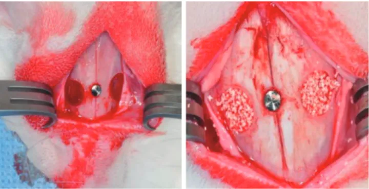
Standardized, bilateral, circular, transosseous defects 6 mm in diameter were made on the maxillary sinus (Figs. 1 and 2).
Preparation of ErhBMP-2 coated BCP
Prior to the surface coating, the ErhBMP-2 was produced at the Cowell Medi Implant (Busan, Korea). Nonglycosylated rhBMP-2 was obtained in the form of inclusion bodies and Group Material Experimental period Defect no.
Control OSTEONTM 4 weeks, 8 weeks 20
Experimental 1 ErhBMP-2 0.05 mg/mL 4 weeks, 8 weeks 10 Experimental 2 ErhBMP-2 0.5 mg/mL 4 weeks, 8 weeks 10 ErhBMP-2: Escherichia coli-expressed recombinant human bone morphogenetic protein 2.
Figure 1. Maxillary sinus defects in the rabbit. Standardized, bilater- al, circular, transosseous windows were prepared on the maxillary sinus using a 6-mm diameter trephine bur with the pin inserted.
Figure 2. Schematic diagram of trephine bur (outer diameter 8.0 mm, inner diameter 6.0 mm, depth 1.5 mm).
8.0 6.0
1.5 1.5
coating was performed to combine HA, one of the constitu- ents of BCP (Osteon, Dentium, Korea), with ErhBMP-2. Ini- tially, the hydroxyl group of hydroxyapitite was linked with 3-aminopropyl triethoxysilane (APTES) (Sigma-Aldrich Co., St. Louis, MO, USA), a silane coupling agent, then followed by linkage with N-succinimidyl 3-maleimidopropionate (N- SMP) (Sigma-Al drich Co.) and then ErhBMP-2. This proce- dure can be sum marized as a three-step reaction procedure:
1) silanization with APTES, 2) cross-linking with N-SMP, 3) combining N-SMP with ErhBMP-2. Two different concen- trations of ErhBMP-2 (0.5 and 0.05 mg/mL) were made after the coating procedure. ErhBMP-2-coated BCP was lyophi- lized. The solution was frozen cooled down to -43°C. The formu lations were maintained at this temperature for 3 hours, after which they were dried in a condenser at -40°C (primary dry ing) and kept in a pressure chamber at 5 mTorr for 2 hours. Secondary drying was performed on a shelf us- ing the follow ing sequence: -20°C for 4 hours, -10°C for 4 hours, 0°C for 2 hours, and 20°C for 20 hours. After the ly- ophilizing procedure, the ErhBMP-2-coated BCP was steril- ized with ethylene oxide gas in a gas sterilizer (Steri-Vac 400B, 3M, St. Paul, MN, USA) at 29°C for 5 hours, which was report- ed as the ideal sterilization protocol to avoid reducing osteo- inductive activity [21].
Surgical procedure
For all of the surgical procedures involved in the study, the animals were first sedated with a mixture of ketamine hy- drochloride (Ketalar, Yuhan, Seoul, Korea) and xylazine (Rum- pun, Bayer Korea Ltd., Seoul, Korea) via an intramuscular in- jection. After the isolation of the surgical site by shaving and sterilizing with a povidone-iodine solution, infiltration anes- thesia with 2% lidocaine (with 1:100,000 epinephrine) was also administrated to the surgical sites on the nasal bone. A straight incision along the sagittal midline on the nasal bone was then made followed by a full-thickness flap including the skin and the elevation of the periosteum laterally. Stan- dardized, bilateral, circular, transosseous windows were pre- pared on the maxillary sinus using a trephine bur 6 mm in diameter under copious irrigation with saline. The pin was inserted at the point where the sagittal midline met the imaginary line that connected the right and left windows in order to indicate the reference point for micro computed to- mography (CT) analysis as well as for preparation of the specimen. The trephined bony disk was carefully removed and the experimental and control treatments with grafting materials were applied to the windows accordingly.
After the grafting materials was placed, the periosteum was re-positioned over the windows on each side followed by su-
absorbable monofilament, B-Braun, Aesculap, Center Valley, PA, USA), which was removed after 7 days. The animals were allowed a healing period of either 4 or 8 weeks postopera- tively and sacrificed accordingly by euthanasia.
Radiographic analysis: Micro-CT
After obtaining block sections including the augmented si- nus and the surrounding bone, each block section was fixed in 10% buffered formalin for 10 days and scanned using mi- cro-CT (SkyScan 1076, SkyScan, Aartselaar, Belgium) at a res- olution of 35 μm (100 kV and 100 μA). The scanned data set were analyzed and the area of interest was reconstructed with a CT analyzer program (SkyScan). After a 3D-reconstructed image of the maxillary sinus and supporting bone was visu- alized using a data viewer program, the CT volume, the max- imum augmented height (MAH), and the deepest depth of the defect (DDD) [16] were measured on the coronally sec- tioned images; MAH was measured linearly on the cross- sectional image. The CT volume was calculated from the per- centage of window closure where new bony formation or re- maining grafting materials could be seen. MAH was mea- sured linearly on the cross-sectional image whereas the DDD was measured from the imaginary line through the original bone to the bottom of the bony crater.
Histological analysis
Following decalcification in 5% formic acid for 14 days, all specimens were embedded in paraffin and sliced coronally into sections about 5 μm thick along the center of the aug- mented sinus. The 2 most-central sections were selected and stained with hematoxylin and eosin (H&E) for the light mi- croscopic examination (BX50, Olympus Co., Tokyo, Japan).
Histometric analysis
Computer-assisted histometric analysis was performed us- ing an automated image-analysis system (Image-Pro Plus, The Proven Solution, Media Cybernetics Inc., Silver Spring, MD, USA). Two parameters were measured and calculated as follows:
Newly formed bone area (NB%)=newly formed area/total augmented bone area×100
Remaining grafts area (RG%)=remaining graft area/total augmented bone area×100
Statistical analysis
The statistical analysis regarding micro-CT data and histo- morphometric measurement of the samples was performed using a SPSS ver. 15.0 (SPSS Inc., Chicago, IL, USA). The Mann- Whitney U test was used to compare differences between
differences according to time of healing. The level of statisti- cal significance was set at P<0.05, and the data were present- ed as means±standard deviation values.
RESULTS
Radiographic and histometric analysis
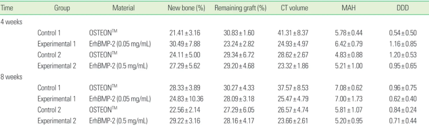
In the 4-week healing groups, new bone formation (%) was greater in experimental group 1 than in the control group 1 (P<0.05). The remaining graft material (%) was less in experi- mental group 1 than in the control group 1 (P<0.05). No sig- nificant difference was found between experimental group 1 and experimental group 2 in new bone formation or remain- ing graft (Table 2).
In the 8-week healing groups, there was no statistical dif- ference in new bone formation, remaining graft, or CT vol-
2. However, new bone formation (%) was greater in experi- mental group 2 than the control group 2 (P<0.05). There was no difference in the remaining graft between experimental group 1 and the control group 1. The CT volume was greater in the control group 1 than experimental group 1 (P<0.05) and the MAH was greater in experimental group 1 than in experimental group 2 (Table 2).
Histologic findings
The histologic findings revealed that both experimental group 1 and group 2 showed a mixture of newly formed bone from the surgically created defects and newly formed bone originating from bone graft materials. As shown in Figs 3-6, newly formed bone from the surgically created defect is par- ticularly distinguishable as it is shown to be more mature in contrast to newly formed bone that originated from bone Table 2. Measurements in standardized rabbit sinus model.
Time Group Material New bone (%) Remaining graft (%) CT volume MAH DDD
4 weeks
Control 1 OSTEONTM 21.41±3.16 30.83±1.60 41.31±8.37 5.78±0.44 0.54±0.50
Experimental 1 ErhBMP-2 (0.05 mg/mL) 30.49±7.88 23.24±2.82 24.93±4.97 6.42±0.79 1.16±0.85
Control 2 OSTEONTM 24.11±5.00 29.34±6.72 28.62±2.67 4.83±0.88 1.20±0.53
Experimental 2 ErhBMP-2 (0.5 mg/mL) 27.29±5.62 29.20±4.68 23.32±1.86 5.21±1.00 0.95±0.65 8 weeks
Control 1 OSTEONTM 28.33±3.89 30.27±4.33 37.57±8.53 7.08±0.62 0.96±0.75
Experimental 1 ErhBMP-2 (0.05 mg/mL) 24.83±10.36 28.09±3.18 25.47±4.79 7.00±1.73 0.62±0.40
Control 2 OSTEONTM 22.56±2.14 27.29±6.05 26.57±4.74 5.81±1.07 0.84±0.24
Experimental 2 ErhBMP-2 (0.5 mg/mL) 29.22±3.16 28.16±4.17 23.66±2.61 5.20±0.95 0.71±0.44 Values are presented as mean±standard deviation.
CT: computed tomography, MAH: maximum augmented height, DDD: deepest depth of the defect, ErhBMP-2: Escherichia coli-expressed recombinant human bone morphogenetic protein 2.
Figure 3. Histologic findings of experimental group 1 (0.05 mg/mL ErhBMP-2) at 4 weeks. (A) There appeared to be a mixture of newly formed bone from the surgically created defect and newly formed bone originating from bone graft materials. Newly formed bone from the surgical- ly created defect is shown to be more mature whereas newly formed bone from the bone graft materials is still undergoing the mineraliza- tion process (×12.5). (B) Defect margin area: The arrowheads indicate the margin of the surgically created defect. The white asterisk indicates newly formed bone assumed to have originated from the defect margin (×50). (C) The Schneiderian membrane: The black asterisk indicates newly formed bone assumed to have originated from graft materials (×50). Note that immature woven bone is evident here in contrast to mature lamellar bone as indicated by the white asterisk in Fig. 3B. (D) Middle area: Immature woven bone can be seen (×50).
A B C D
Figure 4. Histologic findings of experimental group 1 (0.05 mg/mL ErhBMP-2) at 8 weeks. (A) When compared to the 4 week healing group, more vascularization is evident in the 8-week healing group (arrowheads). The surgically created defect was almost completely closed. The particle size of the graft materials is dramatically reduced (white asterisk). Note that mature lamellar bone (black asterisk) is evident in the newly formed bone found in between graft materials (×12.5). (B) Defect margin area: both inflammatory cells and blood vessels can be ob- served, as indicated by the arrows and the arrowhead, respectively. The black asterisk indicates matured lamellar bone (×50). (C) The Sch- neiderian membrane: The Schneiderian membrane was also thicker than that of the 4 week healing group (×50). (D) Middle area: More ma- ture lamellar bone is identified in newly formed bone found in between the graft materials (×50).
A B C D
Figure 5. Histologic findings of experimental group 2 (0.5 mg/mL ErhBMP-2) at 4 weeks. (A) The histologic findings were similar to those observed in experimental group 1 (0.05 mg/mL ErhBMP-2) at 4 weeks. Note that surgically created defect was completely closed (×12.5). (B) Defect margin area (×50). (C) The Schneiderian membrane (×50). (D) Middle area (×50).
A B C D
Figure 6. Histologic findings of experimental group 2 (0.5 mg/mL ErhBMP-2) at 8 weeks. (A) The histologic findings were similar to those observed in experimental group 1 (0.05 mg/mL ErhBMP-2) at 8 weeks. More vascularization (arrowheads) and matured lamellar bone (black asterisk) are also evident here (×12.5). (B) Defect margin area both inflammatory cells and blood vessels can be observed, as indicated by the arrow and the arrowhead, respectively. The black asterisk indicates matured lamellar bone (×50). (C) The Schneiderian membrane (×50). (D) Middle area (×50).
A B C D
graft materials, which can be seen to be still undergoing the mineralization process.
In general, when compared to the 4-week healing group,
more vascularization is evident in the 8-week healing group in both experimental group 1 and group 2 (Figs. 4 and 6) The Schneiderian membrane was also slightly thickened with the
8-week healing group. The particle size of graft materials was also dramatically reduced. Instead, mature lamellar bone was identified in between the graft materials. It was also noted that the surgically created defects were almost completely closed in the 8-week healing group.
DISCUSSION
The effectiveness of bone substitutes and dental implants has been evaluated through various sinus models [20-23].
Asai et al. [20] introduced the rabbit sinus model to under- stand the mechanism of new bone formation in humans be- cause the composition and mechanical properties of bone tissue in humans and rabbits are known to be very similar to each other [24]. Based on this study, researchers have used this rabbit sinus model to evaluate histomorphometric chang- es of the bone after sinus grafting, assessing the effectiveness of various bone substitute materials. The purpose of the present study was to determine whether BCP with two dif- ferent concentrations of ErhBMP-2 enhances new bone for- mation in a standardized rabbit sinus model and to evaluate the concentration-dependent response of ErhBMP-2. In the present study, the standardized sinus model with the same shape and size of window was used, as introduced by De Souza Nunes et al. [25] to provide precisely the same condi- tions to the control and the experimental groups and to min- imize procedural errors. The degree of anatomical variation between individuals was also smaller in the rabbit sinus mod- el than in larger animals, thereby enabling a consistent re- production of defects in different individuals [16], as evidenced by the present histologic findings. According to Roberts and Breznak [26], the rabbit metabolic rate is known to be three to four times faster than that of humans. Thus, the 4-week healing group and 8-week healing group were selected on the assumption that a 4-week healing period and 8-week healing period correspond to a 3 to 4 months healing period and 6 to 8 months healing period in humans, respectively.
The surgical sites were exposed by an extraoral approach, which enabled good visibility and accessibility to the surgical site as well as preservation of the Schneiderian membrane through delicate drilling and careful removal of the window.
Therefore, taking the aforementioned factors into account, the standardized sinus augmentation model used in this study can be considered to be a valid study model in which various bone graft materials can be evaluated.
In order to evaluate the volumetric change in new bone, 3D volumetric measurement of the augmented bone could have led to more accurate results; however, we used a two-dimen- sional section of the window region for the comparison of
group. To minimize the error from this procedure, linear measurements including MAH, DDD, and CT volume were performed at the two-dimensional section of the window re- gion using micro-CT.
From histomorphometric analysis, it was found that in the 4-week healing groups, new bone formation was statistically significantly greater in experimental group 1 than control group 1. Based on this result, it can be interpreted that in ex- perimental group 1, more graft materials were substituted with new bone. In other words, bone graft materials coated with even low concentration of ErhBMP-2 (0.05 mg/mL) showed better osteoinductive potential than bone graft ma- terials alone. Although the difference is not statistically sig- nificant, bone graft materials coated with a high concentra- tion of ErhBMP-2 (0.5 mg/mL) also showed better osteoin- ductive potential than the control group. Thus, these findings demonstrate that ErhBMP induces rapid bone formation and remodeling at an earlier stage, and this is consistent with findings of a previous study [2] that used the rat calvarial de- fect model. On the other hand, in the 4-week healing groups, the remaining graft was statistically significantly less in ex- perimental group 1 than control group 1. This can be inter- preted as showing that more of the bone graft material in ex- perimental group 1 had been substituted with new bone, thereby showing less remaining graft material. Considering the fact that the amount of graft material applied to each group was the same, there might be no difference between experimental group 1 and group 2 in osteoinductive poten- tial. This result is also supported by the results of the 8-week healing groups where MAH was statistically significantly greater in experimental group 1 than group 2, which can be interpreted as showing that the total amount of bone includ- ing new bone and graft materials increased to some extent.
However, since there was no significant difference in CT volume between experimental group 1 and group 2, it is dif- ficult to conclude that a difference in osteoinductive poten- tial between two groups is evident.
In the 8-week healing groups, CT volume was statistically significantly greater in control group 1 than experimental group 1. However, there was not any difference between ex- perimental group 1 and control group 1 in new bone forma- tion and remaining graft material. This can be interpreted as showing that replacement of biomaterial with newly formed bone does not necessarily occur in a 1:1 ratio, which is in agreement with previous research [27]. In other words, the volume of bone produced in this case was greater than the volume of biomaterial absorbed. In the 8-week healing groups, the MAH in experimental group 1 was greater than experimental group 2. However, there was no significant dif-
mental group 1 is difficult to interpret as ‘more new bone formation’ in experimental group 1 than group 2.
Nevertheless, as new bone formation was statistically sig- nificantly greater in experiment group 2 than control group 2, it seems that bone graft materials coated with a high concen- tration of ErhBMP-2 (0.5 mg/mL) show better ongoing os- teoinductive potential even in that late healing period. This could lead to an assumption that, although no significant difference between the low concentration of ErhBMP-2 (0.05 mg/mL) and high concentration of ErhBMP-2 (0.5 mg/mL) in osteoinductive potential was found, the high concentration of ErhBMP-2 (0.5 mg/mL) can be more effective when long- term osteoinductive potential is needed.
In conclusion, within the limitations of this study, while BCP coated with ErhBMP-2 significantly enhanced osteoin- ductive potential, a concentration-dependent response to os- teoinductive potential was not found.
CONFLICT OF INTEREST
Kyung-Hee Choi who is one of co-authors works for Cow- ell R&D Institute as a researcher.
ACKNOWLEDGEMENTS
This research was supported by Basic Science Research Program through the National Research Foundation of Ko- rea (NRF) funded by the Ministry of Education, Science and Technology (2010-0007829).
REFERENCES
1. Bessho K, Konishi Y, Kaihara S, Fujimura K, Okubo Y, Iizu- ka T. Bone induction by Escherichia coli-derived recom- binant human bone morphogenetic protein-2 compared with Chinese hamster ovary cell-derived recombinant human bone morphogenetic protein-2. Br J Oral Maxillo- fac Surg 2000;38:645-9.
2. Lee JH, Kim CS, Choi KH, Jung UW, Yun JH, Choi SH, et al. The induction of bone formation in rat calvarial defects and subcutaneous tissues by recombinant human BMP-2, produced in Escherichia coli. Biomaterials 2010;31:3512-9.
3. Long S, Truong L, Bennett K, Phillips A, Wong-Staal F, Ma H. Expression, purification, and renaturation of bone morphogenetic protein-2 from Escherichia coli. Protein Expr Purif 2006;46:374-8.
4. Vallejo LF, Brokelmann M, Marten S, Trappe S, Cabrera- Crespo J, Hoffmann A, et al. Renaturation and purification of bone morphogenetic protein-2 produced as inclusion
erichia coli. J Biotechnol 2002;94:185-94.
5. Choi KH, Moon K, Kim SH, Yun JH, Jang KL, Cho KS. Pu- rification and biological activity of recombinant human bone morphogenetic protein-2 produced by E. coli expres- sion system. J Korean Acad Periodontol 2008;38:41-50.
6. Walker DH, Wright NM. Bone morphogenetic proteins and spinal fusion. Neurosurg Focus 2002;13:e3.
7. Wong DA, Kumar A, Jatana S, Ghiselli G, Wong K. Neuro- logic impairment from ectopic bone in the lumbar canal:
a potential complication of off-label PLIF/TLIF use of bone morphogenetic protein-2 (BMP-2). Spine J 2008;8:1011-8.
8. Kaneko H, Arakawa T, Mano H, Kaneda T, Ogasawara A, Nakagawa M, et al. Direct stimulation of osteoclastic bone resorption by bone morphogenetic protein (BMP)-2 and expression of BMP receptors in mature osteoclasts. Bone 2000;27:479-86.
9. Smucker JD, Rhee JM, Singh K, Yoon ST, Heller JG. In- creased swelling complications associated with off-label usage of rhBMP-2 in the anterior cervical spine. Spine (Phila Pa 1976) 2006;31:2813-9.
10. Bae JH, Kim YK, Kim SG, Yun PY, Kim JS. Sinus bone graft using new alloplastic bone graft material (Osteon)-II: clin- ical evaluation. Oral Surg Oral Med Oral Pathol Oral Ra- diol Endod 2010;109:e14-20.
11. Kim S, Jung UW, Lee YK, Choi SH. Effects of biphasic cal- cium phosphate bone substitute on circumferential bone defects around dental implants in dogs. Int J Oral Maxil- lofac Implants 2011;26:265-73.
12. Zhu X, Fan H, Li D, Xiao Y, Zhang X. Protein adsorption and zeta potentials of a biphasic calcium phosphate ce- ramic under various conditions. J Biomed Mater Res B Appl Biomater 2007;82:65-73.
13. Truumees E, Herkowitz HN. Alternatives to autologous bone harvest in spine surgery. Univ Pa Orthop J 1999;12:
77-88.
14. Le Nihouannen D, Guehennec LL, Rouillon T, Pilet P, Bil- ban M, Layrolle P, et al. Micro-architecture of calcium phosphate granules and fibrin glue composites for bone tissue engineering. Biomaterials 2006;27:2716-22.
15. Le Nihouannen D, Saffarzadeh A, Gauthier O, Moreau F, Pilet P, Spaethe R, et al. Bone tissue formation in sheep muscles induced by a biphasic calcium phosphate ceram- ic and fibrin glue composite. J Mater Sci Mater Med 2008;
19:667-75.
16. Choi Y, Yun JH, Kim CS, Choi SH, Chai JK, Jung UW. Sinus augmentation using absorbable collagen sponge loaded with Escherichia coli-expressed recombinant human bone morphogenetic protein 2 in a standardized rabbit sinus model: a radiographic and histologic analysis. Clin Oral
17. Jung JH, Yun JH, Um YJ, Jung UW, Kim CS, Choi SH, et al.
Bone formation of Escherichia coli expressed rhBMP-2 on absorbable collagen block in rat calvarial defects. Oral Surg Oral Med Oral Pathol Oral Radiol Endod 2011;111:
298-305.
18. Kim JW, Choi KH, Yun JH, Jung UW, Kim CS, Choi SH, et al. Bone formation of block and particulated biphasic cal- cium phosphate lyophilized with Escherichia coli-derived recombinant human bone morphogenetic protein 2 in rat calvarial defects. Oral Surg Oral Med Oral Pathol Oral Ra- diol Endod 2011;112:298-306.
19. Park JC, So SS, Jung IH, Yun JH, Choi SH, Cho KS, et al. In- duction of bone formation by Escherichia coli-expressed recombinant human bone morphogenetic protein-2 us- ing block-type macroporous biphasic calcium phosphate in orthotopic and ectopic rat models. J Periodontal Res 2011;46:682-90.
20. Asai S, Shimizu Y, Ooya K. Maxillary sinus augmentation model in rabbits: effect of occluded nasal ostium on new bone formation. Clin Oral Implants Res 2002;13:405-9.
21. Ijiri S, Yamamuro T, Nakamura T, Kotani S, Notoya K. Ef- fect of sterilization on bone morphogenetic protein. J Or- thop Res 1994;12:628-36.
changes in the sinus membrane after maxillary sinus aug- mentation in goats. J Oral Maxillofac Surg 1998;56:1170-6.
23. Scala A, Botticelli D, Rangel IG Jr, de Oliveira JA, Okamoto R, Lang NP. Early healing after elevation of the maxillary sinus floor applying a lateral access: a histological study in monkeys. Clin Oral Implants Res 2010;21:1320-6.
24. Wang X, Mabrey JD, Agrawal CM. An interspecies com- parison of bone fracture properties. Biomed Mater Eng 1998;8:1-9.
25. De Souza Nunes LS, De Oliveira RV, Holgado LA, Nary Filho H, Ribeiro DA, Matsumoto MA. Immunoexpression of Cbfa-1/Runx2 and VEGF in sinus lift procedures using bone substitutes in rabbits. Clin Oral Implants Res 2010;
21:584-90.
26. Roberts EG, Breznak N. Bone Physiology and Metabolism.
In; Mish CE, editor. Contemporary implant dentistry. Or- lando: Mosby Year Book; 1994. p.557-98.
27. Frenken JW, Bouwman WF, Bravenboer N, Zijderveld SA, Schulten EA, ten Bruggenkate CM. The use of straumann bone ceramic in a maxillary sinus floor elevation proce- dure: a clinical, radiological, histological and histomor- phometric evaluation with a 6-month healing period. Clin Oral Implants Res 2010;21:201-8.