대한소화기학회지 2010;55:1-3 □ IMAGE OF THE MONTH □ DOI: 10.4166/kjg.2010.55.1.1
연락처: 이오영, 133-792, 서울시 성동구 행당동 17 한양대학교 의과대학 내과학교실
Tel: (02) 2290-8343, Fax: (02) 2298-9183 E-mail: leeoy@hanyang.ac.kr
Correspondence to: Oh Young Lee, M.D.
Division of Gastroenterology, Department of Internal Medi- cine, Hanyang University College of Medicine, 17, Haeng- dang-dong, Seongdong-gu, Seoul 133-792, Korea
Tel: +82-2-2290-8343, Fax: +82-2-2298-9183 E-mail: leeoy@hanyang.ac.kr
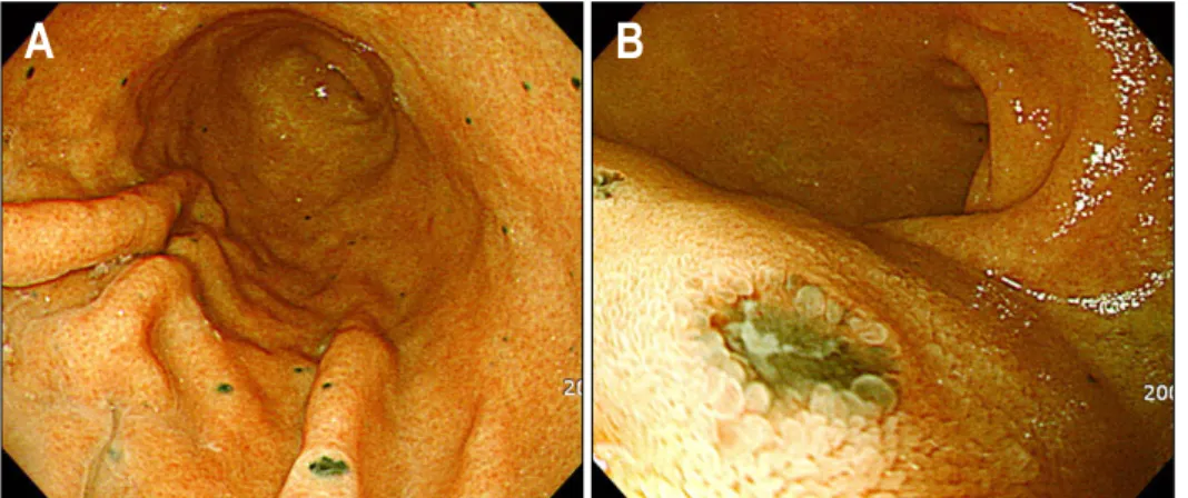
Fig. 1. (A) Endoscopic findings.
Multiple melanotic nodules were scattered on the mucosal surfaces of lower and mid body of the stomach. (B) Slightly depressed mucosae with a pigmented base were seen in the bulb of the duodenum.
간과 위장관에 전이된 악성 흑색종 1예
한양대학교 의과대학 내과학교실, 소화기 센터
이강녕ㆍ이오영
A Case of Malignant Melanoma Metastasized to Liver and Gastrointestinal Tract
Kang Nyeong Lee, M.D. and Oh Young Lee, M.D.
Department of Internal Medicine, Digestive Disease Center, Hanyang University College of Medicine, Seoul, Korea
증례: 70세 여자 환자가 복부 불편감과 흑색변을 주증상 으로 내원하였다. 2주 전부터 흑색변이 시작되었으며, 심와 부 동통과 식후 팽만감으로 식욕 부진을 호소하였다. 내원 시 혈압은 130/90 mmHg, 맥박 112회/분, 호흡수 20회/분, 체 온은 36.7oC였다. 신체 검사에서 심와부에 압통이 있었으며 우상복부에 종물이 촉지되었다. 과거력에서 20년 전에 좌측 하지에 악성 흑색종을 진단 받고 넓은 지역 절단술을 시행 받은 후 추적 검사 없이 지내다가 내원 6개월 전에 재발하 여 무릎 하 하지절단술을 시행 받았다. 말초혈액검사에서 백혈구 8,500/mm3, 혈색소 11.1 g/dL, 혈소판 237,000/mm3였
고, AST 277 IU/L, ALT 61 IU/L, 총 빌리루빈 5.1 mg/dL, ALP 820 IU/L, GGT 742 IU/L, LDH 844 IU/L, INR 1.10였다.
상부위장관 내시경에서 위체부와 저부 및 십이지장 구부에 다수의 작은 반점 모양의 흑색의 색소 침착이 관찰되었고 (Fig. 1), 대장내시경검사에서는 다수의 대장 용종이 발견되 었다. 복부 전산화단층촬영에서 전 간에 표적 모양의 병변 들이 산재되어 있었다(Fig. 2). 색소 침착이 있는 위점막을 생검한 조직에서 멜라닌이 침착된 세포들이 고유판에 둥지 형태를 이루고 있었고 HMB와 S-100을 이용한 면역조직화 학염색에서 양성 소견을 보였다(Fig. 3). 대장의 용종에서 시
2 대한소화기학회지: 제55권 제1호, 2010
Fig. 2. Abdominal CT scan with oral and intravenous contrast.
Multiple hypodense target-like lesions were scattered throughout the entire liver.
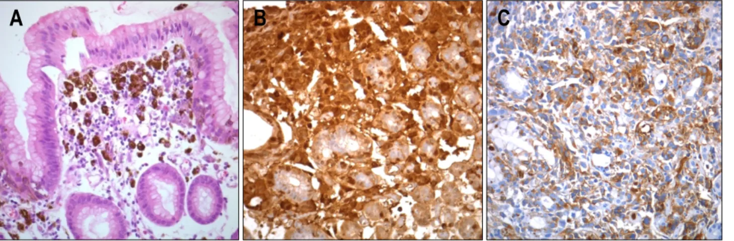
Fig. 3. (A) Pathologic findings. Malignant melanoma cells infiltrated the lamina propria of the stomach in a nesting fashion (H&E stain,
×400). (B) Pathologic findings. Cells infiltrated in the lamina propria of gastric mucosa showed a stong positive immunohistochemical stains for S-100 protein suggesting a metastatic malignant melanoma (S-100 IHC stain, ×400). (C) Pathologic findings. HMB im- munohistochemical stains demonstrated metastatic malignant melanoma cells scattered around the glandular structures of gastric mucosa (HMB-45 IHC stain, ×400).
행한 조직 검사는 선종성 용종이었다. 상부 및 하부 위장관 검사에서 출혈 병소를 찾지 못하여 추가 검사를 계획했으나 환자가 검사 및 치료를 포기하여 자의 퇴원하였다. 진단은?
진단: 악성 흑색종의 위장관 및 간 전이
악성 흑색종은 피부, 눈, 위장관 등의 멜라닌세포에서 기 원하는 악성 종양으로 모든 장기에 전이될 수 있다. 최근 전 세계적으로 발생률은 증가 추세에 있으며 대개 남자가 여자 에 비해 1.5배 더 높은데, 우리나라에서는 아직 드물게 발생 하나 향후 우리나라에서도 서구와 같이 발생률이 증가할 가 능성이 높다. 다른 장기의 암이 소화관으로 전이되는 경우
는 드물지만 악성 흑색종으로 사망한 환자의 부검 결과 약 60%에서 소화관 전이가 있었던 것으로 보고되고 있어 악성 흑색종은 소화관으로 전이되는 가장 흔한 종양 중의 하나로 생각한다.1 소화관 중 가장 전이 빈도가 높은 장기는 소장이 고, 그 외에도 대장, 위, 식도에도 전이되는 것으로 알려져 있다.2-4
소화관 전이 시의 증상은 구역, 구토, 복통, 체중 감소 및 빈혈 등의 증상을 보이지만,3,5,6 악성 흑색종 환자의 약 1-4%만이 임상적으로 분명한 소화관 침범을 보이는 것으로 알려져 있고,1 평균적으로 증상이 발생하기까지는 4년 이상 의 기간이 필요하다고 한다.7
악성 흑색종의 위 전이는 부검례에서 약 20-40%로 보고 되는데, 비특이적인 증상으로 나타나기 때문에 진단이 늦어 지기 쉽다. 위로 전이된 악성 흑색종의 내시경 소견은 색소 가 있는 병변이 가장 흔하며 주로 세 가지 형태로 분류할 수 있었다는 보고가 있다. 정상 주름위에 산재된 흑색의 궤 양성 결절의 형태, 궤양을 동반하는 점막하종양의 형태, 색 소와 괴사를 동반하는 종괴의 형태가 그것이다. 그러나, 무 색소 병변으로 나타날 수도 있어서 주의를 요한다.8 소장 전이는 소화관 전이 중 가장 흔하며,3 출혈, 충수돌 기염의 증상 및 징후, 체중 감소, 장폐쇄, 흡수 장애 등으로 나타난다.9-11 주로 장막층과 장간막을 통해 전이되기 때문에 진단을 위해서는 내시경 검사보다는 소장 조영술이 보다 용 이한 것으로 알려져 있다. 방사선 검사 소견은 벽결절, 표적 병변, 궤양성 종괴 등의 형태를 보이고, 내시경상은 색소 침 착을 갖는 궤양성 결절이나 궤양을 동반한 점막하병변의 형 태를 보인다.
간 전이는 악성 흑색종의 약 30-40%에서 발생하나 심한
이강녕 외 1인. 간과 위장관에 전이된 악성 흑색종 1예 3
간기능 손상을 일으키는 예는 흔치 않고 드물게 급성 간부 전이나 간 파열 사례가 보고되었다. 악성 흑색종 환자에서 LDH가 상승하면 간 전이가 있음을 시사하는데,12,13 LDH는 예후와도 관련이 있는 것으로 알려져 있다.14,15
한편, 악성 흑색종이 가장 드물게 전이되는 소화관은 대 장으로 알려져 있으며 대장 전이는 용종성 병변이나 궤양성 병변 혹은 점막하결절 형태로 나타나거나 단일 병변으로 나 타나 장중첩증을 유발하기도 한다.16,17
내시경 소견에서 소화관으로 전이된 악성 흑색종 병변이 색소 침착을 띠는 경우가 대부분으로 보고하고 있으며8,18 색소 침착이 없는 경우도 S-100 단백이나 HMB-45를 이용한 면역조직화학염색으로 멜라닌을 확인하여 진단할 수 있다.
이번 증례는 약 20년 전에 치료한 악성 흑색종이 재발 및 위장관으로 전이를 일으킨 사례로 악성 흑색종이 우리나라 에서는 아직 드물지만 전 세계적으로 증가 추세에 있고 원 발 부위를 적절히 치료하더라도 언제든지 전이될 가능성이 있으며 위장관에 전이되는 가장 흔한 암 중 하나이기 때문 에 내시경검사를 시행하는 중에 색소를 동반하는 병변이 있 을 때는 반드시 생검과 특수 염색을 통해 악성 흑색종을 진 단하는 노력이 필요하다. 또한, 이번 증례와 같이 악성 흑색 종의 위장관 전이는 위장관 출혈이나 심와부 동통 등 소화 성 궤양 양상으로 발현할 수 있어 임상적인 주의가 요구된 다. 그러나 악성 흑색종의 소화관 전이 병변에 대한 치료 효 과는 아직 미흡하며 향후 소화관 전이를 예방하고 전이 병 변을 적절히 치료할 수 있는 방법에 대한 연구가 필요할 것 으로 생각한다.
참고문헌
1. Patel JK, Didolkar MS, Pickren JW, Moore RH. Metastatic pattern of malignant melanoma. A study of 216 autopsy cases. Am J Surg 1978;135:807-810.
2. Kadakia SC, Parker A, Canales L. Metastatic tumors to the upper gastrointestinal tract: endoscopic experience. Am J Gastroenterol 1992;87:1418-1423.
3. Caputy GG, Donohue JH, Goellner JR, Weaver AL. Metasta- tic melanoma of the gastrointestinal tract. Results of surgical management. Arch Surg 1991;126:1353-1358.
4. Ollila DW, Essner R, Wanek LA, Morton DL. Surgical re- section for melanoma metastatic to the gastrointestinal tract.
Arch Surg 1996;131:975-980.
5. Ihde JK, Coit DG. Melanoma metastatic to stomach, small bowel, or colon. Am J Surg 1991;162:208-211.
6. Reintgen DS, Thompson W, Garbutt J, Seigler HF. Radiolo- gic, endoscopic and surgical considerations of malignant mel- anoma metastatic to the small intestine. Curr Surg 1984;41:
87-89.
7. Blecker D, Abraham S, Furth EE, Kochman ML. Melanoma in the gastrointestinal tract. Am J Gastroenterol 1999;94:3427- 3433.
8. Nelson RS, Lanza F. Malignant melanoma metastatic to the upper gastrointestinal tract. Endoscopic and radiologic correla- tions, form and evolution of lesions, and value of directed bi- opsy in diagnosis. Gastrointest Endosc 1978;24:156-158.
9. Wilson BG, Anderson JR. Malignant melanoma involving the small bowel. Postgrad Med J 1986;62:355-357.
10. Raymond AR, Rorat E, Goldstein D. An unusual case of ma- lignant melanoma of the small intestine. Am J Gastroenterol 1984;79:689-692.
11. Benisch BM, Abramson S, Present DH. Malabsorption and metastatic melanoma. Mt Sinai J Med 1972;39:474-477.
12. Campora E, Repetto L, Giuntini P, et al. LDH in the fol- low-up of stage I malignant melanoma. Eur J Cancer Clin Oncol 1988;24:277-278.
13. Muss HB, Richards F 2nd, Barnes PL, Willard VV, Cowan RJ. Radionuclide scanning in patients with advanced malig- nant melanoma. Clin Nucl Med 1979;4:516-518.
14. Garg R, McPherson TA, Lentle B, Jackson F. Usefulness of an elevated serum lactate dehydrogenase value as a marker of hepatic metastases in malignant melanoma. Can Med Assoc J 1979;120:1114, 1116.
15. Finck SJ, Giuliano AE, Morton DL. LDH and melanoma.
Cancer 1983;51:840-843.
16. McClenathan JH. Metastatic melanoma involving the colon.
Report of a case. Dis Colon Rectum 1989;32:70-72.
17. Watson RGK, McMullin JP. Intussusception of the colon sec- ondary to gastrointestinal metastatic melanoma. Br J Surg 1981;68:667.
18. Branum GD, Seigler HF. Role of surgical intervention in the management of intestinal metastases from malignant mela- noma. Am J Surg 1991;162:428-431.