<접수일:2007년 3월 30일, 심사통과일:2007년 6월 8일>
※통신저자:최 효 진
인천시 남동구 구월동 1198번지 가천의과학대학교 길병원 류마티스내과
Tel:032) 460-8218, Fax:032) 469-4320, E-mail:hjsalom@hanmail.net
시신경척수염(Neuromyelitis Optica)이 동반된 전신홍반루푸스 1예
가천의과학대학교 길병원 내과학교실 류마티스내과
오병천ㆍ김금하ㆍ최효진ㆍ백한주
= Abstract =
A Case of Systemic Lupus Erythematous
Associated with Neuromyelitis Optica (Devic's Syndrome)
Pyung Chun Oh, M.D., Geum Ha Kim, M.D., Choi Hyo Jin, M.D., Han Joo Baek, M.D.
Division of Rheumatology, Department of Internal Medicine, Gil Medical Center, Gachon University of Medicine and Science, Incheon, Korea
Neuromyelitis optica (NMO) is an idiopathic inflammatory demyelinating disease, characterized by optic neuritis and myelitis. NMO is a very uncommon and serious neurologic manifestation of systemic lupus erythematous (SLE). We report a 28-year-old man with NMO as neuropsy- chiatric manifestation of SLE. He was diagnosed as lupus nephritis at 16-year-old. He had optic neuritis at three years and seven months ago. Oral prednisolone was tapered off according to the improved eye symptoms. Two months later, he visited rheumatology clinics for urinary distur- bance and paresthesia on both feet. A spinal magnetic resonance imaging revealed increased signal intensity in T2-weighted images from second to sixth cervical level and from eleventh to twelfth thoracic level. We diagnosed neuromyelitis optica and treated with intravenous cyclophos- phamide therapy monthly for three times. He was discharged without any neurological deficits and has been followed up.
Key Words: Systemic lupus erythematous, Neuromyelitis optica, Devic's syndrome
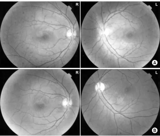
Fig. 1. (A) Fundus photography as the frist episode of optic neuritis. Left fundus photography shows the hyperemia, edematous change and blurring around optic disc and right fundus photography shows normal optic disc. (B) Fundus photography as the second episode of optic neuritis.
Right fundus photography shows the hyperemia around optic disc and left fundus photography shows the pale optic disc.
서 론
전신홍반루푸스는 다기관을 침범하는 전신성 염증 성 자가면역질환으로 매우 다양한 임상 양상을 보인 다. 신경계를 침범하는 경우는 60%까지 보고 되고 있으며 인지 기능 장애, 두통, 다발성 신경병증, 뇌 혈관계 질환 등이 발생할 수 있다 (1,2). 시신경척수 염(neuromyelitis optica, Devic's syndrome)은 뇌의 백 질기능장애 없이 시신경과 척수신경만을 침범하는 염증성 탈수초성 질환이다. 시신경척수염의 원인에 대해 명확히 밝혀져 있지 않지만 전신홍반루푸스, 쇼그렌증후군, 다발성 경화증과 같은 면역질환과 동 반되어 나타날 수 있으며 세균성(특히 결핵), 바이러
스성 감염과 연관되어 나타날 수 있다 (3). 국내에서 보고된 시신경척수염은 대부분 특발성이었으며 전신 홍반루푸스의 초기 증상으로 발현된 1예 (4)와 폐결 핵 이후 발생한 1예 (5)가 보고되었다. 저자들은 루 푸스 신염 환자에서 양측 시신경염과, 배뇨장애로 발현한 횡단 척수염으로 시신경척수염 1예를 진단하 였기에 보고하는 바이다.
증 례
환 자: 28세 남자 주 소: 배뇨 장애
현병력: 13년 전 부종과 단백뇨를 주소로 내원하여 신장 조직 검사와 항핵항체 양성, 항dsDNA 항체 상
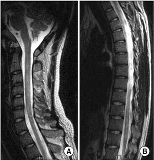
Fig. 2. Sagittal T2-weighted magnetic resonance image of the cervical and thoracic spine. (A) High signal intensity from C2 to C6, (B) high signal intensity and cord swelling from T11 to T12.
승, 림프구감소 소견으로 루푸스 신염(class IV)을 진 단받고 cyclophosphamide 치료 후 호전되었다. 이 후 자의로 치료를 중단하고 지내다가 3년 전 좌측 시력 이 감소하여 내원하였다. 당시 시행한 안저검사에서 좌안 시신경유두부위의 충혈과 부종이 있었고 (그림 1A), 시신경유발전위검사에서 좌안의 P100 신경전도 속도가 지연되어 있었다. 좌측 시신경염 진단 하에 스테로이드 충격 요법 시행 후 시력 저하는 호전되 었다. 경구 스테로이드 요법으로 추적 관찰 중, 내원 7개월 전 우측 시력 감소를 호소하여 시행한 안저검 사에서 우안 시신경유두부위의 충혈 소견과 좌안 시 신경유두 창백 소견을 보였다(그림 1B). 시신경유발 전위검사에서 우안의 P100 신경전도속도가 지연되어 있었고 좌안은 정상이었다. 우측 시신경염 진단 하 에 cyclophosphamide 충격 요법을 시행 하였다. 우안 의 시력은 호전되었고, 이 후 경구 프레드니솔론과 경구 azathioprine을 투여하였다. 증상 재발이 없어 경구 프레드니솔론을 감량하였으며, 중단 2달 뒤 갑 자기 배뇨장애와 양측 발의 이상 감각을 주소로 내 원하였다.
과거력: 1년 전 양측 대퇴골두에 무혈성 골괴사가 발생하여 인공 고관절 치환술을 시행받았다.
이학적 소견: 내원 당시 의식은 명료하였으며 혈 압, 맥박, 체온, 호흡수 등의 활력증후는 정상이었다.
흉부와 복부 진찰에서 특이 소견은 없었다. 신경학 적 검사에서 뇌신경의 이상 소견은 없었다. 양측 상, 하지 근력 감소는 없었으나, 심부 건 반사는 양측 하지에서 증가되어 있었고 양측 발의 이상 감각이 있었다.
검사 소견: 혈액 검사에서 혈색소 14.0 g/dL, 백혈 구 4,630/mm3, 혈소판 166,000/mm3, 적혈구 침강속도 는 6 mm/hr이었다. 생화학검사에서 총단백 6.6 g/dL, 알부민 4.4 g/dL, AST 14 U/L, ALT 6 U/L, 크레아티 닌 0.9 mg/dL, 혈당 93 mg/dL이었고 소변 검사는 정 상이었다. 혈청 검사에서 항핵항체 양성, 항dsDNA 항체 7.4 IU/mL (normal<7), C3 120 mg/dL (86∼
160), C4 28.6 mg/dL (17∼45)이었고, C 반응 단백 0.14 mg/dL (0.01∼0.5)이었다. 뇌척수액 검사에서 색 깔은 무색 투명하였고, 백혈구수 3/μL, 단백 46.6 mg/dL (10∼60), 알부민 18.79 mg/dL (13.4∼23.7), 포 도당 54 mg/dL (40∼70), Ig G 2.0 mg/dL (68∼1,620)
이었으며 단크론성 IgG 대역(oligoclonal IgG band)은 음성이었다. 환자가 특별한 호흡기계 증상을 호소하 지 않았고 흉부 단순 촬영 소견도 정상이어서 결핵 에 관한 검사는 시행하지 않았다.
양측 하지 신경전도검사와 정중신경 및 후경골신 경 유발전위검사는 정상이었다. 요역동학검사에서 배 뇨근 과반사(detrusor hyperreflexia), 배뇨근 괄약근실 조(detrusor-sphincter dyssynergia) 소견이 관찰되었다.
방사선 소견: 척수 자기공명영상에서 경부척수(C2
∼C6)와 흉부척수(T11∼T12)에 걸쳐 T2 영상에서 고 신호 강도를 보이고 척수의 종창과 일부 조영 증강 되는 소견을 보였다(그림 2).
치료 및 경과: 전신성 홍반성 루푸스에 동반된 시 신경척수염으로 진단하였고 cyclophosphamide 충격 요법을 시행하였다. 코르티코스테로이드 장기 사용 으로 인한 양측 대퇴골두의 무혈성 골괴사 기왕력과 달모양 얼굴 등의 부작용으로 인해 스테로이드 치료 는 시행하지 않았고, 시신경염 진단 후 복용하였던 경구 azathioprine (100 mg/일)은 지속적으로 투약하 였다. 4주 간격으로 cyclophosphamide 충격 요법을 2 회 더 시행하여 배뇨 장애와 이상 감각의 호전을 보
였으며, 현재 증상의 악화 없이 추적 관찰 중이다.
고 찰
시신경척수염(Neuromyelitis optica, NMO)은 전신홍 반루푸스의 심각한 합병증 중의 하나로, 뇌의 백질 기능장애 없이 시신경과 척수신경만을 침범하는 염 증성 탈수초성 질환이다. 시신경척수염 환자의 25%
는 시신경염과 급성 척수염이 동시에 발병하고 수년 간 재발이 없으나, 나머지 75%는 수개월 또는 수년 의 간격을 두고 따로 발생하며 자주 재발하는 경향 을 보인다 (3). Wingerchuk 등에 의하면 시신경염과 척수염이 있으면서 다음 3개(3개 이상의 척수분절 침범, 다발성 경화증과 맞지 않는 자기공명영상 소 견, NMO-IgG 양성) 중 2개 이상일 때 시신경척수염 을 진단할 수 있다 (6). 또한 Mandler 등의 기준을 보면 시신경과 척수신경을 침범하면서 NMO- IgG 양성이거나, NMO-IgG 음성이더라도 임상 양상과 척 수 및 뇌 자기공명영상 소견을 바탕으로 진단할 수 있다 (3). 본 환자는 좌측 시신경염이 발생한지 약 3 년 후 횡단척수염이 병발한 증례로, 시신경과 척수 신경 이 외의 다른 중추신경계통 이상이 없고 3개 이상의 척수 분절을 침범한 점 등에 의거하여 시신 경척수염을 진단하였다. 위의 두 진단 기준에 포함 되는 NMO-IgG 검사는 시행하지 못했다.
시신경척수염의 발생 기전은 명확히 알려져 있지 않지만 NMO-IgG 및 다른 자가항체의 생성, 보체 시 스템의 활성화 등 체액성 면역과 관련이 있다 (7,8).
시신경척수염의 전형적인 병리 소견은 과도한 탈수 초화와 괴사, 공동화 및 중간 크기 동맥의 유리질화 (hyalinization)이다 (7). Lucchinetti 등은 면역글로블린 과 C9 신항원(보체 매개성 조직 손상의 표지자), 활 성화된 대식세포가 뇌의 손상된 혈관 주위에서 발견 됨을 보고하였다 (8).
Mok 등은 횡단척수염이 동반된 루푸스 환자의 항 dsDNA 항체 양성률(40%)이 척수병증 이 외의 활성 루푸스에서 보이는 항dsDNA 항체 양성률(75%)보다 낮음을 보고하였다 (9). 또한 횡단척수염 환자 10명 중 단지 3명에서만 보체 저하가 관찰되었다 (9). 횡 단척수염을 포함한 루푸스의 신경계 침범은 다른 계 통의 침범 보다 항dsDNA 항체, 보체와 관련성이 적
은 것을 알 수 있으며 (10), 이로 인해 증상 발현 후 루푸스 신경 합병증의 진단까지 시간이 지연되거나 오진될 수 있다. 본 환자에서도 추적 관찰 중 항 dsDNA 항체와 보체 및 적혈구 침강속도와 C 반응 단백이 정상이어서 배뇨장애가 발생하기 전까지 횡 단척수염을 의심하기 어려웠다.
시신경척수염의 치료는 아직까지 확립되지 않았 다. 대개 시신경척수염은 빠르게 진행하여 운동 기 능, 감각 기능, 자율신경계 기능의 장애를 초래하게 되며 적절히 치료하지 않으면 신경학적 후유증이 남 게 된다. 급성 발작기에는 고용량 스테로이드를 사 용할 수 있으며 스테로이드에 반응이 없는 경우 정 주 면역글로블린을 사용 할 수 있다 (3). 한 후향적 인 연구에서 스테로이드에 반응하지 않는 심한 시신 경척수염 환자 10명을 대상으로 혈장분리반출술(pla- smapheresis)을 시행하여 6명의 환자에서 임상적 호 전이 있었다 (11). Mandler 등은 처음 진단된 시신경 척수염 환자 7명을 대상으로 프레드니솔론과 경구 azathioprine을 병합하여 사용한 후, 18개월 추적관찰 기간 중 급성 발작이 없었다고 보고하였다 (12). 또 한 Barile-Fabris 등은 루푸스에 의한 신경계 합병증 치료에 cyclophosphamide 치료가 코르티코스테로이드 정주 요법보다 효과가 좋다고 보고한 바 있다 (13).
본 환자에서는 코르티코스테로이드 장기 사용으로 인한 양측 대퇴골두의 무혈성 골괴사 기왕력과 달모 양 얼굴, 코르티코스테로이드와 azathioprine 병합 요 법에도 불구하고 발생한 횡단척수염인 점 등으로, 코르티코스테로이드 충격요법 대신 cyclophosphamide 충격 요법을 시행하였다. 또한 시신경염 진단 후 복 용하였던 경구 azathioprine을 지속적으로 투약하였 다. 본 증례는 cyclophosphamide 충격 요법을 4주 간 격으로 3회 시행하였으며, 배뇨 장애와 이상 감각이 호전되어 비교적 양호한 경과를 밟았다.
전신홍반루푸스의 합병증으로 나타난 시신경척수 염은 다발성 경화증과 구분해야 한다. 두 질환을 감 별할 수 있는 특이적 진단 표지자는 없지만 임상 양 상, 면역병리학적 기전, 신경방사선 소견, 뇌척수액 결과를 종합하여 감별 가능하며 조기에 진단하여 치 료하는 것이 시신경척수염으로 인한 사망률을 감소 시킨다 (14). 즉 시신경척수염은 대개 시신경, 시신 경교차부(optic chiasm), 척수를 침범하며, 다발성 경
화증에서 보이는 대뇌나 뇌간, 소뇌의 백질 이상 소 견은 보이지 않는다. 시신경척수염에서의 척수 침범 은 3개 분절 이상에서 관찰되며 척수 자기공명영상 에서 척수의 종창과 불균일한 조영 증강을 특징으로 한다. 이에 반해 다발성 경화증은 대개 두 분절 미 만의 척수를 침범하고 축면 자기공명영상에서 쐐기 모양의 병변을 보이며 균일하거나 링 모양의 조영 증강을 보인다 (15). 본 환자는 뇌 침범의 임상 양상 이 없고 척수 자기공명영상에서 3개 이상의 연속된 척추분절에 걸쳐 병변이 있었으며 부분적으로 조영 증강되는 소견과 뇌척수액에서 단크론성 대역 (oligoclonal band)이 없는 점으로 보아 다발성 경화 증 보다 시신경척수염으로 진단할 수 있었다.
요 약
시신경염과 횡단척수염은 전신성 홍반성 루푸스의 심각한 합병증이며 다른 뇌의 백질기능 장애 없이 시신경염과 횡단척수염이 동반된 경우를 시신경척수 염이라 한다. 저자들은 양측 시신경염의 기왕력이 있으면서 양측 발의 이상 감각과 배뇨 장애를 주소 로 내원한 전신성 홍반성 루푸스 환자에서 시신경척 수염을 진단하고 cyclophosphamide 충격 요법을 시행 한 후 호전된 증례를 경험하였기에 보고하는 바이 다.
REFERENCES
1) Handly JG. Neuropsychiatric lupus. Rheum Dis Clin N Am 2005;31:273-98.
2) Kovacs B, Lafferty TL, Brent LH, DeHoratius RJ.
Transverse myelopathy in systemic lupus erythema- tosus: an analysis of 14 cases and review of the litera- ture. Ann Rheum Dis 2000;59:120-4.
3) Mandler RN. Neuromyelitis optica-Devic's syndrome, update. Autoimmun Rev 2006;5:537-43.
4) 안태범, 윤병우. 반복적인 척수염과 시신경염을 보인 전 신성홍반성낭창 1예. 대한신경과학회지 2000; 18: 657- 60.
5) 정상준, 박기형, 김희태, 김명호. 폐결핵이후 발생한 시속
신경척수염 1예. 대한신경과학회지 2000; 18: 85-8.
6) Wingerchuk DM, Lennon VA, Pittock SJ, Lucchinetti CF, Weinshenker BG. Revised diagnostic criteria for neuromyelitis optica. Neurology 2006;66:1485-9.
7) Minagar A, Alexander JS, Fowler MR, Long AC, Kelley RE. Devic disease: clinical course, pathophy- siology, and management. Pathophysiology 2002;9:
33-40.
8) Lucchinetti CF, Mandler RN, McGavern D, Bruck W, Gleich G, Ransohoff RM, et al. A role for humoral mechanisms in the pathogenesis of Devic's neurom- yelitis optica. Brain 2002;125:1450-61.
9) Mok CC, Lau CS, Chan EY, Wong RW. Acute transverse myelopathy in systemic lupus erythemato- sus: clinical presentation, treatment, and outcome. J Rheumatol 1998;25:467-73.
10) Hagiwara N, Toyoda K, Uwatoko T, Yasumori K, Ibayashi S, Okada Y. Successful high dose glucocor- ticoid treatment for subacute neuromyelitis optica with systemic lupus erythematosus. Intern Med 2005;
44:998-1001.
11) Keegan M, Pineda AA, McClelland RL, Darby CH, Rodriguez M, Weinshenker BG. Plasma exchange for severe attacks of CNS demyelination: predictors of response. Neurology 2002;58:143-6.
12) Mandler RN, Ahmed W, Dencoff JE. Devic's neuromyelitis optica: a prospective study of seven patients treated with prednisone and azathioprine.
Neurology 1998;51:1219-20.
13) Barile-Fabris L, Ariza-Andraca R, Olguin-Ortega L, Jara LJ, Fraga-Mouret A, Miranda-Limon JM, et al.
Controlled clinical trial of IV cyclophosphamide versus IV methylprednisolone in severe neurological manifestations in systemic lupus erythematosus. Ann Rheum Dis 2005;64;620-5.
14) Lennon VA, Wingerchuk DM, Kryzer TJ, Pittock SJ, Lucchinetti CF, Fujihara K, et al. A serum autoantibody marker of neuromyelitis optica: distinc- tion from multiple sclerosis. Lancet 2004;364:2106- 12.
15) Protti A, Erminio C, Piccolo I, Spreafico C, Colombo F, Ghezzi A. An unusual case with relapsing neuromyelitis optica associated with undifferentiated connective tissue disease. Neurol Sci 2004;25(suppl 4):S383-5.