DOI:10.4078/jkra.2009.16.3.248
<접수일:2009년 5월 7일, 수정일 (1차:2009년 7월 2일, 2차:2009년 7월 9일), 심사통과일:2009년 7월 10일>
※통신저자:김 진 석
제주도 제주시 아라1동 1753-3 제주대학교 의과대학 내과학교실
Tel:064) 717-1457, Fax:064) 717-1468, E-mail:[email protected]
항인지질항체 음성인 전신홍반루푸스에 동반된 콩팥동맥 혈전증 1예
제주대학교 의과대학 내과학교실1, 영상의학교실2
강동구
1ㆍ김봉수
2ㆍ장은희
1ㆍ신상엽
1ㆍ김진석
1= Abstract =
Renal Artery Thrombosis in a Patient with Systemic Lupus Erythematosus without Antiphospholipid Antibody Syndrome: A Case Study
Dong Gu Kang1, Bong Soo Kim2, Eun Hee Jang1, Sang Yop Shin1, Jinseok Kim1
Departments of Internal Medicine1 and Radiology2, College of Medicine, Jeju National University, Jeju, Korea
Systemic lupus erythematosus (SLE) is a multisystemic inflammatory autoimmune disease caused by various autoantibodies and immune complexes. SLE and antiphospholipid antibodies are associated with thrombotic manifestations. However, renal artery thrombosis which causes renal artery occlusion is uncommon even in SLE patients with antiphospholipid antibodies. A 27-year-old woman with SLE suddenly developed left flank pain and generalized edema. From the laboratory workup, the woman was negative for antiphospholipid antibody and nephrotic- range proteinuria was detected. Computed tomography revealed renal artery thromboembolism and multiple renal infarctions with parenchymal perfusion defects in the left kidney. Renal biopsy showed WHO classification III and V lupus nephritis. Left flank pain, generalized edema and proteinuria were resolved and the thromboembolism resolved itself after a high dose of steroid and anticoagulation therapy. In SLE patients, sudden onset of unexplained flank pain is considered as a possible symptom of renal vessel thromboembolism even if the antiphospholipid antibody is negative.
Key Words: Systemic lupus erythematosus, Antiphospholipid antibodies, Renal infarction
서 론
전신홍반루푸스는 다양한 자가항체와 면역 복합체 에 의해 전신적인 증상이 발생하는 자가면역질환이 다. 전신홍반루푸스 환자에서는 죽상경화증 및 심혈 관계 질환의 빈도가 높은 것으로 알려져 있다. 문헌 에 따르면 인종에 따라 차이는 있지만 전신홍반루푸 스 환자에서 전반적인 동맥과 정맥 혈전증의 빈도는 16/1,000인년(patient-years), 13/1,000인년으로 보고되 고 있다 (1). 전신홍반루푸스 환자에서 항인지질항체 증후군이 동반된 경우에는 동맥 및 정맥 혈전증, 유 산, 신경 질환, 혈소판 감소증의 빈도가 더 높은 것 으로 알려져 있으며 (2), 전신홍반루푸스 환자의 약 30∼50%에서 항인지질항체를 가지고 있고 그 중에 서 약 1/3이 항인지질항체 증후군으로 진행한다 (3).
그러나 항인지질항체가 양성인 경우라도 콩팥동맥의 협착을 유발하는 콩팥동맥 혈전증이 보고된 경우는 흔하지 않다 (4). 국내에서 항인지질항체가 음성인 전신홍반루푸스 환자에서 혈전증에 의한 콩팥동맥 협착은 보고된 바가 없으며 외국의 보고도 드물어 소아환자에서는 Tusugawa 등의 보고가 유일하며 (4) 성인 환자에서는 Suwabe 등의 보고가 있다 (5). 본 저자들은 항인지질항체가 음성인 전신홍반루푸스 환 자에서 발생한 콩팥동맥 혈전증 1예를 경험하였기에 보고하는 바이다.
증 례
27세 여자 환자가 내원 3주전부터 발생한 전신 부 종과 1주전부터 발생한 왼쪽 옆구리 통증을 주소로 본원 내과 외래로 내원하였다. 환자는 본원 내원 전 인근 병원에 내원하여 급성신우신염과 신증후군 의 증으로 입원 치료 일주일 후 증상 호전되어 퇴원하 였다. 그러나 퇴원 이틀 뒤인 본원 내원 2일전 다시 왼쪽 옆구리 통증이 악화되고 전신 부종이 발생하여 정밀검사 위해 본원 내과로 전원 되었다.
환자는 4년 전 반복적인 안면 홍조, 간헐적인 구 강 궤양과 레이노 현상으로 전신홍반루푸스를 진단 받았으며 경구 프레드니솔론과 항말라리아제를 복용 하다가 치료를 임의로 중단하고 지내왔고 그 이후
특이 약물 복용병력은 없었다.
입원 당시 혈압은 120/90 mmHg, 심장 박동수는 80 회/분, 체온은 36.4oC, 체중은 42 kg였으며 좌측 늑골 척추 각 타진 시 통증을 호소하였고 양 하지에 중등 도의 함몰 부종이 있었다. 흉부청진에서 호흡음 및 심음은 정상이었고 복부의 이학적 검사도 정상이었다.
내원 당시의 혈액검사 소견은 백혈구 6,600/uL (림 프구 10.2%), 혈색소 9.3 g/dL, 혈소판 169,000/uL, PT 1.77 INR (정상: 0.88∼1.20 INR), aPTT 43 sec (정상: 20∼36 sec), 혈청 알부민 1.8 g/dL, AST 124 IU/L, ALT 57 IU/L, ALP 646 IU/L, 혈액뇨소질소 24.1 mg/dL, 혈중 크레아티닌 1.2 mg/dL, C 반응 단 백(hs-CRP) 4.85 mg/dL, FDP test 5.0∼20.0 ug/mL (정상: <5 ug/mL), D-dimer<0.5 ug/mL (정상: <0.5 ug/mL), anti-dsDNA antibody 114.65 IU/mL (정상: 0∼
5.3 IU/mL), anti-SS-A/Ro antibody는 양성 이었고 an- ti-Sm antibody, anti-RNP antibody, anti-SS-B/La anti- body는 음성이었으며, C3 62 mg/dL (정상: 90∼180 mg/dL), C46 mg/dL (정상: 10∼40 mg/dL), VDRL 정성 검사 음성이었다. 루푸스 항응고인자 확진 검사는 음성이었고, IgG/IgM 항카디오리핀항체, IgG/IgM Anti-beta2 glycoprotein-I antibody 모두 음성으로 항인 지질항체 증후군의 증거는 없었다. 소변검사에서 요 단백 +++, 혈뇨 +++, 무작위 요단백 627 mg/dL, 무 작위 요중 크레아티닌 121.5 mg/dL으로 무작위 요단 백과 요중 크레아티닌 분획(random urine protein- creatinine ratio)으로 계산해 보았을 때 단백뇨는 약 5.1 g/day였다. 흉부 단순촬영에서 좌측 늑막삼출액 이 보였고, 복부 컴퓨터 단층 촬영에서 왼쪽 콩팥실 질의 관류 장애 및 콩팥동맥의 혈전증을 시사하는 다발성 콩팥동맥 충만 결손이 관찰되었다(그림 1A).
환자의 SLEDAI (SLE disease activity index)는 단백 뇨 4점, 혈뇨 4점, 늑막삼출액 2점, 보체 감소 2점, anti-dsDNA 증가 2점, 탈모 2점으로 총 16점이었다.
입원 후 저분자량 헤파린으로 항응고요법을 시행 하면서 환자의 왼쪽 옆구리 통증은 호전되었고 하지 부종과 단백뇨를 조절하기 위해 ramipril, furosemide 복용 및 간헐적인 알부민 보충을 시행하였다. 입원 1주 후 추적 관찰을 위해 시행한 콩팥 동맥 컴퓨터 단층 혈관 조영술에서 왼쪽 콩팥실질의 다발성 관류 장애는 호전을 보였으며 콩팥 동맥 혈전증은 사라진
Fig. 1. 27-year-old woman with multiple left renal infarctions. (A) Contrast-enhanced computed tomography image during corticomedullary phase shows multiple parenchymal perfusion defects in the left kidney compatible with renal infarctions. Multiple filling defects (arrows) in left main and segmental renal arteries suggest the thromboembolism. (B) Follow-up contrast-enhanced maximal intensity projection image obtained one week later shows the thromboembolism is no longer present in the left main and segmental renal arteries as well as a decreased extent of parenchymal loss.
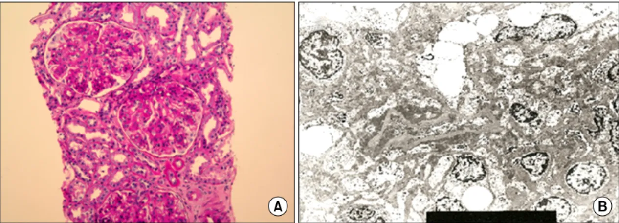
Fig. 2. 27-year-old woman with lupus nephritis WHO class III+V. (A) Light microscopy shows moderate hypercellular mesangial tissue and focal endocapillary proliferation. Capillary loops are focally thickened with subendothelial deposition (H&E stain, ×400). (B) Electron microscopy shows diffuse effacement of epithelial foot processes and wide spread subepithelial electron-dense deposition (×5,000).
소견을 보였다(그림 1B). 그러나 환자의 체중은 입 원 당시 42 kg에서 48 kg으로 증가하였고, 단백뇨도 진행하는 양상을 보였으며 무작위 요단백과 요중 크 레아티닌 분획은 20.8이었다. 저자들은 입원 1주 후 부터 고용량의 프레드니솔론(1 mg/kg)을 경구 투여 하기 시작하였고 입원 2주 후 초음파 유도 하 우측 콩팥 조직검사를 시행하였다. 콩팥 조직검사의 결과
는 광학 현미경 사진에서 사구체의 일부 분절에서 국소적인 모세혈관 내 증식 및 내피 밑 침착을 보이 고 전자 현미경 사진에서 미만성 발 세포의 소실 및 광범위한 상피밑 전자밀도침착 소견을 보여 WHO classification III와 V, ISN (international society of neph- rology)/RPS (renal pathology society) classification III A/C와 V가 동반된 루푸스 신염 소견을 보였다(그림 2).
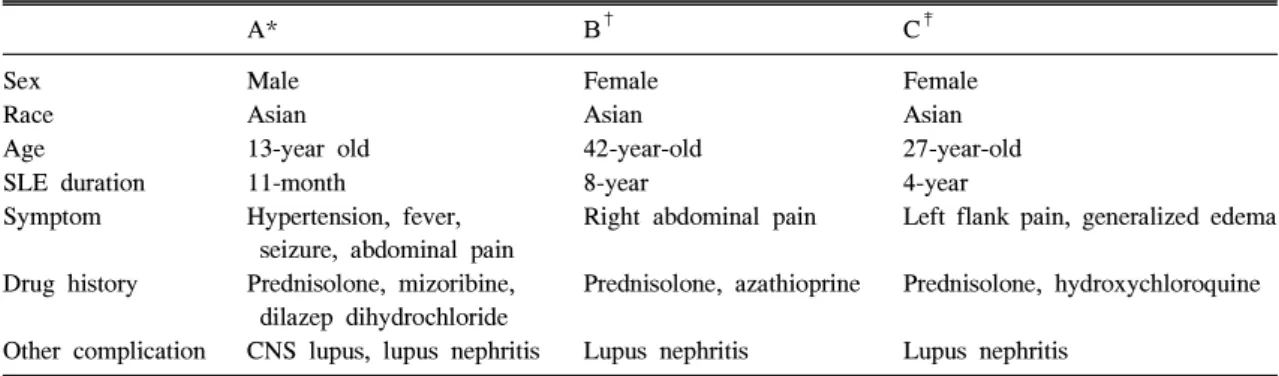
Table 1. Comparison of previous renal infarction cases of SLE patients negative for antiphospholipid antibodies to the current case
A* B† C‡
Sex Male Female Female
Race Asian Asian Asian
Age 13-year old 42-year-old 27-year-old
SLE duration 11-month 8-year 4-year
Symptom Hypertension, fever, seizure, abdominal pain
Right abdominal pain Left flank pain, generalized edema
Drug history Prednisolone, mizoribine, dilazep dihydrochloride
Prednisolone, azathioprine Prednisolone, hydroxychloroquine
Other complication CNS lupus, lupus nephritis Lupus nephritis Lupus nephritis
*Tsugawa K, Tanaka H, Kudo M, Nakahata T. Renal artery thrombosis in a pediatric case of systemic lupus erythematosus without antiphospholipid antibodies. Pediatr Nephrol 2005; 20(11):1648-50., †Suwabe H, Moriuchi J, Hoshina Y. Renal and cerebral infarctions in a patient with systemic lupus erythematosus without antiphospholipid antibodies. Ryumachi 1993;33(4):335-40., ‡Current case
콩팥 조직검사 시행 후 경구 프레드니솔론은 지속 적으로 사용하였고 저 분자량 헤파린은 경구 와파린 으로 변경하였다. 입원 1달 뒤 전신 부종이 호전되 어 퇴원하였으며 현재 외래에서 추적 관찰하면서 프 레드니솔론 용량 감량 중이다.
고 찰
전신홍반루푸스는 원인이 알려지지 않은 전신질환 으로 기본적인 병태 생리는 면역 복합체의 침착, 보 체의 활성화, 염증 반응의 결과로 생각되며 (6) 일반 적으로 심혈관계 질환의 발생의 빈도가 높은 것으로 알려져 있다. Mok 등은 625명의 전신홍반루푸스 환 자를 대상으로 관찰하여 인종에 따라 60개월 뒤의 동맥질환의 이환율은 5.1%에서 8.5%, 정맥질환의 이 환율은 3.7%에서 10.3%로 보고하였으며, 동맥 질환 은 뇌경색 및 일과성 허혈 발작이 대부분 (65%)이었 고 정맥 질환은 심부정맥 혈전증 또는 폐동맥 혈전 증이 대부분 (80%)이었다 (1). 전신홍반루푸스 환자 에서 관상동맥 질환을 일으키는 위험인자로는 진단 당시 고령, 긴 유병 기간, 긴 프레드니솔론 사용기 간, 고혈압의 병력, 폐경, 고지혈증 등이 있다 (7).
전신홍반루푸스 환자에서 항인지질항체가 양성인 경우에는 혈전증의 발병률이 높다는 것은 잘 알려진 사실로, 문헌에 의하면 콩팥 혈전증이 있는 전신홍 반루푸스 환자는 일반적으로 항인지질항체가 양성인 소견을 보인다 (8, 9). Laskin 등의 보고에 의하면 전
신홍반루푸스 환자에서 루푸스 항응고인자가 양성인 경우에는 혈전증의 빈도가 40%였으나 루푸스 항응 고인자가 음성인 경우에는 혈전증의 빈도가 12%였 고, 항카디오리핀항체가 양성인 경우에는 혈전증의 빈도가 40%였으며 항카디오리핀항체가 음성인 경우 에는 혈전증의 빈도는 18% 였다 (10). 그리고 Annexin V라는 anti-thrombotic plasmaprotein이 혈관 내피세포 에 결합하는 것을 항인지질항체가 저해함으로 인해 전신홍반루푸스 환자에서 심혈관 질환이 높아진다고 알려져 있다 (3). 따라서 전신홍반루푸스에서 항인지 질항체의 존재는 그렇지 않은 경우에 비해 혈전증의 빈도를 높인다고 추정할 수 있다. 전신홍반루푸스 환자에서 콩팥 혈전증이 동반된 경우에 일반적으로 동맥 혈전증보다 정맥 혈전증이 많이 발생하며 (11) 콩팥 동맥 침범이 있는 경우에도 주로 소동맥에서 혈전증이 관찰되었다. 콩팥 동맥 혈전증이 콩팥 정 맥 혈전증보다 드문 이유는 혈관간세포에 의한 다량 의 산화질소와 프로스타글란딘 I2의 합성이 혈소판 활성화를 억제하여 사구체 모세혈관의 혈전증 발생 을 예방하기 때문이라고 보고된 바 있다 (12).
전신홍반루푸스 환자에서 항인지질항체 외에 심혈 관계 질환을 유발하는 다른 요소로는 염증반응, 스 테로이드 치료에 의한 과다응고, 고혈압, 이상지질혈 증, 지질의 산화 등을 생각해 볼 수 있다. 최근에는 C 반응 단백 및 염증 반응이 죽상경화증의 위험인 자로 생각되고 있고, 죽상경화증 부위에 활성화된 대식세포와 T 세포가 많다는 점에서 죽상경화증이
염증성 질환으로 이해되고 있다 (13). 또한 ESR, α1 antitrypsin, C반응 단백, TNF-α 등의 급성기 반응 물 질들이 전신홍반루푸스 환자의 심혈관 질환과 관련 이 있는 것으로 알려져 있고, 전신홍반루푸스 환자 에서 볼 수 있는 특징적인 이상지질혈증인 고밀도지 질단백의 감소와 중성지방의 상승, 정상 또는 약간 상승한 저밀도지질단백은 TNF-α에 의한 간의 지질 합성 촉진 및 lipoprotein lipase의 억제에 의한 것으 로 설명된다 (3).
본 증례의 환자는 전신홍반루푸스로 진단 후에 일 시적으로 프레드니솔론 및 항말라리아제를 복용하다 가 추적 소실되었으며 이로 인해 질병의 활성도가 높아져 있었던 상태로 생각된다. 이전에 보고되었던 항인지질항체가 음성인 전신홍반루푸스 환자에서 발 생한 콩팥 경색 증례와 비교해 보면 모두 루푸스 신 염이 동반되었으며 복부 또는 옆구리의 통증이 있었 고 Tusugawa 등의 보고 (4)에서는 소아환자에서 발 생한 콩팥 경색이었으며 중추신경계 합병증도 동반 되어있었다(표 1). 결론적으로 본 증례는 항인지질항 체 음성인 전신홍반루푸스 환자에서도 콩팥 동맥 혈 전증이 발생할 수 있음을 보여주며, 상기 고찰에서 언급한 내용을 종합해볼 때 본 증례의 환자에서는 높은 질병의 활성도로 인한 전신적인 염증 반응이 혈전증의 발생에 기여를 했을 것으로 추정된다. 이 에 전신홍반루푸스의 지속적인 치료로 질병의 활성 도를 떨어뜨리는 것이 혈전성 합병증의 발생을 예방 하는데 도움이 될 것이라고 생각되며, 전신홍반루푸 스 환자에서 갑작스러운 원인 불명의 옆구리 통증이 있을 때는 콩팥 혈전증과 콩팥 경색의 가능성에 대 한 임상적인 주의가 필요하다.
요 약
저자들은 갑작스러운 옆구리 통증을 주소로 내원 한 전신홍반루푸스 환자에서 컴퓨터 단층촬영검사소 견을 바탕으로 콩팥 동맥 혈전에 의한 콩팥 경색을 진단하였으나 혈액검사 소견에서 항인지질항체 증후 군의 증거는 없었던 증례를 경험하였기에 문헌 고찰 과 함께 보고하는 바이다.
참고문헌
1) Mok CC, Tang SS, To CH, Petri M. Incidence and risk factors of thromboembolism in systemic lupus erythematosus: a comparison of three ethnic groups.
Arthritis Rheum 2005;52:2774-82.
2) Appel GB, Pirani CL, D'Agati V. Renal vascular complications of systemic lupus erythematosus. J Am Soc Nephrol 1994;4:1499-515.
3) Frostegard J. SLE, atherosclerosis and cardiovascular disease. J Intern Med 2005;257:485-95.
4) Tsugawa K, Tanaka H, Kudo M, Nakahata T. Renal artery thrombosis in a pediatric case of systemic lupus erythematosus without antiphospholipid antibodies.
Pediatr Nephrol 2005;20:1648-50.
5) Suwabe H, Moriuchi J, Hoshina Y. Renal and cerebral infarctions in a patient with systemic lupus erythe- matosus without antiphospholipid antibodies. Ryu- machi 1993;33:335-40.
6) Hall S, Buettner H, Luthra HS. Occlusive retinal vas- cular disease in systemic lupus erythematosus. J Rheumatol 1984;11:846-50.
7) Nikpour M, Urowitz MB, Gladman DD. Premature atherosclerosis in systemic lupus erythematosus. Rheum Dis Clin North Am 2005;31:329-54.
8) Asherson RA, Derksen RH, Harris EN, Bouma BN, Gharavi AE, Kater L, et al. Chorea in systemic lupus erythematosus and "lupus-like" disease: association with antiphospholipid antibodies. Semin Arthritis Rheum 1987;16:253-9.
9) Hernandez D, Dominquez ML, Diaz F, Fernandez ML, Lorenzo V, Rodriquez A, et al. Renal infarction in a severely hypertensive patient with lupus ery- thematosus and antiphospholipid antibodies. Nephron 1996;72:298-301.
10) Laskin CA, Clark CA, Spitzer KA. Antiphospholipid syndrome in systemic lupus erythematosus: is the whole greater than the sum of its parts? Rheum Dis Clin North Am 2005;31:255-72.
11) Soltesz P, Veres K, Lakos G, Kiss E, Muszbek L, Szeqedi G. Evaluation of clinical and laboratory features of antiphospholipid syndrome: a retrospective study of 637 patients. Lupus 2003;12:302-7.
12) Raij L, Shultz PJ. Endothelium-derived relaxing fac- tor, nitric oxide: effects on and production by me- sangial cells and the glomerulus. J Am Soc Nephrol 1993;3:1435-41.
13) Ross R. Atherosclerosis--an inflammatory disease. N Engl J Med 1999;340:115-26.