http://dx.doi.org/10.4078/jrd.2014.21.2.77
77
<Received:April 22, 2013, Revised (1st: June 3, 2013, 2nd: June 16, 2013), Accepted:June 17, 2013>
Corresponding to:Seung-Geun Lee, Division of Rheumatology, Department of Internal Medicine, Pusan National University School of Medicine, 179, Gudeok-ro, Seo-gu, Busan 602-739, Korea. E-mail:[email protected]
pISSN: 2093-940X, eISSN: 2233-4718
Copyright ⓒ 2014 by The Korean College of Rheumatology
This is a Free Access article, which permits unrestricted non-commerical use, distribution, and reproduction in any medium, provided the original work is properly cited.
A Case of Tjalma Syndrome Coincidentally Accompanied by an Ovarian Teratoma Successfully Treated with Intravenous
Immunoglobulin-G Adjunctive Therapy
Eun-Kyoung Park1, Seung-Geun Lee1, Ik Soo Byon2, Sun-Hee Lee1, Seong-Jun Lee1, Yoon-Jeong Nam1, Ji-Hye Park1, Geun-Tae Kim3, Young-Eun Park4, Seong-Hu Park5,
Joung-Wook Lee6, Jun-Hee Lee7, Seung-Hoon Baek7
Department of Internal Medicine, Pusan National University School of Medicine1, Busan, Department of Ophthalmology, Pusan National University Yangsan Hospital2, Yangsan, Division of Rheumatology, Department of Internal Medicine, Kosin University College of Medicine3,
Malgeunsem Hospital4, Changwon, Young-do Hospital5, Busan St. Mary’s Medical Center6, Department of Internal Medicine, Ilsin Christian Hospital7, Busan, Korea
Tjalma or pseudo-pseudo Meigs’ syndrome is a clinical con- dition that is characterized with ascites, pleural effusion, and increased serum CA-125 levels in patients with systemic lu- pus erythematosus (SLE) without the presence of ovarian tumor. On the other hand, Meigs’ and pseudo-Meigs’ syn- dromes represent the same manifestations with ovarian tumor. In this case report, we present a 43-year-old SLE pa-
tient suffering from Tjalma syndrome with the coexistence of incidental ovarian teratoma, who was successfully treated with intravenous immunoglobulin-G adjunctive therapy af- ter inadequate response to surgical excision of the ovarian tumor, steroid, and cyclophosphamide pulse therapy.
Key Words. Systemic lupus erythematosus, Intravenous im- munoglobulin, Ascites, Pleural effusion
Introduction
Meigs’ and pseudo-Meigs’ syndromes are paraneoplastic conditions characterized by ascites, pleural effusion, and in- creased CA-125 serum levels accompanied by ovarian tumor with resolution of the ascites and hydrothorax after removal of the ovarian lesion (1,2). Meigs’ syndrome is associated with benign ovarian fibromas (1). On the other hand, pseu- do-Meigs’ syndrome occurs in the presence of ovarian ter- atoma, ovarian carcinoma or leiomyoma. In patients with sys- temic lupus erythematosus (SLE), there is a similar condition referred to as Tjalma or pseudo-pseudo Meigs’ syndrome, which has the systemic manifestations of Meigs’ syndrome, but occurs in the absence of an ovarian neoplasm (3). To date, 6 cases of Tjalma syndrome have been reported and im- munosuppressive therapy was shown to effectively treat all of these cases (3-8). However, there are few reports of the coex-
istence of an incidental ovarian tumor with the efficacy of in- travenous immunoglobulin-G (IVIG) adjunctive therapy re- fractory to conventional therapy in Tjalma syndrome.
Here, we present a case of Tjalma syndrome coincidentally accompanied by an ovarian teratoma that was successfully treated with IVIG adjunctive therapy.
Case Report
A 43-year-old woman was admitted to our academic rheuma- tology clinic with complaints of generalized edema, facial rash, and arthralgia lasting for 5 months. She also complained of abdominal distension, a weight gain of 4 kg over 1 month, and generalized weakness. On physical examination, prominent as- cites, bilateral pitting edema of the lower extremities, and swe- lling of proximal interphalangeal joints were detected. Derma- tological examination revealed an erythematous malar rash.
Figure 1. Chest radiograph showing right pleural effusion (arrow).
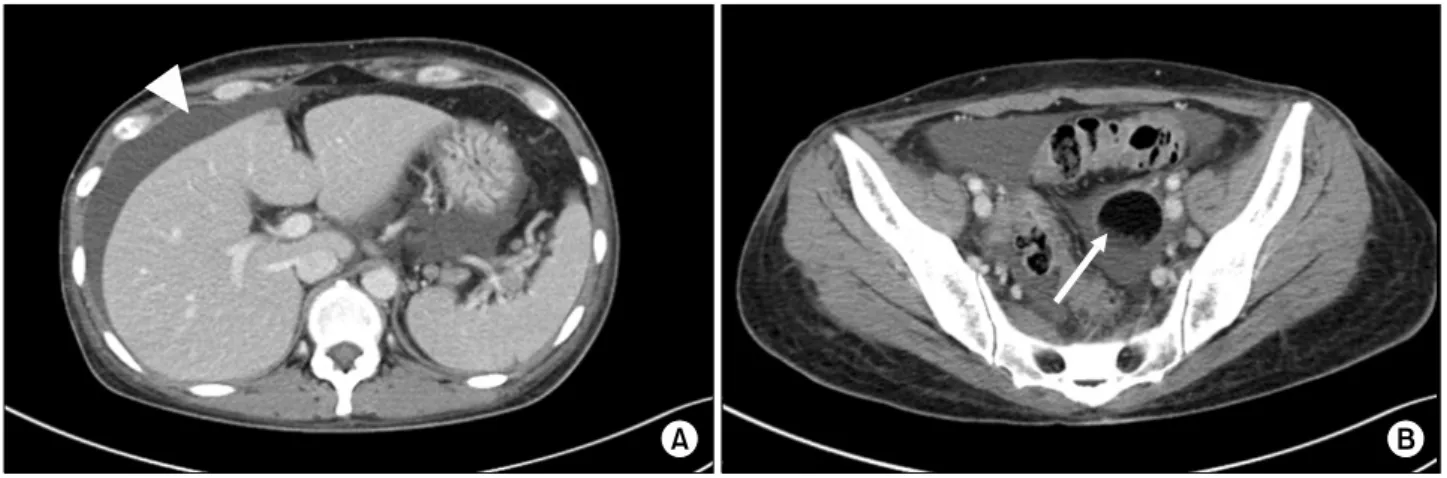
Figure 2. Abdominal computed tomography showing ascites (arrowhead in A) and 4.3 cm sized ovarian tumor (arrow in B).
On the first day of admission (day 0), laboratory findings were as follows: white blood cell count (WBC), 4,900/mm3 (lymphocytes 872/mm3); hemoglobin, 13.6 g/dL; platelet count, 208,000/mm3; erythrocyte sedimentation rate, 43 mm/hr; C-re- active protein, 0.44 mg/dL; serum creatinine, 0.58 mg/dL; se- rum total protein, 4.6 g/dL; serum albumin, 1.8 g/dL; serum lactate dehydrogenase (LDH), 415 IU/L (normal range 218∼
472 IU/L); anti-nuclear antibody (ANA), 1:1,280 positive (speckled pattern); anti-double stranded DNA IgG, 37.2 IU/mL (normal range 0∼20 IU/mL); C3, 32.0 mg/dL (normal range 90∼180 mg/dL); C4, 5.4 mg/dL (normal range 10∼40 mg/dL); and serum CA-125, 544.4 U/mL (normal range 0∼35 U/mL); positive for anti-RNP Ab, anti-Smith Ab, anti-Ro, an- ti-histone Ab. Rheumatoid factor, anti-cyclic citrullinated pep- tide Ab, anti-La, anti-cardiolipin IgG and IgM, and lupus anti- coagulant were negative. Urine dipstick protein value was neg- ative and urine protein/creatinine ratio was 298.7 mg/g. Taken together, she was diagnosed with SLE (malar rash, arthritis, lymphopenia, positive ANA, and anti-Smith Ab).
Chest radiography showed a right pleural effusion (Figure 1).
Thoracentesis was performed on the right side and fluid analy- sis showed the following results: total protein, 1.6 g/dL; albu- min, 1.0 g/dL; LDH, 169 IU/L; and adenosine deaminase (ADA), 10.8 IU/L (normal range 4.3∼20.3 IU/L). Staining and cultures for acid-fast bacilli (AFB) were negative. Pleural effusion was consistent with a transudate by Light’s criteria.
Abdominal and pelvic computed tomography (CT) scanning showed a 3.6-cm left ovarian teratoma, uterine myoma, and ascites (Figure 2). However, no definite peritoneal or bowel wall thickening or liver cirrhosis, which can cause ascites, was noted. A blood interferon gamma release assay was negative.
No abnormality was found on colonoscopy and gastroscopy.
Ascitic fluid analysis showed a WBC of 105/mm3 (2% neu- trophils, 34% lymphocytes, and 64% other cells) and CA-125
levels of 302.2 U/mL; the ADA range was normal, and stain- ing and cultures for AFB were negative. Cytologic examina- tion of the ascitic fluid was negative for malignancy and the serum-to-ascites albumin gradient was 0.6 g/dL. On echo- cardiography, her ejection fraction was 65% and there was no abnormality except for mild eccentric mitral regurgitation.
Fecal alpha-1 antitrypsin (AAT) clearance was 24.7 mL/day, which was within the normal range.
As the patient had left ovarian teratoma, ascites, pleural effu- sion, and increased serum and ascites CA-125 levels, we ini- tially suspected pseudo-Meigs’ syndrome incidentally accom- panied by SLE. She consulted a gynecologist for removal of the ovarian teratoma in order to clarify her diagnosis. However, the operation was postponed because of her general medical problems including hypoalbuminemia and ascites. Medical treatment was started with prednisolone at 0.5 mgㆍkg-1ㆍday-1, 400 mg of hydroxychloroquine for SLE, and diuretics for as-
Table 1. Changes in serum CA-125 and albumin concentrations according to therapy
Day 0 98 102 109 141 143 148 150 154 156 162 178 197
Serum albumin (mg/dL) Serum CA-125 (U/mL) Treatment
1.4
544.4 1.5
2,266
Oophorectomy 1.5
1,910 1.8
2,951
MPT for 3 days
Cyclopho- sphamide
1.6
2,783 1.5
IVIG for 5 days
2
1,710 3.4
455.1 3.8
33.6
MPT: methylprednisolone pulse therapy, IVIG: intravenous immunoglobulin-G.
cites and pleural effusion. She was discharged on day 22 and medications were maintained in the outpatient department. Her symptoms had not worsened for about 2 months.
Ninety-eight days after her first visit to our clinic (day 98), the patient was readmitted because her peripheral pitting ede- ma had worsened with an additional weight gain of 5 kg.
Furthermore, dyspnea had newly developed. A follow-up CT scan showed increased ascites and pleural effusion, and her serum CA-125 levels had increased to 2,266 U/mL (Table 1).
Left salphingo-oophorectomy and uterine myomectomy were performed on day 102. A mature teratoma was diagnosed on pathologic examination of the ovarian specimen. Despite elim- ination of the ovarian tumor, her pleural effusion and ascites did not resolve, and serum CA-125 levels further increased to 2,951 U/mL on day 141 (Table 1). Thus, we assumed that persistent pleural effusion and ascites and increased levels of CA-125 were not caused by the teratoma (pseudo-Meigs’ syn- drome) but by an autoimmune disease such as SLE (Tjalma syndrome). Thereafter, pulse methylprednisolone therapy on 3 consecutive days given as a 1g intravenous bolus followed by oral prednisolone at 1 mgㆍkg-1ㆍday-1 was started on day 143. However, her ascites and pleural effusion persisted.
Subsequently, on day 148, pulse therapy with 750 mg of cy- clophosphamide was added, but her ascites and hypo- albuminemia worsened (Table 1). Thus, we thought that the patient was refractory to this cytotoxic agent and recommend additional treatment. But, she firmly refused other im- munosuppressive agents such as mycophenolate mofetil or azathioprine due to fear of adverse events. Based on previous reports of favorable response of various autoimmune con- ditions to IVIG, therapy with IVIG was discussed with the patient. Her serum IgA levels were 216.3 mg/dL (normal range 70∼400 mg/dL); after obtaining the patient’s consent, an IVIG infusion (2 g/kg over 5 consecutive days) was per- formed on day 156. After IVIG administration, her pleural ef- fusion and ascites dramatically resolved. In addition, serum al- bumin levels increased to 4.2 g/dL, and serum CA-125 levels normalized to 33 U/mL on day 197 (Table 1). Since the IVIG
infusion, she has remained in remission and is being followed up in the outpatient department maintaining low dose pre- dnisolone (2.5 mg/day) and hydroxychloroquine only.
Discussion
We believe that this report represents a case of Tjalma syn- drome and implies that SLE was the cause of the patient’s ascites, pleural effusion, and elevated CA-125 levels. Initially, an ovarian tumor was indeed found on abdominal CT, leading us to diagnose Meigs’ or pseudo-Meigs’ syndrome. However, there was no improvement in the patient’s systemic findings and serum CA-125 levels after removal of the ovarian tumor, which was histologically confirmed as a well-differentiated teratoma. It is well known that after removal of the causative tumor in pseudo-Meigs’ syndrome, ascites invariably im- proves, and recurrence is rare (1,2). We therefore concluded that the teratoma was a coincidental finding and not the direct cause of her signs. To our knowledge, this is the first reported case of a Tjalma syndrome incidentally accompanied by ovar- ian teratoma. Although Bes et al. (7) reported Tjalma syn- drome with the coexistence of cystic formation in the right adnexal region, this was a misdiagnosis of ovarian malignancy rather than a true ovarian tumor.
Among other diagnostic possibilities, such as constrictive pericarditis, liver cirrhosis, tuberculous peritonitis, and neph- rotic syndrome were excluded by echocardiography, abdomi- nal CT scan, microbiologic testing for tuberculosis, and uri- nalysis findings, respectively. Although protein-losing enter- opathy was also a consideration, this condition was excluded for the following reasons. First, AAT clearance was within the normal range (nuclear radiology to quantitate enteric protein loss was impossible in our tertiary hospital). Second, ascites had an exudative nature rather than transudative features, im- plying that these were not caused by hypoalbuminemia. Third, she had no diarrhea or dyslipidemia and no abnormality on colonoscopy. Acute or chronic lupus peritonitis was another diagnostic consideration. However, acute lupus peritonitis usu- ally presents with severe abdominal pain and evidence of
bowel wall inflammation on imaging, which were not ob- served in this case. In addition, lupus peritonitis usually does not cause clinically significant ascites (9).
CA-125, a useful biomarker for evaluation of gynecologic tu- mors, is expressed in mesenchymal cell and increases as a re- sult of interaction between mesothelial cell and cytokines such as vascular endothelial growth factor (VEGF) and fibroblast growth factor (10). CA-125 may be elevated by renal involve- ment of increased disease activity in SLE, but this finding re- mains controversial (11). Recently, Yang et al. (11) showed a significant relationship between serositis and elevate serum CA-125 in SLE patients. However, they did not report any patient with massive pleural effusion and ascites in combina- tion with high CA-125 levels. Therefore, we assume that CA-125 elevation in our patient is not caused by “simple” se- rositis but a manifestation of Tjalma syndrome which is a dis- tinctive clinical entity in SLE.
Treatment of Tjalma syndrome has yet to be established as a result of the limited number of reports of this condition. In previous reports, patients with Tjalma syndrome in whom ste- roids and other immunosuppressive agents including cyclo- phosphamide, mycophenolate mofetil, and azathioprine have been employed have been successfully treated with no relapse (3-8). In this case, we initiated IVIG treatment 1 week after steroid and cyclophosphamide pulse therapy as she did not show recovered systemic signs and serum CA-125 levels de- spite treatment with these immunosuppressive drugs. Just after administration of IVIG infusion over 5 consecutive days, a rapid improvement of all clinical and serologic markers was observed. Because of the relatively short time interval (1 week) between the initiation of immunosuppressive agents and IVIG, it is difficult to determine whether IVIG treatment sole- ly led to clinical remission in this patient. However, we infer that IVIG could have played an adjunctive role in the clinical improvement of her condition considering both the onset of clinical improvement after IVIG treatment and the pharmaco- kinetics of IVIG. Once administered, IVIG causes an initial sharp rise in serum concentrations followed by a rapid waning for 1∼4 days (12), which suggests that the effects of IVIG therapy can be observed rapidly, as in the current case.
There exist many uncertainties as to how immunoglobulin acts although numerous reports of its effectiveness in various autoimmune diseases (12). It is assumed that it would work through Fc receptor blockade, inhibition of complement depo- sition, enhancement of regulatory T cells, inhibition or neu- tralization of cytokines and growth factors, accelerated clear- ance of autoantibodies, modulation of adhesion molecules and cell receptors, and activation of regulatory macrophages
through the FcγRIIb receptor (12). IVIG also showed an- ti-VEGF antibody activity via stimulating the production of IL-12 which has an anti-angiogenic cytokine and inhibiting VEGF mRNA expression (13). We speculated that anti-VEGF antibody activity of IVIG would play a role in our patient, considering that VEGF can stimulate CA-125 expression.
Summary
In conclusion, this case suggests that the combination of as- cites, pleural effusion, and elevated CA-125 in patients with SLE should indicate the possibility of Tjalma syndrome, even in the presence of ovarian tumor. Additionally, IVIG ad- junctive therapy can be effective for the management of Tjalma syndrome.
Acknowledgements
No potential conflict of interest relevant to this article is reported. We thank late professor Sung-Il Kim who devoted himself to patient care, research, and education in the Division of Rheumatology, Department of Internal Medicine, Pusan National University School of Medicine (1963∼2011). This work was supported by the year 2013 clinical research grant from Pusan National University Hospital.
References
1. Meigs JV, Cass JW. Fibroma of the ovary with ascites and hydrothorax: with a report of seven cases. Am J Obstet Gynecol 1937;33:249-66.
2. Nagakura S, Shirai Y, Hatakeyama K. Pseudo-Meigs' syndrome caused by secondary ovarian tumors from gas- trointestinal cancer. A case report and review of the literature. Dig Surg 2000;17:418-9.
3. Widra EA, Armstrong J. Pseudo-Meigs' syndrome and lupus. Int J Gynaecol Obstet 1995;49:193-4.
4. Schmitt R, Weichert W, Schneider W, Luft FC, Kettritz R. Pseudo-pseudo Meigs' syndrome. Lancet 2005;366:
1672.
5. Tjalma WA. Ascites, pleural effusion, and CA 125 ele- vation in an SLE patient, either a Tjalma syndrome or, due to the migrated Filshie clips, a pseudo-Meigs syndrome.
Gynecol Oncol 2005;97:288-91.
6. Ural UM, Kiliç A, Güngör T, Ozdal B, Mollamahmutoğlu L. Tjalma's or pseudo-pseudo-Meigs' syndrome: a case report. Clin Exp Dermatol 2008;33:363-4.
7. Bes C, Dağlı Ü, Memedoğlu P, Soy M. A rare form of SLE: pseudo-pseudo meigs syndrome and hydrocephalus.
Rheumatol Int 2013;33:2175-6.
8. Dalvi SR, Yildirim R, Santoriello D, Belmont HM.
Pseudo-pseudo Meigs' syndrome in a patient with sys- temic lupus erythematosus. Lupus 2012;21:1463-6.
9. Prasad S, Abujam B, Lawrence A, Aggarwal A. Massive ascites as a presenting feature of lupus. Int J Rheum Dis
2012;15:e15-6.
10. Abramov Y, Anteby SO, Fasouliotis SJ, Barak V. The role of inflammatory cytokines in Meigs' syndrome.
Obstet Gynecol 2002;99:917-9.
11. Yang Z, Liang Y, Li C, Zhong R. Serum CA125 ele- vation is independently associated with serositis in SLE patients. Clin Exp Rheumatol 2012;30:93-8.
12. Bayry J, Negi VS, Kaveri SV. Intravenous immunoglobulin therapy in rheumatic diseases. Nat Rev Rheumatol 2011;
7:349-59.
13. Damianovich M, Blank M, Raiter A, Hardy B, Shoenfeld Y. Anti-vascular endothelial growth factor (VEGF) spe- cific activity of intravenous immunoglobulin (IVIg). Int Immunol 2009;21:1057-63.