http://dx.doi.org/10.3988/jcn.2014.10.2.125 J Clin Neurol 2014;10(2):125-132
Introduction
Obsessive-compulsive disorder (OCD) is a prevalent psychi- atric disease that is characterized by disabling obsessions about intrusive unwanted thoughts and images, and/or com- pulsions expressed as ritualized repetitive behaviors.1 OCD has a lifetime prevalence of 2–3% in the general population,2 and typically 10–20% of affected people exhibit marked malad-
justments in professional and social functioning even after treatments that include behavioral therapy and medications.3,4
Possible mechanisms underlying OCD are related to the functional dysregulation of the frontal-cingulate-thalamic- limbic circuit.5 Several psychiatric and neurologic disorders are thought to be associated with OCD or obsessive-compul- sive symptoms (OCS) due to the involvement of this circuit.
People with epilepsy (PWE) can also exhibit obsessional per- sonalities linked to particular types of epilepsy.6 Uncontrolled temporal lobe epilepsy (TLE) was first reported with OCS and OCD.7 The frequency of OCD or OCS in controlled or uncon- trolled TLE patients, which has ranged from 11% to 34.5%,
Obsessive-Compulsive Symptoms and Their Impacts on Psychosocial Functioning in People with Epilepsy
Ji-Hye Seo,a* Won-Kee Lee,b* Sung-Pa Parka
aDepartment of Neurology and bCenter of Biostatics, School of Medicine, Kyungpook National University, Daegu, Korea
Received July 4, 2013 Revised October 28, 2013 Accepted October 30, 2013 Correspondence Sung-Pa Park, MD, PhD Department of Neurology, School of Medicine,
Kyungpook National University, 680 Gukchaebosang-ro, Jung-gu, Daegu 700-842, Korea Tel +82-53-420-5769 Fax +82-53-422-4265 E-mail [email protected]
*These authors contributed equally as first authors in this study.
Background and PurposezzObsessive-compulsive symptoms (OCS) in people with epilepsy (PWE) have not been studied systematically. We evaluated the severity, predictors, and psycho- social impact of OCS in PWE.
MethodszzWe recruited PWE who visited our epilepsy clinic and age-, gender-, and education- matched healthy controls. Both PWE and healthy controls completed the Maudsley Obsessional- Compulsive Inventory (MOCI), which measures OCS. PWE also completed the Beck Depres- sion Inventory (BDI) and the Quality of Life in Epilepsy Inventory-31 (QOLIE-31). We examined the severity of OCS in PWE relative to healthy controls. Predictors of OCS and the QOLIE-31 score were measured by regression analyses. A path analysis model was constructed to verify interrelations between the variables.
ResultszzThe MOCI total score was significantly higher in PWE than in healthy controls (p=
0.002). OCS were found in 20% of eligible patients. The strongest predictor of the MOCI total score was the BDI score (β=0.417, p<0.001), followed by EEG abnormality (β=0.194, p<0.001) and etiology (β=0.107, p=0.031). Epileptic syndrome, the side of the epileptic focus, and action mechanisms of antiepileptic drugs did not affect the MOCI total score. The strongest predictor of the QOLIE-31 overall score was the BDI score (β=-0.569, p<0.001), followed by seizure con- trol (β=-0.163, p<0.001) and the MOCI total score (β=-0.148, p=0.001). The MOCI total score directly affected the QOLIE-31 overall score and also exerted indirect effects on the QOLIE-31 overall score through seizure control and the BDI score.
ConclusionszzOCS are more likely to develop in PWE than in healthy people. The development of OCS appears to elicit psychosocial problems directly or indirectly by provoking depression
or uncontrolled seizures. J Clin Neurol 2014;10(2):125-132
Key Wordszz obsessive-compulsive symptom, MOCI, predictor, epilepsy, quality of life, depression.
Open Access
cc This is an Open Access article distributed under the terms of the Cre- ative Commons Attribution Non-Commercial License (http://creative- commons.org/licenses/by-nc/3.0) which permits unrestricted non-com- mercial use, distribution, and reproduction in any medium, provided the ori- ginal work is properly cited.
was higher than that of healthy controls (0–3%).7-11 It has re- cently been reported that idiopathic frontal lobe epilepsy and idiopathic generalized epilepsy (GE) are also associated with a higher risk of developing OCS, with frequencies of 11.8%
and 16.2%, respectively.11 Although these studies demonstrat- ed a relationship between OCD or OCS and specific epilepsy syndromes, the participating subjects were not representative of the general population of PWE. Rather, the studies dealt with patients who were confined to a specific epilepsy syn- drome7-10 or included a high proportion of subjects with un- controlled epilepsy (UCE; nearly 50%) and who had normal neuroimaging results.11 Therefore, PWE with diverse etiolo- gies and epileptic syndromes still need to be investigated in order to obtain an accurate understanding of the pathogenic mechanisms of OCS.
Observed risk factors for OCD or OCS in PWE were male gender, older age, longer duration of illness, TLE, the number of antiepileptic drugs (AEDs), and uncontrolled seizures with AEDs.9-11 However, one study found no relationship between OCD and age, gender, duration of disease, or seizure frequen- cy.8 Depression and/or anxiety are commonly related psycho- pathologies with OCS.8-11 The side of the epileptic focus as a risk factor for OCS is controversial. A right-hemisphere focus in TLE had initially been suggested to predispose to OCD.12 However, other studies demonstrated an equal contribution between the right- and left-sided foci to OCD or OCS.7,8 Three recent studies found that a left-sided focus in TLE or other epilepsy types was a risk factor for OCD or OCS.9-11 A role of AEDs as a risk factor for OCS has not yet been reported. Pa- tients with TLE taking carbamazepine (CBZ) were more likely to develop OCD than those with idiopathic GE taking valpro- ate (VPA).8 PWE taking CBZ had a higher risk of developing OCS than healthy controls, but their risk did not differ from that of PWE taking VPA.11 However, these studies have a lim- itation that they did not discriminate between the impacts of AEDs and epilepsy syndrome on OCD or OCS. Although many risk factors for OCD or OCS have been suggested, their relative importance to determining OCS was not reported.
Identifying decisive factors may help to minimize OCD or OCS and understand its prognosis.
Various psychiatric symptoms including depression and anxiety are the strongest predictors of the quality of life (QOL) in PWE.13,14 The QOL is higher in subjects with drug-refrac- tory epilepsy but no depression or anxiety symptoms than in subjects with seizure freedom exhibiting depression or anxi- ety symptoms.13 Depression symptoms reportedly contribute more to the QOL than do adverse events associated with AEDs in seizure-free patients with monotherapy.14 Although OCS is commonly associated with depression, anxiety, and markedly impairment of professional and social functioning,
the impact of OCS on QOL in PWE is poorly understood.
The above-described situation prompted us to measure the severity of OCS in PWE who visited our epilepsy clinic with diverse etiologies and epileptic syndromes. We also evaluated predictors of OCS among various demographic and clinical characteristics and investigated the impact of OCS on QOL.
Methods
Subjects
We included consecutive PWE who took AEDs and attended our epilepsy clinic between May 1, 2011 and March 31, 2012.
Our clinic takes care of patients aged 15 years or older with various types of seizure control, etiologies, and epileptic syn- dromes; this population is similar to the overall community- based epilepsy population. Subjects were invited to partici- pate in this study if they were aged 17 years or older, had a current diagnosis of epilepsy, had been taking one or more AEDs for at least 1 year, and had the ability to provide in- formed consent and to agree with the study protocol. Subjects who had insufficient information in their medical records, who had mental retardation or serious medical, neurologic, or psychiatric disorders that prevented them from understand- ing the questionnaire and cooperating with the study, or who refused to complete questionnaires were excluded. We recruit- ed age-, gender- and education-matched healthy controls to compare the severity of OCS with PWE. These healthy con- trols were patients’ caregivers, parents, children, relatives, or friends who visited our clinic and had no medical, neurologic, or psychiatric diseases.
Study design
This case-control study was approved by the institutional re- view board of Kyungpook National University Hospital, and all subjects gave written informed consent before participat- ing in the study. Each patient was interviewed by a trained epi- leptologist (S.P.P.) who also reviewed the medical charts to collate demographic and clinical information in a computer- ized database. The information collected included age, gender, education, socioeconomic status, seizure type, etiology, epi- lepsy syndrome, age at onset, duration of epilepsy, side of the epileptic focus, EEG abnormality, MRI abnormality, concur- rent medical diseases, family history of epilepsy, history of fe- brile convulsion, duration of AED intake, number of AEDs, AED action mechanism, seizure control, and depression symp- toms as measured by the Beck Depression Inventory (BDI).
Epilepsy was diagnosed according to the International League Against Epilepsy classification of seizures and epileptic syn- dromes.15,16 We divided the etiology into idiopathic and symp- tomatic epilepsy. We divided epileptic syndromes into three
groups: TLE, extraTLE, and GE; where extraTLE included epilepsy syndromes with epileptic attacks originating from the frontal, parietal, or occipital lobe. We also divided seizure control into two groups: well-controlled epilepsy (WCE) and UCE. WCE was defined as freedom from seizures during the preceding year, while UCE was defined as experiencing a sei- zure during the preceding year despite an adequate intake of tolerated AEDs. We divided AEDs according to their action mechanisms into sodium-channel blockers and other types, because CBZ was reported to be an OCS-inducing drug.
AEDs such as CBZ, phenytoin (PHT), oxcarbazepine (OXC), lamotrigine (LTG), topiramate, and zonisamide were included as sodium-channel blockers according to the review of Peruc- ca and Mula.17 EEG abnormality was considered to be pres- ent when there were interictal epileptiform discharges (IEDs) on EEG recordings. The IED location was categorized into temporal or frontal IEDs, other focal IEDs, and generalized IEDs. According to the occurrence of OCS by the involve- ment of the frontal-cingulate-thalamic-limbic circuit, we ex- amined the degree of OCS between temporal or frontal IEDs and other focal or generalized IEDs. The side of the epileptic focus was determined based on EEG and MRI findings. If the epileptic focus was generalized or ambiguous, we classified it as being generalized or unknown, respectively. Socioeconom- ic factors were classified as follows: having or not having a job; earning at least one million Korean won (KRW) per month (equivalent to US$ 900 per month) or less than one million KRW per month; having or not having a driving li- cense; and being married, or divorced, bereaved, and unmar- ried.
Eligible patients completed reliable and validated self-re- port questionnaires, including Korean versions of the Maud- sley Obsessional-Compulsive Inventory (MOCI),18 the BDI,19 and the Quality of Life in Epilepsy Inventory-31 (QOLIE-31).20 Healthy controls also completed the MOCI.
Questionnaires
The MOCI is a 30-item questionnaire that evaluates OCS.18 It employed a dichotomous response format, and the total scores range from 0 to 30. It had good internal consistency (Cronbach’s α=0.8) and was consistently correlated with the BDI score (r=0.37). This version of the MOCI employed fac- tor analysis with orthogonal rotation (a linear transformation used to facilitate interpretation of results) to test whether in- dividual MOCI items segregate into separate subscales of OCS. Only subscales with eigenvalues ≥1.0 were retained in the analysis, which yielded 22 items classified into 4 sub- scales: checking (6 items), tidiness (6 items), doubting (6 items, including 1 item that was duplicated in checking), and fear of contamination (4 items). Checking and doubting sub-
scales represented compulsive behaviors, and tidiness and fear of contamination represented obsessive behaviors. We measured the MOCI total score and subscale scores. We deter- mined that the cutoff of the MOCI total score for indicating the presence of OCS was 12, based on two standard deviations from the mean total score in healthy controls recruited from our study, because the original study did not suggest it.
The BDI is the most commonly used self-rating scale for depression.19 Participants rate 21 items on a scale from 0 to 3 according to how they feel at the time. The following cutoff scores are used: 0–16, normal; 17–20, mild depression; 21–23, moderate depression; and 24–63, severe depression. Subjects who score more than 16 points are considered to have depres- sion. Cronbach’s α value is 0.8.
The QOLIE-31 is a 31-item, self-administered question- naire specifically designed to measure QOL in PWE.20 It consists of subscales addressing seizure worry, overall QOL, emotional well-being, energy-fatigue, cognitive functioning, medication effects, and social functioning. An overall score for the seven subscales is also calculated. Higher QOLIE-31 scores are indicative of better QOL. Cronbach’s α values range from 0.69 to 0.86.
Statistical analysis
Data for continuous variables are expressed as mean±SD val- ues, and those for categorical variables are expressed as fre- quencies. The independent-samples t test and Fisher’s exact test were applied to compare variables between PWE and healthy controls. We used the Mann-Whitney U test for cate- gorical independent variables and Spearman’s correlation anal- ysis for continuous independent variables to determine the relationship between demographic and clinical variables and the MOCI total score. Variables that were found to be signifi- cantly correlated with the MOCI total score were then includ- ed in a multiple linear regression analyses with stepwise se- lection. The probabilities of entry and exit were 0.05 and 0.1, respectively. Collinearity was addressed by performing col- linearity statistical analysis. Predictors of the QOLIE-31 over- all score were obtained as the same manner, and the MOCI to- tal score were added as an independent variable. Findings from the linear regression analyses were used to construct a struc- tural equation model to test the interrelations between vari- ables and the QOLIE-31 overall score. The model fit was eval- uated using path analysis, a method that estimates the relative importance of different paths of the independent variables onto the dependent variables. An acceptable model fit was defined as having a nonsignificant chi-square (χ2) value, Normed Fit Index (NFI) of ≥0.9, Comparative Fit Index (CFI) of ≥0.9, Goodness of Fit Index (GFI) of ≥0.9, and Root Mean- square Residual (RMR) of ≤0.05. Structural equation model-
ing was used to estimate the total effect of each predictor, in order to establish a linear model to predict the QOLIE-31 overall score with these interrelations accounted for. Except for the structural equation model, all statistical analyses were conducted with SPSS (version 19.0, IBM Inc.). Linear struc- tural relationship 8.8 for Windows (Scientific Software Inter- national, Inc., Lincolnwood, IL, USA) for the path and struc-
tural equation modeling components of the analysis. The level of statistical significance was set at 0.05.
Results
In total, 386 PWE were initially enrolled in the study. Among them, 86 were excluded because of their refusal to complete
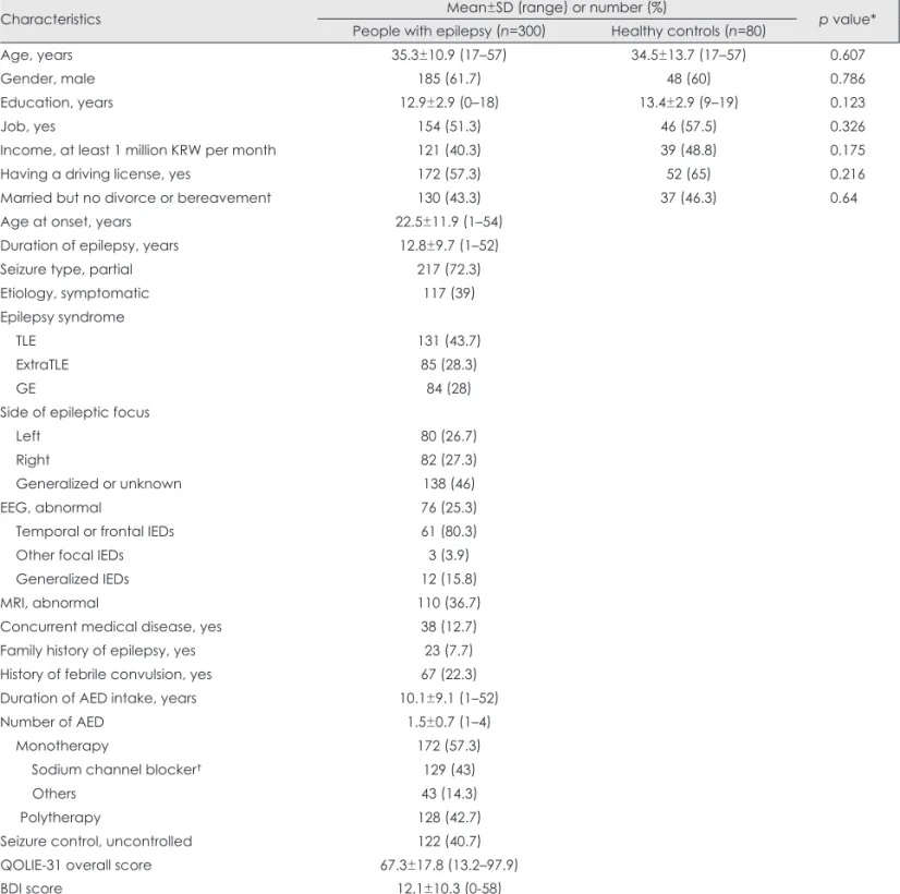
Table 1. Demographic and clinical characteristics of eligible subjects
Characteristics Mean±SD (range) or number (%)
p value*
People with epilepsy (n=300) Healthy controls (n=80)
Age, years 35.3±10.9 (17–57) 34.5±13.7 (17–57) 0.607
Gender, male 185 (61.7) 48 (60) 0.786
Education, years 12.9±2.9 (0–18) 13.4±2.9 (9–19) 0.123
Job, yes 154 (51.3) 46 (57.5) 0.326
Income, at least 1 million KRW per month 121 (40.3) 39 (48.8) 0.175
Having a driving license, yes 172 (57.3) 52 (65) 0.216
Married but no divorce or bereavement 130 (43.3) 37 (46.3) 0.64
Age at onset, years 22.5±11.9 (1–54)
Duration of epilepsy, years 12.8±9.7 (1–52)
Seizure type, partial 217 (72.3)
Etiology, symptomatic 117 (39)
Epilepsy syndrome
TLE 131 (43.7)
ExtraTLE 85 (28.3)
GE 84 (28)
Side of epileptic focus
Left 80 (26.7)
Right 82 (27.3)
Generalized or unknown 138 (46)
EEG, abnormal 76 (25.3)
Temporal or frontal IEDs 61 (80.3)
Other focal IEDs 3 (3.9)
Generalized IEDs 12 (15.8)
MRI, abnormal 110 (36.7)
Concurrent medical disease, yes 38 (12.7)
Family history of epilepsy, yes 23 (7.7)
History of febrile convulsion, yes 67 (22.3)
Duration of AED intake, years 10.1±9.1 (1–52)
Number of AED 1.5±0.7 (1–4)
Monotherapy 172 (57.3)
Sodium channel blocker† 129 (43)
Others 43 (14.3)
Polytherapy 128 (42.7)
Seizure control, uncontrolled 122 (40.7)
QOLIE-31 overall score 67.3±17.8 (13.2–97.9)
BDI score 12.1±10.3 (0-58)
*The independent-samples t test and Fisher’s exact test were applied, †AEDs such as carbamazepine, phenytoin, oxcarbazepine, la- motrigine, topiramate, and zonisamide were included.
AED: antiepileptic drug, BDI: Beck Depression Inventory, EEG: electroencephalography, extraTLE: extratemporal lobe epilepsy, GE:
generalized epilepsy, IEDs: interictal epileptiform discharges, KRW: Korean won, MRI: magnetic resonance imaging, QOLIE-31: Quality of Life in Epilepsy Inventory-31, TLE: temporal lobe epilepsy.
the questionnaires (n=22), inability to complete questionnaires due to mental retardation (n=20) or serious diseases (n=10), insufficient information about epileptic attacks in their medi- cal records (n=5), young age (n=23), and having received less than 1 year of AED treatment (n=6). Therefore, 300 PWE (age:
35.3±10.9 years; 61.7% males) and 80 healthy controls (age:
34.5±13.7 years; 60% males) were included. Demographic and clinical characteristics of eligible subjects are summa- rized in Table 1. Age, gender, education level, and socioeco- nomic status (including job, income, having a driving license, and marital status) did not differ between PWE and healthy controls. Among 300 PWE, 217 patients (72.3%) had partial seizures. Symptomatic epilepsy was present in 117 patients (39%). The most common epilepsy syndrome was TLE (43.7%). The frequencies of left and right epileptic focuses were similar. EEG abnormality was present in 76 patients (25.3%), and the frequency of temporal or frontal IEDs was higher than those of other focal or generalized IEDs. MRI abnormality was found in 110 patients (36.7%), with etiolo- gies of hippocampal sclerosis (n=36), trauma (n=25), brain anomaly (n=19), vascular injury (n=17), infection (n=10), tu- mor (n=2), and unknown (n=9). UCE was present in 122 pa- tients (40.7%). Concurrent medical diseases were present in 38 patients (12.7%), comprising diabetes and other endocri- nologic disorders (n=12), cerebrovascular disease and other neurologic disorders (n=11), hypertension and other cardio- vascular disorders (n=10), brain and systemic tumors (n=4), gastrointestinal disorders (n=4), autoimmune disorders (n=3), renal diseases (n=2), and other diseases (n=3). The duration of AED intake and number of AEDs were 10.1±9.1 years (range: 1–52 years) and 1.5±0.7 (range: 1–4), respectively.
The QOLIE-31 overall score and BDI score were 67.3±17.8 (range: 13.2–97.9) and 12.1±10.3 (range: 0–58), respectively.
The severities of OCS in PWE and healthy controls are compared in Table 2. The MOCI total score was significantly higher in PWE than in healthy controls (p=0.002). Subscale scores of checking and doubting were also significantly higher in PWE than in healthy controls (p=0.01 and p<0.001,
respectively). The presence of OCS, defined by a MOCI total score of 13 or above, was found in 60 patients (20%). Check- ing and doubting scores did not vary with the epileptic syn- drome.
The correlations between demographic and clinical vari- ables and the MOCI total score are summarized in Table 3.
The variables found to be significantly associated with the MOCI total score were gender (p=0.011), job (p=0.001), in- come (p=0.001), having a driving license (p=0.001), seizure type (p=0.04), etiology (p<0.001), EEG abnormality (p<
0.001), MRI abnormality (p=0.001), number of AEDs (p<
0.001), seizure control (p<0.001), and the BDI score (p<0.001).
That is, subjects with female gender, no job, lower income, no driving license, partial seizures, symptomatic etiology, abnormal EEG and MRI findings, larger number of AEDs, UCE, and depression were more likely to develop OCS. Epi- lepsy syndrome, the side of the epileptic focus, the location of IEDs, and the AED action mechanism did not affect OCS.
Predictors of the MOCI total score by stepwise linear re- gression analyses are listed in Table 4. The strongest predic- tor was the BDI score (β=0.453, p<0.001), followed by EEG abnormality (β=0.194, p<0.001) and etiology (β=0.107, p=
0.031). Stepwise regression produced a three-variable model that explained 29.6% of the variance in the MOCI total score.
According to the standardized β, the contribution of the BDI score to OCS was 2.34 times greater than that of EEG abnor- mality and 4.23 times greater than that of etiology. Tolerance was greater than (1-adjusted R2) and variance inflation fac- tors were greater than 10 for all four variables, suggesting that they exerted independent effects without redundancy.
Predictors of the QOLIE-31 overall score are listed in Ta- ble 5. The strongest predictor was the BDI score (β=-0.569, p<0.001), followed by seizure control (β=-0.163, p<0.001) and the MOCI total score (β=-0.148, p=0.001). Stepwise re- gression produced a three-variable model that explained 53.9% of the variance in the QOLIE-31 overall score. Accord- ing to the standardized β, the contribution of the BDI score to QOL was 3.49 times greater than that of seizure control and Table 2. OCS between people with epilepsy and healthy controls
Mean±SD (range) or number (%)
p value*
People with epilepsy (n=300) Healthy controls (n=80)
Checking 2.4±1.7 (0–6) 1.8±1.4 (0–5) 0.010
Tidiness 0.8±1.2 (0–6) 0.7±1.1 (0–4) 0.896
Doubting 2.3±1.7 (0–6) 1.1±1.3 (0–5) <0.001
Fear of contamination 0.6±0.9 (0–4) 0.6±0.8 (0–3) 0.719
MOCI total score 8.1±5.2 (0–27) 6.0±3.4 (0–15) 0.002
Frequency of OCS† 60 (20)
*Mann-Whitney U test was applied, †Patients who had a score above two standard deviations from the mean total score of MOCI in healthy controls were included.
MOCI: Maudsley Obsessional-Compulsive Inventory, OCS: obsessive-compulsive symptoms.
3.84 times greater than that of the MOCI total score. Collin- earity statistical analysis indicated that these variables exert- ed independent effects without redundancy.
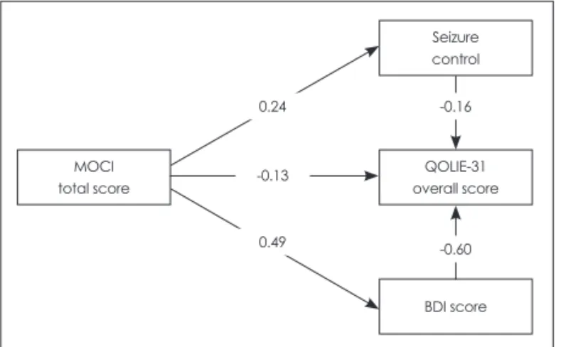
Complex interrelations between predictors and the QO- LIE-31 overall score are illustrated by the refined path analy-
sis model in Fig. 1. According to predefined criteria, the final model provided an acceptable fit to the data (χ2=23.91, p<
0.001; NFI=0.950, CFI=0.950, GFI=0.970, and RMR=
0.080). The MOCI total score, seizure control, and BDI score were found to exert direct effects on the QOLIE-31 overall score. The MOCI total score also exerted indirect effects on the QOLIE-31 overall score through seizure control and the BDI score.
Discussion
This study found that the severity of OCS was significantly higher in PWE than in healthy controls. One fifth of PWE were found to have OCS. Greater depression symptoms, IEDs on EEG recordings, and symptomatic etiology were closely related to the development of OCS, whereas the epileptic syn- drome, side of the epileptic focus, location of IEDs, and AED action mechanism were not predictors of the development of OCS. The development of OCS appears to elicit psychosocial problems directly or indirectly by provoking depression or uncontrolled seizures.
Table 3. Variables and correlation with MOCI total score
Variable p value (r/z)*
Age 0.744 (0.019)
Gender 0.011 (-2.545)
Education 0.105 (-0.094)
Job 0.001 (-3.401)
Income 0.001 (-3.223)
Having a driving license 0.001 (-3.179)
Being married 0.159 (-1.407)
Age at onset 0.129 (-0.088)
Duration of epilepsy 0.066 (0.106)
Seizure type 0.04 (-2.059)
Etiology <0.001 (-3.596)
Epilepsy syndrome
TLE versus extraTLE 0.41 (-0.824)
TLE versus GE 0.059 (-1.887)
ExtraTLE versus GE 0.391 (-0.859)
Side of epileptic focus, left versus right 0.86 (-0.176)
EEG abnormality <0.001 (-4.283)
Temporal or frontal IEDs versus other focal or generalized IEDs
0.865 (-0.17)
MRI abnormality 0.001 (-3.249)
Concurrent medical disease 0.346 (-0.942) Family history of epilepsy 0.606 (-0.515) History of febrile convulsion 0.257 (-1.133)
Duration of AED intake 0.178 (0.078)
Number of AED <0.001 (0.259)
Monotherapy, sodium channel blocker versus others
0.945 (-0.069)
Seizure control <0.001 (-5.779)
BDI score <0.001 (0.452)
*Spearman’s correlation analysis and Mann-Whitney U test were applied. r for continuous variables and z for nominal variables.
AED: antiepileptic drug, BDI: Beck Depression Inventory, EEG:
electroencephalography, extraTLE: extratemporal lobe epi- lepsy, GE: generalized epilepsy, IEDs: interictal epileptiform dis- charges, MOCI: Maudsley Obsessional-Compulsive Inventory, MRI: magnetic resonance imaging, TLE: temporal lobe epilepsy.
Table 4. Predictors to determine MOCI total score by stepwise linear regression analyses
Variable Standardized
coefficients (beta) p value Adjusted R2
BDI score 0.453 <0.001 0.296
EEG abnormality 0.194 <0.001
Etiology 0.107 0.031
BDI: Beck Depression Inventory, EEG: electroencephalogra- phy, MOCI: Maudsley Obsessional-Compulsive Inventory.
Table 5. Predictors to determine QOLIE-31 overall score by step- wise linear regression analyses
Variable Standardized
coefficients (beta) p value Adjusted R2
BDI score -0.569 <0.001 0.539
Seizure control -0.163 <0.001
MOCI total score -0.148 0.001
BDI: Beck Depression Inventory, MOCI: Maudsley Obsessional- Compulsive Inventory, QOLIE-31: Quality of Life in Epilepsy In- ventory-31.
MOCI total score
Seizure control
QOLIE-31 overall score
BDI score -0.16
-0.60 0.24
-0.13
0.49
Fig. 1. Interrelations between clinical variables and the Quality of Life in Epilepsy Inventory-31 (QOLIE-31) overall score by a re- fined path analysis model. An arrow indicates a direct relationship from one variable to another. Numbers denote standardized re- gression coefficients (β weights) for each path. Negative coeffi- cients indicate that when the predictor variable score increases by one standard deviation, the QOLIE-31 overall score decreases by the number of standard deviations equal to the value of the coefficient. BDI: Beck Depression Inventory, MOCI: Maudsley Obsessional-Compulsive Inventory.
We included patients whose clinical constituents were slightly different from those of the general epilepsy popula- tion,21,22 but they had diverse etiologies and epileptic syn- dromes, as indicated by the clinical history and EEG and MRI findings. Therefore, the results of the present study might lead to a better understanding of the pathogenic mechanisms of OCS. The frequency of OCS in our patients resembled the data obtained in other studies.7-11 Among subscales, checking and doubting were significantly increased, as also found by Isaacs et al.7 The OCS of PWE appear to be oriented around compulsive behaviors rather than obsessive behaviors.
Male gender, older age, longer duration of illness, TLE, larger number of AEDs, and uncontrolled seizures with AEDs as well as depression were previously proposed as risk fac- tors for OCS in PWE.9-11 However, the previous studies did not measure the major predictors of OCS among these vari- ables. We found that female gender, no job, lower income, not having a driving license, partial seizures, symptomatic etiolo- gy, abnormal EEG and MRI findings, larger number of AEDs, UCE, and depression as risk factors for OCS, with depres- sion, IEDs on EEG recordings, and symptomatic etiology be- ing major predictors. This is the first study to find that EEG abnormality is a strong predictor of OCS. As there are no stud- ies to elucidate the short-term or long-term effect of IEDs on psychopathology, we hypothesize abnormal electrical dis- charges in the frontal-cingulate-thalamic-limbic circuit are related to the occurrence of OCS. We also found that the spe- cific epilepsy syndrome, the side of the epileptic focus, and the location of IEDs were not risk factors for OCS. Differenc- es in study design may produce different results, and future studies based on the video-EEG monitoring should attempt to clarify the exact epilepsy syndrome or epileptic focus. We also reported that AEDs that block sodium channels did not increase OCS more than AEDs having other action mecha- nisms. CBZ, PHT, OXC, and LTG have a mood-stabilizing ef- fect,17 and hence these agents are not thought to provoke OCS.
The final goal in the management of PWE is to improve psychosocial functioning. The strongest predictors of QOL are depression and anxiety, followed by seizure control.13 We found OCS to be another important risk factor for QOL. The contribution of OCS to QOL was comparable to that of sei- zure control. Furthermore, the development of OCS appears to elicit psychosocial problems directly or indirectly by pro- voking depression or uncontrolled seizures. It is therefore sug- gested that clinicians should determine whether OCS are pres- ent when patients visit an epilepsy clinic, especially when they have depression, IEDs on EEG recordings, or symptomatic etiology. Unfortunately, brief and self-administered screen- ing tools for detecting OCS–specifically designed for PWE in a busy clinical setting–have not yet been developed. A vali-
dated screening tool for OCS in PWE should be developed as soon as possible in order to improve their QOL and to mini- mize AED intractability and depression.
This study was subject to some limitations. First, since the study did not employ structured interviews for the diagnosis of OCS, such as the Structured Clinical Interview for DSM- IV axis I disorders23 and the Mini-International Neuropsychi- atric Interview,24 we could not estimate the frequency of OCD.
The Yale Brown Obsessive Compulsive Scale25 is a popular questionnaire for measuring OCS. However, since that ques- tionnaire takes a long time to complete, we instead used MOCI as a screening test. Second, we did not elucidate the relation- ship between OCS and anxiety in PWE. Anxiety is a frequent psychiatric disorder accompanying PWE,26 and OCS fre- quently manifest in people who have anxiety disorders.27 Therefore, future studies should clarify the impact of OCS on QOL in relation to anxiety disorders. Third, since we deter- mined the presence of epileptic syndrome based on the clini- cal history, EEG findings, and MRI findings, some of our di- agnoses of epileptic syndrome may have been incorrect. To solve this problem, further studies that include PWE who complete video-EEG monitoring or who experience seizure freedom after epilepsy surgery are needed to clarify the exact location of the epileptic focus. Fourth, we did not investigate the impact of OCS on AED compliance. We found that OCS were likely to elicit uncontrolled seizures. Although OCS and uncontrolled TLE are reported linked to each other, OCS as a risk factor for uncontrolled seizures has not been proven, and patients with OCS are actually likely to have better sei- zure control than those without OCS due to higher AED com- pliance associated with their own frequent checking or doubt- ing behavior. Therefore, a longitudinal study involving patients with newly diagnosed epilepsy should clarify whether OCS are likely to produce a favorable outcome in seizure control or to be a predictor of uncontrolled seizures.
Conflicts of Interest
The authors have no financial conflicts of interest.
Acknowledgements
The authors thank Ju-Hui Lee, a neuropsychologist, for helping in the completion of a self-report health questionnaire.
REFERENCES
1. Calvocoressi L, Libman D, Vegso SJ, McDougle CJ, Price LH. Global functioning of inpatients with obsessive-compulsive disorder, schizo- phrenia, and major depression. Psychiatr Serv 1998;49:379-381.
2. Kessler RC, Berglund P, Demler O, Jin R, Merikangas KR, Walters EE. Lifetime prevalence and age-of-onset distributions of DSM-IV disorders in the National Comorbidity Survey Replication. Arch Gen Psychiatry 2005;62:593-602.
3. Stein DJ. Obsessive-compulsive disorder. Lancet 2002;360:397-405.
4. Skoog G, Skoog I. A 40-year follow-up of patients with obsessive-
compulsive disorder [see commetns]. Arch Gen Psychiatry 1999;
56:121-127.
5. Modell JG, Mountz JM, Curtis GC, Greden JF. Neurophysiologic dys- function in basal ganglia/limbic striatal and thalamocortical circuits as a pathogenetic mechanism of obsessive-compulsive disorder. J Neuropsychiatry Clin Neurosci 1989;1:27-36.
6. Tizard B. The personality of epileptics: a discussion of the evidence.
Psychol Bull 1962;59:196-210.
7. Isaacs KL, Philbeck JW, Barr WB, Devinsky O, Alper K. Obsessive- compulsive symptoms in patients with temporal lobe epilepsy. Epilep- sy Behav 2004;5:569-574.
8. Monaco F, Cavanna A, Magli E, Barbagli D, Collimedaglia L, Cantel- lo R, et al. Obsessionality, obsessive-compulsive disorder, and tempo- ral lobe epilepsy. Epilepsy Behav 2005;7:491-496.
9. Ertekin BA, Kulaksizoğlu IB, Ertekin E, Gürses C, Bebek N, Gökyiğit A, et al. A comparative study of obsessive-compulsive disorder and other psychiatric comorbidities in patients with temporal lobe epilepsy and idiopathic generalized epilepsy. Epilepsy Behav 2009;14:634-639.
10. de Oliveira GN, Kummer A, Salgado JV, Portela EJ, Sousa-Pereira SR, David AS, et al. Psychiatric disorders in temporal lobe epilepsy: an overview from a tertiary service in Brazil. Seizure 2010;19:479-484.
11. Hamed SA, Elserogy YM, Abd-Elhafeez HA. Psychopathological and peripheral levels of neurobiological correlates of obsessive-compulsive symptoms in patients with epilepsy: a hospital-based study. Epilepsy Behav 2013;27:409-415.
12. Kulaksizoglu IB, Bebek N, Baykan B, Imer M, Gürses C, Sencer S, et al. Obsessive-compulsive disorder after epilepsy surgery. Epilepsy Be- hav 2004;5:113-118.
13. Park SP, Song HS, Hwang YH, Lee HW, Suh CK, Kwon SH. Differ- ential effects of seizure control and affective symptoms on quality of life in people with epilepsy. Epilepsy Behav 2010;18:455-459.
14. Kwon OY, Park SP. What is the role of depressive symptoms among other predictors of quality of life in people with well-controlled epilep- sy on monotherapy? Epilepsy Behav 2011;20:528-532.
15. Proposal for revised clinical and electroencephalographic classifica- tion of epileptic seizures. From the Commission on Classification and Terminology of the International League Against Epilepsy. Epilepsia
1981;22:489-501.
16. Proposal for classification of epilepsies and epileptic syndromes. Com- mission on Classification and Terminology of the International League Against Epilepsy. Epilepsia 1985;26:268-278.
17. Perucca P, Mula M. Antiepileptic drug effects on mood and behavior:
molecular targets. Epilepsy Behav 2013;26:440-449.
18. Min BB, Won HT. Reliability and validity of the Korean translations of Maudsley Obsessional-Compulsive Inventory and Pauda Inventory.
Korean J Clin Psychol 1999;18:163-182.
19. Rhee MK, Lee YH, Jung HY, Choi JH, Kim SH, Kim YK, et al. A standardization study of Beck Depression Inventory 2: Korean version (K-BDI): validity. Korean J Psychopathol 1995;4:96-104.
20. Yoo HJ, Lee SA, Heo K, Kang JK, Ko RW, Yi SD, et al. The reliability and validity of Korean QOLIE-31 in patients with epilepsy. J Korean Epilepsy Soc 2002;6:45-52.
21. Annegers JF, Hauser WA, Elveback LR. Remission of seizures and re- lapse in patients with epilepsy. Epilepsia 1979;20:729-737.
22. Hitiris N, Mohanraj R, Norrie J, Sills GJ, Brodie MJ. Predictors of pharmacoresistant epilepsy. Epilepsy Res 2007;75:192-196.
23. First MB, Spitzer RL, Gibbon M, Williams JB. Structured Clinical Interview for DSM-V-TR Axis I Disorders. Non-Patient Edition. New York: Biometrics Research Department, 2001.
24. Jones JE, Hermann BP, Barry JJ, Gilliam F, Kanner AM, Meador KJ.
Clinical assessment of Axis I psychiatric morbidity in chronic epilepsy:
a multicenter investigation. J Neuropsychiatry Clin Neurosci 2005;
17:172-179.
25. Mataix-Cols D, Fullana MA, Alonso P, Menchón JM, Vallejo J. Con- vergent and discriminant validity of the Yale-Brown Obsessive-Com- pulsive Scale Symptom Checklist. Psychother Psychosom 2004;73:
190-196.
26. Tellez-Zenteno JF, Patten SB, Jetté N, Williams J, Wiebe S. Psychiatric comorbidity in epilepsy: a population-based analysis. Epilepsia 2007;
48:2336-2344.
27. Murphy DL, Timpano KR, Wheaton MG, Greenberg BD, Miguel EC.
Obsessive-compulsive disorder and its related disorders: a reappraisal of obsessive-compulsive spectrum concepts. Dialogues Clin Neurosci 2010;12:131-148.