Emergent Double-barrel Bypass Shortly after Intravenous Administration of Recombinant Tissue Plasminogen
Activator for Acute Ischemic Stroke
Joon-Ho Choi, Hyun-Seok Park
Department of Neurosurgery, Busan-Ulsan Regional Cardio-Cerebrovascular Center, Medical Science Research Center, College of Medicine, Dong-A University, Busan, Korea
Although intravenous recombinant tissue plasminogen activator (IV rt-PA) is effective in many cases of acute ischemic stroke, the neurologic symp- toms can worsen after IV rt-PA because of sustained vessel occlusion. For such cases, several reperfusion modalities are available, including intra-arterial thrombolysis (IAT), carotid endarterectomy, and superficial temporal artery-mid- dle cerebral artery (STA-MCA) bypass. Invasive procedures, such as major surgery, should be generally avoided within 24 hours after the admin- istration of IV rt-PA. A 66-year-old man with no previous medical history developed left hemiparesis. A computed tomography scan revealed no acute lesion and he received IV rt-PA within 1.5 hours after symptom onset.
Emergent magnetic resonance imaging showed significant diffusion-perfusion mismatch. He received IAT 2 hours after IV rt-PA administration, but IAT failed because of total occlusion of the cervical internal carotid artery. We initially planned to perform STA-MCA bypass the next morning because he had received IV rt-PA, but, 8 hours after IV rt-PA administration, his hemiparesis worsened from motor grade 3/4 to motor grade 1/2. Because of the large perfusion defect in both MCA divisions, double-barrel STA-MCA bypass was performed 10 hours after IV rt-PA administration. His symp- toms rapidly improved after surgery and his modified Rankin Scale score 3 months later was grade 0. We suggest that emergent double-barrel by- pass can be a viable option in patients who have perfusion defects of both MCA divisions in acute ischemic stroke after IV rt-PA administration.
J Cerebrovasc Endovasc Neurosurg.
2016 September;18(3):258-263 Received : 29 February 2016 Revised : 14 June 2016 Accepted : 15 September 2016 Correspondence to Hyun-Seok Park Department of Neurosurgery, Busan-Ulsan Regional Cardiocerebrovascular Center, College of Medicine, Dong-A University, 26 Daesingong won-ro, Seo-gu, Busan 49201, Korea Tel : 82-51-240-5241
Fax : 82-51-242-6714 E-mail : [email protected]
ORCID : http://orcid.org/0000-0002-9936-671X
This is an Open Access article distributed under the terms of the Creative Commons Attribution Non- Commercial License (http://creativecommons.org/li- censes/by-nc/3.0) which permits unrestricted non- commercial use, distribution, and reproduction in any medium, provided the original work is properly cited.
Keywords Acute stroke, Cerebral infarctions, STA-MCA bypass, Cerebral revascularization, Tissue plasminogen activator, Reperfusion
INTRODUCTION
Superficial temporal artery-middle cerebral artery (STA-MCA) bypass has been generally used for stroke prevention in patients with chronic cerebrovascular insufficiency such as in Moyamoya disease and athe- rosclerotic occlusion.5)19) A recent randomized controlled trial, the Carotid Occlusion Surgery Study (COSS),
concluded that STA-MCA bypass did not provide an overall benefit for stroke prevention in patients with symptomatic atherosclerotic internal carotid artery (ICA) occlusion.14) However, patients in the COSS un- derwent surgery during the chronic stage, and it re- mains unknown whether STA-MCA bypass can pro- vide any beneficial effects in the acute stage of ische- mic stroke. Emergent STA-MCA bypass is not usually
A B C
D E
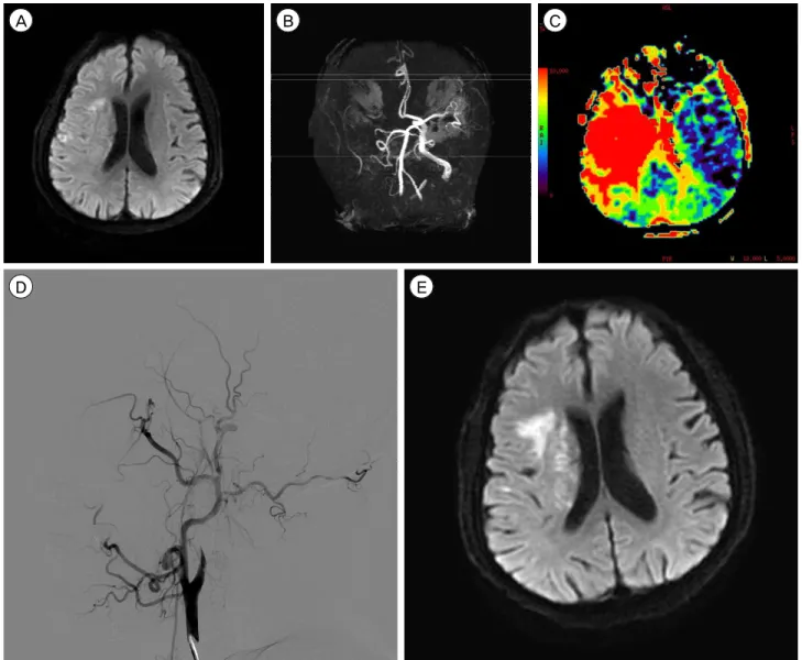
Fig. 1. (A) The initial diffusion-weighted image (DWI) reveals small acute infarcts in the cortex, subcortex and corona radiata of the right hemisphere. (B) The magnetic resonance (MR) angiography shows occlusion of the right internal carotid artery (ICA) and mid- dle cerebral artery (MCA). (C) The MR perfusion (T max map) reveals large perfusion defects in the right MCA whole territory. (D) Endovascular treatment was performed 2 hours after the administration of intravenous recombinant tissue plasminogen activator (IV rt-PA), but recanalization failed because of complete cervical ICA occlusion. (E) Eight hours after the administration of IV rt-PA, left side motor weakness of the patient worsened from motor grade 3/4 to motor grade 1/2, and the immediate follow-up DWI showed an increased cerebral infarction.
performed for acute ischemic stroke in most hospitals.
Intravenous recombinant tissue plasminogen activator (IV rt-PA) is effective for acute ischemic stroke in many cases, but the clinical symptoms of some patients can worsen after the initiation of IV rt-PA because of sus- tained vessel occlusion. For such cases, several re- perfusion modalities are available, including mechan- ical thrombectomy, angioplasty and stenting, carotid endarterectomy (CEA), and extracranial to intracranial
bypass. Invasive procedures, such as major surgery, should generally be avoided within 24 hours after the administration of IV rt-PA. Here, we report a case in which emergent double-barrel STA-MCA bypass was performed shortly after the administration of IV rt-PA.
CASE REPORT
A 66-year-old man presented with cerebral in-
A B C D
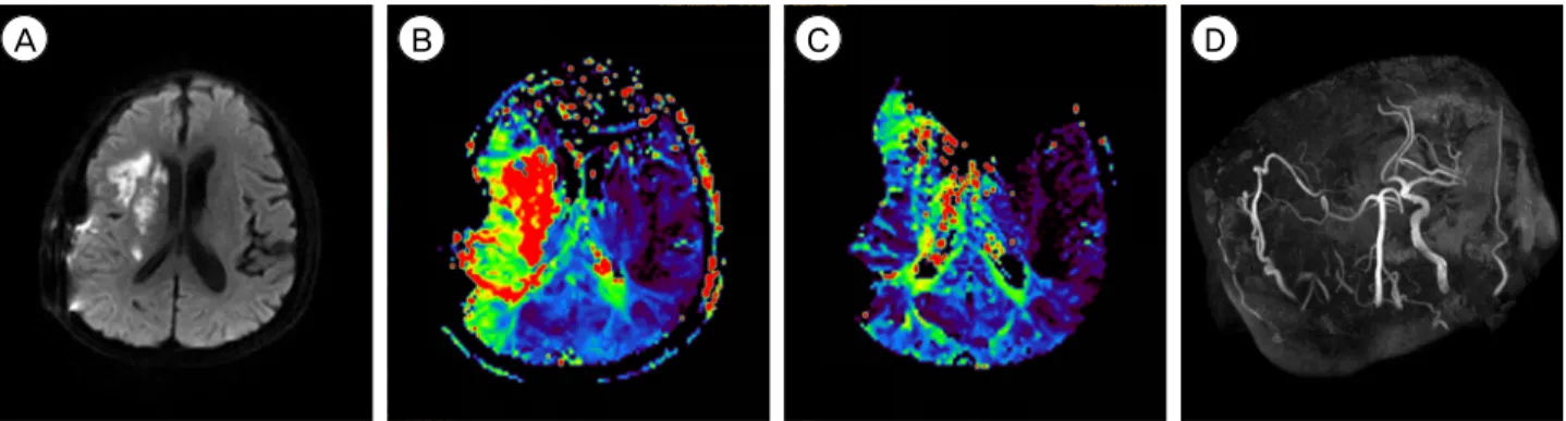
Fig. 2. (A) The diffusion-weighted image (DWI) on postoperative day 7 reveals a slight increase in cerebral infarction. (B) The post- operative 7-day magnetic resonance (MR) perfusion shows improved perfusion status, but some perfusion defects in the right internal border zone are still noted. However, his symptom rapidly improved after surgery. (C) The 1-year follow-up MR perfusion reveals more improved perfusion status. (D) The 1-year follow-up MR angiography demonstrates that the right superficial temporal artery is much thicker than its contralateral counterpart. At 3-month follow-up, the patient's mRS score was 0, and he remained well at his 1.5-year follow-up visit. mRS = modified Rankin scale.
farction in the right MCA territory that manifested as left hemiparesis and dysarthria. The patient's National Institutes of Health Stroke Scale (NIHSS) score was 6 points at the time of admission. A computed tomog- raphy (CT) scan was performed immediately and re- vealed no acute lesion. According to the treatment protocol for acute ischemic stroke, the patient received emergent IV rt-PA (0.6 mg/kg) from a neurologist at our hospital 1 hour and 20 minutes after the onset of the event. Diffusion-weighted image (DWI) of mag- netic resonance imaging (MRI) showed an acute ische- mic lesion in the cortex, subcortex and corona radiata of the right hemisphere (Fig. 1A), and the DWI lesion volume was 1.7 mL. MR angiography (MRA) revealed occlusion of the right ICA (Fig. 1B). MR perfusion demonstrated decreased cerebral blood flow (CBF), slightly increased cerebral blood volume (CBV), and a large perfusion defect in the right MCA territory on the Tmax map (Fig. 1C), and the Perfusion-weighted image (Tmax > 6s) lesion volume was 128.6 mL. Because of the significant diffusion-perfusion mismatch, the patient received endovascular treatment from an in- terventionist 2 hours after the administration of IV rt-PA, but recanalization failed because of total cer- vical ICA occlusion (Fig. 1D). We initially planned to perform STA-MCA bypass the next morning because of his mild symptom and because he had received IV
rt-PA. However, 8 hours after the administration of IV rt-PA, his left hemiparesis worsened from motor grade 3/4 to motor grade 1/2, and his NIHSS score was 14 at that time. The immediate follow-up MRI showed an increased cerebral infarction on DWI (Fig.
1E), and the DWI lesion volume was 6.4 mL. The op- eration was begun 10 hours after the administration of IV rt-PA to prevent further progression of the cere- bral infarction. A successful double-barrel bypass us- ing the frontal branch and parietal branch of the STA with a small craniotomy was achieved, as demonstrated by intraoperative microvascular Doppler sonography.
Revascularization was accomplished 13.5 hours after the administration of IV rt-PA. The operation was un- eventful without any difficulty associated with hemo- stasis, and the estimated total blood loss was less than 50 mL. An immediate postoperative CT revealed no hemorrhage and no additional cerebral infarction lesions. CT angiography revealed good patency of the bypass grafts. Oral administration of the antiplatelet and vasodilating agent cilostazol (200 mg/day) was started on postoperative day 1 to improve CBF and maintain patency of the bypass graft. After surgery, his symptom rapidly improved and his NIHSS score was 4 on postoperative day 3. MRI was performed on postoperative day 7, and revealed a slight increase in cerebral infarction (Fig. 2A) and improved perfusion
status, but a perfusion defect in the right internal bor- der zone (Fig. 2B) was still noted. The DWI lesion volume was 14.3 mL and the Tmax > 6s lesion vol- ume was 61.9 mL. The postoperative course was un- eventful and he fully recovered, his modified Rankin scale was grade 0 at the 3-months follow-up. The 1-year follow-up MRI revealed that his perfusion sta- tus had improved further (Fig. 2C) and MR angiog- raphy revealed that the STA diameters had become much thicker than the contralateral diameters (Fig.
2D). He remained well at his 1.5-year follow-up visit and continued to take cilostazol (200 mg/day).
DISCUSSION
Utilization of IV rt-PA has been the standard treat- ment for acute ischemic stroke. Although IV rt-PA is easily administered and is effective in some cases, the neurologic symptoms can be worsened after IV rt-PA administration if the occluded vessels do not respond to it. Bhatia et al. reported that a low rate (21.25%) of acute recanalization was observed with IV rt-PA administration in acute major vessel occlusion.2) Endovascular recanalization has been currently favored to treat major vessel occlusions unresponsive to IV rt-PA. Although endovascular treatment (EVT) has been developing and higher recanalization rates have been reported in recent trials, the rates of unsuccessful re- canalization (TICI, Thrombolysis in Cerebral infarction scale ≤ 2a) were reported to be 34.3% in the REVASCAT trial16) and 41.3% in the MR CLEAN trial.12) The causes of ischemic stroke are heterogenous. An embolus re- sponds well to thrombectomy devices, but in situ throm- bosis does not respond well to them. Atherosclerotic occlusion may cause EVT failure and require addi- tional balloon angioplasty or stenting. The natural his- tory of acute cervical ICA occlusion with serious neu- rological symptom is unfavorable.1) Cervical ICA occlu- sion is a difficult lesion to treat in acute ischemic stroke, and Choi et al. reported that the successful re- canalization rate (TICI ≥ 2b) of EVT was 36.4% in
acute ischemic stroke according to cervical ICA occlu- sion and the rate of symptomatic intracranial hemor- rhage (ICH) that resulted in death was 11.8%.3) McPherson et al. reported that early CEA can be per- formed safely in patients after IV rt-PA administration for acute ischemic stroke.11) However, in our case, digital subtraction angiography (DSA) showed total occlusion of the cervical ICA, which cannot be treated with CEA. Recent studies have demonstrated the ben- efits of using emergent STA-MCA bypass in patients with acute ischemic stroke.7)8)10)13)15) We suggest that STA-MCA bypass should be considered an alternative treatment when other reperfusion therapies are ineligible.
IV rt-PA exerts a prolonged fibrinolytic effect for more than 24 hours despite its short circulatory half-life (4-5 minutes).18) Therefore, it is recommended that in- vasive procedures be avoided within 24 hours after the administration of IV rt-PA. There have been rare reports of emergent STA-MCA bypass performed after IV rt-PA administration.9)17) Although a major concern was bleeding during or after surgery, it was relatively easy to achieve satisfactory hemostasis during surgery and a postoperative CT revealed no hemorrhage. Furthermore, in previous reports and in our case emergent STA-MCA bypass caused no re- perfusion hemorrhage, likely owing to low-flow bypass.
In our case, a remnant perfusion defect was found on follow-up MRI on postoperative day 7, and cerebral infarction had slightly increased. However, the patient showed remarkable improvement immediately after the bypass surgery. Our case suggests that the addi- tional low-flow blood supply provided by an STA in the ischemic penumbra can minimize the progression of ischemia. The amount of flow increases over time as the bypass graft vessel matures, which is beneficial in acute ischemic stroke because it minimizes the risk of cerebral hyperperfusion syndrome.
Modern procedural descriptions of STA-MCA bypass mostly include the use of a single-branch approach.
The parietal branch of the STA is usually anasto- mosed to either a superior division or an inferior divi-
sion M4 branch, based on the gross suitability of the recipient. However, each patient develops different collateralization, which results in regional differences in perfusion. Single-barrel bypass rarely improves the perfusion status of both superior and inferior divi- sions of the MCA because a single branch anasto- mosis cannot irrigate both divisions of the MCA via retrograde filling through M1.4) Hayashi et al. de- scribed that patients had an ischemic event in the ear- ly postoperative period as a result of an insufficient increase in blood flow following the single-barrel STA-MCA bypass surgery.6) We believe that dou- ble-barrel bypass is a more suitable method for pa- tients who have perfusion defects of both MCA divi- sions in acute ischemic stroke because the penumbra states are highly unstable in the acute stage of cere- bral infarction.
CONCLUSION
This is the first report of emergent double-barrel STA-MCA bypass performed 10 hours after IV rt-PA administration in acute ischemic stroke. We suggest that emergent double-barrel bypass can be a viable option in patients who have perfusion defects of both MCA divisions in acute ischemic stroke after IV rt-PA administration.
ACKNOWLEDGMENTS
This work was supported by the Dong-A University Research Fund.
Disclosure
The authors report no conflict of interest concerning the materials or methods used in this study or the findings specified in this paper.
REFERENCES
1. Adams HP Jr, Bendixen BH, Leira E, Chang KC, Davis PH, Woolson RF, et al. Antithrombotic treatment of is-
chemic stroke among patients with occlusion or severe stenosis of the internal carotid artery: A report of the Trial of Org 10172 in Acute Stroke Treatment (TOAST).
Neurology. 1999 Jul;53(1):122-5.
2. Bhatia R, Hill MD, Shobha N, Menon B, Bal S, Kochar P, et al. Low rates of acute recanalization with intra- venous recombinant tissue plasminogen activator in is- chemic stroke: real-world experience and a call for action. Stroke. 2010 Oct;41(10):2254-8.
3. Choi JY, Lee JI, Lee TH, Sung SM, Cho HJ, Ko JK.
Emergent recanalization with stenting for acute stroke due to athero-thrombotic occlusion of the cervical internal carotid artery : a single center experience. J Korean Neurosurg Soc. 2014 Jun;55(6):313-20.
4. Duckworth EA, Rao VY, Patel AJ. Double-barrel bypass for cerebral ischemia: technique, rationale, and prelimi- nary experience with 10 consecutive cases. Neurosurgery.
2013 Sep;73(1 Suppl Operative):ons30-8; discussion ones37-8.
5. Garrett MC, Komotar RJ, Starke RM, Merkow MB, Otten ML, Sciacca RR, et al. The efficacy of direct extracranial- intracranial bypass in the treatment of symptomatic he- modynamic failure secondary to athero-occlusive disease: a systematic review. Clin Neurol Neurosurg. 2009 May;111(4):
319-26.
6. Hayashi T, Shirane R, Fujimura M, Tominaga T. Postoperative neurological deterioration in pediatric moyamoya disease:
watershed shift and hyperperfusion. J Neurosurg Pediatr.
2010;6(1):73-81.
7. Horiuchi T, Nitta J, Ishizaka S, Kanaya K, Yanagawa T, Hongo K. Emergency EC-IC bypass for symptomatic athe- rosclerotic ischemic stroke. Neurosurg Rev. 2013 Oct;36(4):
559-64; discussion 564-5.
8. Hwang G, Oh CW, Bang JS, Jung CK, Kwon OK, Kim JE, et al. Superficial temporal artery to middle cerebral artery bypass in acute ischemic stroke and stroke in progress.
Neurosurgery. 2011 Mar;68(3):723-9; discussion 729-30.
9. Ishishita Y, Kimura T, Morita A. Urgent superficial tem- poral artery to middle cerebral artery bypass shortly after intravenous rt-PA. Br J Neurosurg. 2012 Oct;26(5):773-5.
10. Lee SB, Huh PW, Kim DS, Yoo DS, Lee TG, Cho KS.
Early superficial temporal artery to middle cerebral artery bypass in acute ischemic stroke. Clin Neurol Neurosurg.
2013 Aug;115(8):1238-44.
11. McPherson CM, Woo D, Cohen PL, Pancioli AM, Kissela BM, Carrozzella JA, et al. Early carotid endarterectomy for critical carotid artery stenosis after thrombolysis ther- apy in acute ischemic stroke in the middle cerebral artery.
Stroke. 2001 Sep;32(9):2075-80.
12. Molina CA, Chamorro A, Rovira A, de Miquel A, Serena J, Roman LS, et al. REVASCAT: a randomized trial of revascularization with SOLITAIRE FR device vs. best medical therapy in the treatment of acute stroke due to anterior circulation large vessel occlusion presenting within eight-hours of symptom onset. Int J Stroke. 2015 Jun;10(4):
619-26.
13. Nussbaum ES, Janjua TM, Defillo A, Lowary JL, Nussbaum LA. Emergency extracranial-intracranial bypass surgery for acute ischemic stroke. J Neurosurg. 2010 Mar;112(3):666-73.
14. Powers WJ, Clarke WR, Grubb RL Jr, Videen TO, Adams HP Jr, Derdeyn CP, et al. Extracranial-intracranial bypass
surgery for stroke prevention in hemodynamic cerebral ischemia: the Carotid Occlusion Surgery Study random- ized trial. JAMA. 2011;306(18):1983-92.
15. Sakai K, Nitta J, Horiuchi T, Ogiwara T, Kobayashi S, Tanaka Y, et al. Emergency revascularization for acute main-trunk occlusion in the anterior circulation. Neurosurg Rev. 2008 Jan;31(1):69-76; discussion 76.
16. Saver JL, Goyal M, Bonafe A, Diener HC, Levy EI, Pereira VM, et al. Stent-retriever thrombectomy after in- travenous t-PA vs. t-PA alone in stroke. N Engl J Med.
2015 Jun;372(24):2285-95.
17. Takeuchi S, Wada K, Arimoto H, Kumagai K, Osada H, Otani N, et al. Emergency superficial temporal artery to middle cerebral artery bypass after intravenous administration of tissue plasminogen activator for stroke. Turk Neurosurg.
2015;25(4):633-7.
18. Tanswell P, Tebbe U, Neuhaus KL, Glasle-Schwarz L, Wojcik J, Seifried E. Pharmacokinetics and fibrin specific- ity of alteplase during accelerated infusions in acute my- ocardial infarction. J Am Coll Cardiol. 1992 Apr;19(5):1071-5.
19. Zipfel GJ, Fox DJ Jr, Rivet DJ. Moyamoya disease in adults: the role of cerebral revascularization. Skull Base.
2005 Feb;15(1):27-41.