교신저자: 이동국, 대구시 남구 대명 4동 3056-6, 705-718, 대구가톨릭대학교 의과대학 신경과학교실 Tel: 053-650-4267, Fax: 053-654-9786, E-mail: [email protected]
Coma is a state of unresponsiveness in which a person is unaware of themself and the environment and cannot be aroused into a state of awareness or respond to the environment. Coma is among the most common and striking problems in general medicine, It accounts for a substantial portion of admissions to emergency wards and occurs on all hospital services. Because coma demands immediate attention, the physician must employ an organized approach. Most causes of coma speedily threaten life. Therefore, they must be promptly identified and treated. Unfortunately, patients with a depressed level of alertness cannot give an account of the events leading to their situation, and often no one who has observed the patient before admission is available to provide such information. Therefore, the physician has to rely on examination of the patient, not only to localize the damaged anatomic structures but also to identify the offending agent. The examination should be thoughtful and well in- formed but not necessarily long, A delay in protecting the airway of a poorly responsive patient may cause severe neurologic damage.
Key Words: Coma, Organized approach
혼수환자의 임상적 접근
이 동 국
대구가톨릭대학교 의과대학 신경과학교실
Clinical Approach to the Patient in Coma
Dong Kuck Lee, M.D.
Department of Neurology, Catholic University of Daegu School of Medicine, Daegu, Korea
서 론
의식이란 각성상태에서 인지기능을 가지고 자기 자신과 주변 환경을 자각하고 있는 상태를 말한다. 이런 의식을 유지하는 중요한 해부학적 구조는 뇌피질과 뇌간의 망상각성계(reticular activating system)이다. 결국 어떤 원인에 의해서든지 시상과 뇌간에 있는 상행 망상체나 양쪽 대뇌반구에 기능이상이 생기면 혼수가 온다. 혼수란 자기 자신과 주변 환경에 대해 알지 못해 외부자극에 대해 전혀 반응이 없는 상태를 말한다.
혼수는 응급실에서 가장 흔히 접하게 되는 증상중의 하나로 응급진단과 신속한 치료가 필요한 분야이다.
그러나 혼수환자를 신속하게 진단 및 치료하기 위해서는 의식수준 평가를 포함한 다양한 신경학적 진찰법
을 반드시 알아야 한다.
본 론
1-10)1. 의식수준의 분류
1) 정상상태: 완전히 각성하여 시간과 장소에 대한 지남력이 있으며 외부 자극에 대해 적절하게 반응하는 상태를 말한다.
2) 혼동(Confusion): 의식이 흐리고 사고력과 이해력이 떨어지며 외부 자극에 대한 반응이 느리고 시간 과 장소에 대한 지남력과 집중력이 떨어지며 주의산만한 상태를 말한다.
3) 섬망(Delirium): 혼동이 더 심해진 상태로 불안, 흥분, 환각, 식별력저하(disorientation), 및 안절부절한 모습을 보이며 망상을 보이기도 하는 상태를 말한다.
4) 둔감(Obtundation): 자는 것 같으며 주변 환경에 대해서는 무관심하지만 다른 사람의 말에는 반응을 하는 상태를 말한다.
5) 혼미(Stupor): 자는 것 같은 상태이지만 강한 자극이나 통증에는 약간 반응을 보인 후 곧 다시 잠에 빠지는 것 같은 상태를 말한다.
6) 혼수(Coma): 의식없이 외부자극에 대해 전혀 반응이 없는 상태를 말한다.
2. 혼수의 원인(Table 1)
혼수의 원인은 다양하지만 크게 대칭적-비구조적, 대칭적-구조적, 및 비대칭적-구조적 원인으로 나눌 수 있다. 대칭적-비구조적 원인으로는 독소, 약물, 대사성, 감염, 정신과적, 및 기타 등이 있고 대칭적-구조적 원인으로는 천막상부 및 하부 원인이 있으며 비대칭적-구조적 원인으로도 천막상부와 하부 원인들이 있다.
3. 혼수환자의 진찰
혼수환자를 진찰하는 첫걸음은 우선 생체징후를 확인한 후 일단 생명을 위협하는 병이 있는지를 알아내 고 치료하는 것이다. 출혈이 있으면 지혈하고 필요하면 삽관(intubation)하여 기도를 유지하며 순환을 유지하 고 위험한 부정맥을 찾기 위해 심전도검사를 한다. 만약 혼수의 원인을 바로 알기 힘들면 혈당검사를 하면 서 50% 포도당을 티아민과 함께 정맥주사한다. 그러나 티아민 부족환자에게 포도당만 주면 Wernicke- Korsakoff증후군이 생길 수 있으므로 주의한다. 만약 아편중독의 가능성이 있으면 Naloxone hydrochloride를 준다. 외상환자이면 내부장기 손상이나 경추 골절의 가능성을 생각하여 적절한 영상의학적 검사를 한다.
일단 응급조치가 끝난 후 다음은 혼수를 일으킨 병의 위치와 원인을 찾는 것이다. 우선 환자의 보호자나 목격자를 통해 그 당시 병력을 알아 본다. 그후 두피, 피부, 손톱, 점막층, 호흡, 안저소견, 동공, 대소변 실금, 경부강직, 및 관절운동 등을 포함한 전신적 및 신경학적 진찰을 신속하게 한다.
1) 운동반응: 우선 전체적으로 호흡, 사지자세와 운동, 자발운동, 경련, 및 근간대경련(myoclonus) 등을
살핀 후 근긴장이 대칭적인지 또는 근육긴장병증(paratonia)이 있는지 본다. 또한 대뇌제거경축(decerebrate
rigidity)과 겉질제거경축(decorticate rigidity)이 있는지 또는 약한 자극에도 적절한 반응을 하는지 아니면 강한
자극에도 전혀 반응이 없는지를 살핀다. 일반적으로는 사지를 쭉 펴고 있는 상태보다는 굽히고 있는 상태가
더 나은 예후를 보인다. 만약 어떤 자극에도 사지운동이 없으면 잠김증후군, 길랑 바레 증후군, 및 경추
외상 등도 고려해야 한다. 혼수환자에서 운동반응, 언어반응, 및 눈뜨기를 점수화하여 그 정도를 평가하는
Table 1. Causes of coma
Ⅰ. Symmetric-nonstructural A Toxins
Lead Thallium Mushrooms Cyanide Methanol Ethylene glycol Carbon monoxide
C Metabolic Hypoxia Hypercapnia Hypernatremia Hyponatremia Hypoglycemia
Hyperglycemic nonketotic coma Diabetic ketoacidosis
Lactic acidosis Hypercalcemia Hypocalcemia Reye encephalopathy Aminoacidemia
Wernicke encephalopathy Porphyria
Hepatic encephalopathy Uremia
Dialysis encephalopathy Hypothyroidism Addisonian crisis
D Infections
Bacterial meningitis Viral encephalitis
Postinfectious encephalomyelitis Syphilis
Sepsis Typhoid fever Malaria
Waterhouse-Friderichsen syndrome
B Drug
Sedatives Barbiturates Other hypnotics Tranquilizers Bromides Alcohol Opiates Paraldehyde Salicylate Psychotropics Anticholinergics Amphetamines Lithium Phencyclidime
Monoamine oxidase inhibitors
E Psychiatric Catatonia
Conversion reaction F Other
Postictal
Diffuse ischemia (myocardial infarction, cardiac heart failure, arrhythmia)
Hypotension Fat embolism
Hypertensive encephalopathy
Ⅱ. Symmetric-structural A Supratentorial
Bilateral internal carotid occlusion Bilateral anterior cerebral artery occlusion Subarachnoid hemorrhage
Thalamic hemorrhage Thauma-contusion, concussion Hydrocephalus
B Infratentorial Basilar occlusion Midline brainstem tumor Pontine hemorrhage
Ⅲ. Asymmetric-structural A Supratentorial
Thrombotic thrombocytopenic purpura Disseminated intravascular coagulation
Nonbacterial thrombotic endocarditis (marantic endocarditis) Subacute bacterial endocarditis
Fat emboli unilateral hemispheric mass (timor, bleed;with herniation) Subdudal Hemorrhage (bilateral subdurals may be symmetric) Intracerebral bleed
Pituitary apoplexy
Massive or bilateral supratentorial infarction Multifocal leukoencephalopathy
Adrenal leukodystrophy Cerebral vasculitis Cerebral abscess Subdudal empyema Thrombophlebitis Multiple sclerosis
Leukoencephalopathy associated with chemotherapy Acute disseminated encephalomyelitis
Creutzfeldt-Jakob disease
B Infratentorial
Brainstem infarction
Brain hemorrhage
Table 2. The Glasgow coma scale Best motor response
Obeys Localizes Withdraws Abnormal flexion Extensor response Nil
Verbal response Oriented
Confusedconversation Inappropriate words Incomprehensible sounds Nil
Eye opening Spontaneous To speech To pain Nil
M6 5 4 3 2 1 V5 4 3 2 1 E4 3 2 1
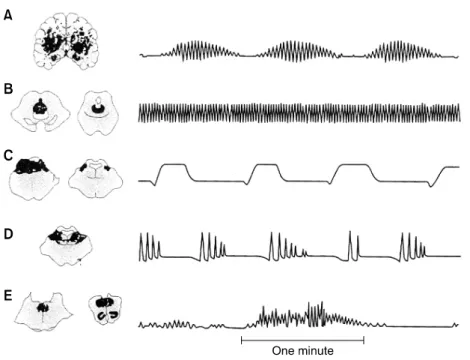
Figure 1. Abnormal respiratory patterns associated with pathologic lesions (shaded areas) at various levels of the brain. The tracings were obtained by chest-abdomen pneumograph; inspiration reads up. (A) Cheyne-Strokes respiration-diffuse forebrain damage. (B) Central neurogenic hyperventilation-lesions of low midbrain ventral to aqueduct of Sylvius and of upper pons ventral to the fourth ventricle. (C) Apneusis-dorsolateral tegmental lesion of middle and caudal pons. (D) Cluster breathing-lower pontine tegmental lesion. (E) Ataxic breathing-lesion of the reticular formation of the dorsomedial part of the medulla.
것이 Glasgow 혼수 척도이다(Table 2).
2) 호흡(Figure 1): Cheyne-Stokes 호흡이란 과호흡과 무호흡이 주기적으로 반복되어 나타나는 것으로 천막 경유탈출(transtentorial herniation)을 포함한 양쪽 대뇌
병, 상부뇌간병, 및 대사성 뇌병증 등에서 나타나지 만 그래도 당장 심각한 상태는 아니다. 그러나 군집 (cluster)호흡은 급성 뇌압상승이나 후두개와(posterior fossa)병에서 나타나는 것으로 당장 심각한 상태를 뜻 한다. 지속적인 과호흡은 대사성 산증, 폐울혈, 간성 뇌병증, 및 진통제 자극 등에 의해 생기고 지속흡입 (apneustic)호흡은 뇌교경색에서 보인다. 연수에 병이 생기면 실조(ataxic)호흡이나 자발호흡은 유지되지만 자율(automatic)호흡은 되지않는 Ondine curse가 보인다.
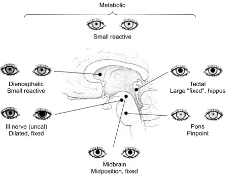
3) 동공(Figure 2): 혼수환자에서 동공의 이상은
교감신경과 부교감신경사이에 불균형이 생긴 것을
뜻한다. 비록 정상인에서도 많은 사람들에서 동공
크기에 약간의 차이가 있지만 혼수환자에서 동공
크기가 차이가 나면 일단 병이 있다고 본다. 그러나
망막이나 시신경은 손상되어도 동공부등(anisocoria)
Figure 2. Pupils in comatose patients.
이 생기지 않는다. 구상회탈출(uncal herniation)이 동안신경을 누르거나 내경동맥 동맥류가 터지면 동공이 커지고 결국 동공반사가 없어지게 된다. 뇌내외병변에 의해 교감신경이 손상되면 호너증후군이 생긴다.
중뇌에 병변이 생기면 동공이 눈의 가운데 위치하면서 동공반사가 없어지고 뇌교출혈이 되면 동공반사는 있으나 심하게 축동된다. 그러나 대사성 병에서는 대부분 동공이상이 생기지 않는다. 만약 광범위한 저산소 -허혈환자에서 동공이 확대고정되면 예후가 나쁘다. 항콜린제와 항파킨슨제도 동공반사를 없앤다. 경련후 에는 한쪽이나 양쪽 동공이 커지고 동공반사가 없어지지만 아편중독에서는 심한 축동이 생긴다. 저체온증 이나 barbiturate중독이 되면 동공반사가 없어진다.
4) 눈꺼풀과 안구운동: 혼수환자가 눈을 감고 있다는 것은 연수 하부가 기능이 있다는 것을 뜻한다. 만약 반신마비가 된 반대쪽으로 안구가 쏠려 있으면 대뇌병변을 생각하고 반신마비된 쪽으로 안구가 쏠리면 뇌교병변, 경련, 또는 시상출혈 등을 생각한다. 안정시 양쪽 안구가 제멋대로 방향이면 각 안근마비, 핵간 안구마비(internuclear ophthalmoplegia), 또는 사시(phoria) 등을 생각한다. 뇌간이 정상이면 안구가 좌우로 산만 하게 움직이며 이런 소견은 뇌경색 또는 소뇌출혈때 볼 수 있다. 만약 경추손상이 없다면 인형안(doll's eye)운 동을 해 보고 양쪽 귀에 교대로 찬물을 넣은 후 안구운동을 살펴 보기도 한다. 시상과 중뇌병변이 있거나 경련 또는 barbiturate 중독이 되면 안구가 하부로 쏠린다. 소뇌나 뇌교병에서는 양쪽 안구가 서로 비스듬하 게 위치(skew deviation)한다. 중뇌병에서는 눈모음(convergence) 안구진탕이 생기고 뇌교병에서는 양쪽 안구가 같이 살짝 밑으로 내려가는 증상(ocular bobbing)을 보인다.
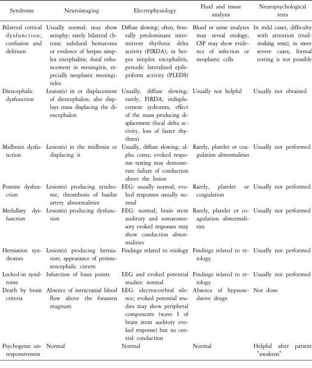
4. 혼수환자의 검사방법(Table 3)
혼수의 원인이 바로 밝혀지지 않으면 신속하게 CT나 MRI검사를 하고 필요하면 뇌척수액검사를 한다.
물론 혈청에서 포도당, Na, Ca, BUN, creatinine, 간기능, 동맥 PH, PO
2, 및 PCO
2도 검사하고 필요하면 혈액과
소변에서 독성물질도 검사한다. 정신과적인 무반응, 대사성 뇌병증, 잠김증후군, 및 경련 등이 의심되면
뇌파검사도 도움이 된다.
Table 3. Useful studies in the evaluation of disorder of level of consciousness
Syndrome Neuroimaging Electrophysiology Fluid and tissue
analysis
Neuropsychological tests Bilateral cortical
dysfunction;
confusion and delirium
Diencephalic dysfunction
Midbrain dysfu- nction
Pontine dysfun- ction Medullary dys-
function
Herniation syn- dromes Locked-in synd-
rome
Death by brain criteria
Psychogenic un- responsiveness
Usually normal; may show atrophy; rarely bilateral ch- ronic subdural hematoma or evidence of herpes simp- lex encephalitis; dural enha- ncement in meningitis, es- pecially neoplastic meningi- tides
Lesion(s) in or displacement of diencephalon; also disp- lays mass displacing the di- encephalon
Lesion(s) in the midbrain or displacing it
Lesion(s) producing syndro- me; thrombosis of basilar artery abnormalities Lesion(s) producing dysfunc-
tion
Lesion(s) producing hernia- tion; appearance of perime- sencephalic cistern Infarction of basis points Absence of intracranial blood
flow above the foramen magnum
Normal
Diffuse slowing; often, fron- tally predominant inter- mittent rhythmic delta activity (FIRDA); in her- pes simplex encephalitis, periodic lateralized epile- ptiform activity (PLEDS) Usually, diffuse slowing;
rarely, FIRDA; indispla- cement sydromes, effect of the mass producing di- splacement (focal delta ac- tivity, loss of faster rhy- thms)
Usually, diffuse slowing; al- pha coma; evoked respo- nse testing may demonst- rate failure of conduction above the lesion EEG: usually normal; evo-
ked responses usually no- rmal
EEG: normal; brain stem auditory and somatosen- sory evoked responses may show conduction abnor- malities
Findings related to etiology
EEG and evoked potential studies: normal
EEG: electrocerebral sile- nce; evoked potential stu- dies may show peripheral components (wave I of brain stem auditory evo- ked response) but no cen- tral conduction
Normal
Blood or urine analyses may reveal etiology;
CSF may show evide- nce of infection or neoplastic cells
Usually not helpful
Rarely, platelet or coa- gulation abnormalities
Rarely, platelet or coagulation
Rarely, platelet or co- agulation abnormali- ties
Findings related to et- iology
Findings related to et- iology
Absence of hypnose- dative drugs
Normal
In mild cases, difficulty with attention (trail- making tests); in more severe cases, formal testing is not possible
Usually not obtained
Usually not performed
Usually not performed
Usually not performed
Usually not performed
Usually not performed Not done
Helpful after patient
"awakens"
5. 혼수환자의 다양한 임상 증상
1) 천막상부(Supratentorial) 병변에 의한 혼수(Table 4): 양측 대뇌손상이나 급성으로 편측에 큰 병변이
생기면 혼수가 생긴다. 그후 병변이 진행되면 뇌실질이 하부로 내려와서 천막경유탈출(transtentorial
herniation)이 일어난다. 천막경유탈출은 구상회(uncal) 탈출과 중심성 탈출로 나눈다. 경막하 혈종에서처럼
Table 5. Coma due to infratentorial structural lesions Discriminating features
Presents with cranial nerve involvement plus contralateral limb involvement Intranuclear ophthalmoplegia
MRI/CT shows lesion Consistent features Asymmetric pupils
Oculomotor nerve involvement Variable features
Abnormal respiratory patterns
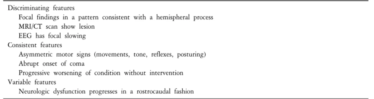
Table 4. Coma due to supratentorial structural lesions Discriminating features
Focal findings in a pattern consistent with a hemispheral process MRI/CT scan show lesion
EEG has focal slowing Consistent features
Asymmetric motor signs (movements, tone, reflexes, posturing) Abrupt onset of coma
Progressive worsening of condition without intervention Variable features
Neurologic dysfunction progresses in a rostrocaudal fashion
구상회 탈출이 생기면 동안신경을 압박하여 그쪽 동공이 커지게 된다. 그후 점차 양쪽 동공이 모두 커지고 고정되며 과호흡이나 실조호흡이 생기며 사지는 굴곡되었다가 차차 신전되며 결국 깊은 혼수상태가 되었 다가 사망하게 된다. 시상출혈에서 처럼 중심성 탈출이 생기면 의식이 빠르게 저하되지만 동공은 정상이거 나 약간 작고 대광반사도 있다. 그후 동공은 가운데 고정되며 구상회 탈출과 비슷한 경과를 보인다. 만약 중뇌부위의 Kernohan notch에 뇌가 압박되면 병변쪽에 반신마비가 생긴다. 천막경유탈출의 주요 원인으로는 외상(경막외, 경막하, 뇌실질 출혈), 혈관(허혈, 출혈), 감염(농양, 육아종), 및 종양(일차성, 전이성) 등이 있다.
2) 천막하부 병변에 의한 혼수(Table 5): 천막하부의 구조적 병변은 뇌간을 직접 압박하거나 파괴한다.
그 결과 뇌가 천막상부로 탈출하면 중뇌를 누르고 대공(foramen magnum)을 통해 하부로 탈출하면 연수를 압박한다. 급성 소뇌편도탈출(tonsillar herniation)이 되면 무호흡과 순환이 마비된다. 천막하부병변에 의한 혼수에서는 사지마비나 감각장애, 축동, 좌우안구운동 소실, 주시 부전, 안근마비, 지속흡입호흡 또는 실조 호흡 등을 보인다. 뇌교출혈에서는 급성 혼수, 대광반사는 있으나 아주 작은 동공, 및 안구운동마비 등을 보인다.
3) 대사성 또는 광범위한 뇌병에 의한 혼수(Table 6): 다양한 원인에 의한 대사성 또는 광범위한 뇌병에 서는 발병 초기에 인지기능과 호흡이상이 생기며 진전, 자세고정불능(asterixis), 및 근간대경련(myoclonus)도 자주 동반된다. 대부분 국소적 이상소견은 적으며 동공도 반응한다. 대사성 혼수를 진단하기 위해서는 동맥 혈 가스분석이 필요하다.
4) 기타
(1) 히스테리와 긴장증(Catatonia): 히스테리성 무반응은 드물지만 과진단되기도 하며 임상적으로 꾀병
과 감별이 힘들 때가 많다. 히스테리성 무반응은 일단 눈을 꼭 감고 있으나 호흡은 정상이거나 빠른 호흡을
Table 6. Toxic/metabolic coma Discriminating features
Symmetric abnormalities on neurologic examination: lack of focal findings Mental status change precedes motor signs
MRI/CT does not show lesion EEG shows diffuse slowing Consistent feature
Abnormal respiratory pattern Pupillary responses intact
Tone is normal or slightly reduced Waxing and waning level of responsiveness Variable features
Laboratory studies abnormal Abnormal movements common
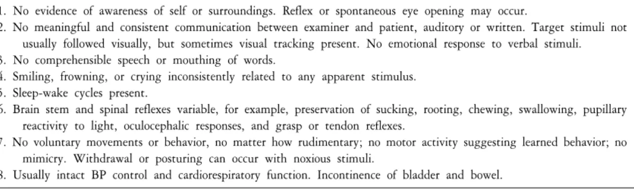
Table 7. Criteria for determination of vegetative state
1. No evidence of awareness of self or surroundings. Reflex or spontaneous eye opening may occur.
2. No meaningful and consistent communication between examiner and patient, auditory or written. Target stimuli not usually followed visually, but sometimes visual tracking present. No emotional response to verbal stimuli.
3. No comprehensible speech or mouthing of words.
4. Smiling, frowning, or crying inconsistently related to any apparent stimulus.
5. Sleep-wake cycles present.
6. Brain stem and spinal reflexes variable, for example, preservation of sucking, rooting, chewing, swallowing, pupillary reactivity to light, oculocephalic responses, and grasp or tendon reflexes.
7. No voluntary movements or behavior, no matter how rudimentary; no motor activity suggesting learned behavior; no mimicry. Withdrawal or posturing can occur with noxious stimuli.
8. Usually intact BP control and cardiorespiratory function. Incontinence of bladder and bowel.
보이고 동공은 정상이다. 눈꺼풀을 벌리려면 저항하지만 가만히 두면 바로 눈을 감는다. 사지는 꼼짝하지 않고 있으나 긴장도는 정상이며 기질적인 병이나 약물에 의한 것이 아니라면 뇌파는 각성상태를 보인다.
정신분열병, 우울증, 또는 독성 정신병 등에서 나타나는 긴장증은 무운동함구증(akinetic mutism), 얼굴 찡그 림, 강직, 흥분, 및 허탈발작(cataplexy) 등을 보인다. 호흡은 정상이거나 빠르고 동공은 크지만 반응은 정상이 며 안구운동도 정상이고 뇌파도 대부분 정상이다.
(2) 감금증후군(Locked-in syndrome): 뇌경색이나 중추 뇌교 수초용해(central pontine myelinolysis)가 뇌 교기저(basis pontis)를 파괴하면 의식과 호흡은 유지되면서 하부 뇌신경과 사지에 마비가 생긴다. 언뜻 보면 아무반응이 없는 것처럼 보이지만 안구를 자발적으로 아래위로 움직이며 가끔은 수평으로 움직이기도 하 고 눈을 깜박거리기도 한다. 따라서 안구운동과 눈 깜빡거림을 통해 간단한 의사소통도 가능하다.
(3) 식물상태(Vegetative state)와 약간 의식이 있는 상태(Minimally conscious state): 식물상태란 내 적이나 외적 각성은 없이 수면-각성 주기와 심호흡기능은 유지되며 자극에 대해서는 간단한 반응은 보이는 상태를 말한다. 혼수에서 살아남은 사람은 2∼4주일사이에 다양한 정도의 회복을 보이며 식물상태에 들어간 경우도 마찬가지 경과를 보인다. 만약 1개월 이상 식물상태가 지속되면 지속적(persistent) 식물상태라고 한다.
대부분의 지속적 식물상태는 시상과 뇌피질하 백질에 심한 손상이 있는 경우에 생긴다(Table 7). 약간 의식
이 있는 상태란 의식은 심하게 저하되었으나 자신이나 외부 환경에 대해 약간 아는 상태를 말한다. 이 상태
는 식물상태처럼 심한 상태에서 호전되는 경과중에 보이는 현상으로 본다. 물론 예후는 식물상태보다 좋다.
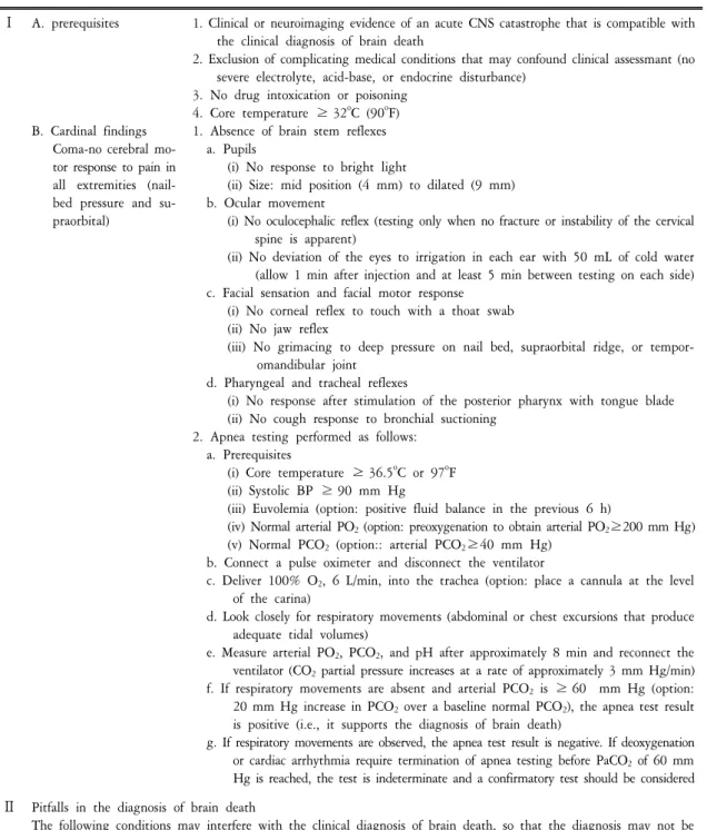
Table 8. Criteria for determination of brain death
Ⅰ A. prerequisites
B. Cardinal findings Coma-no cerebral mo-
tor response to pain in all extremities (nail- bed pressure and su- praorbital)
1. Clinical or neuroimaging evidence of an acute CNS catastrophe that is compatible with the clinical diagnosis of brain death
2. Exclusion of complicating medical conditions that may confound clinical assessmant (no severe electrolyte, acid-base, or endocrine disturbance)
3. No drug intoxication or poisoning 4. Core temperature ≥ 32
oC (90
oF) 1. Absence of brain stem reflexes a. Pupils
(i) No response to bright light
(ii) Size: mid position (4 mm) to dilated (9 mm) b. Ocular movement
(i) No oculocephalic reflex (testing only when no fracture or instability of the cervical spine is apparent)
(ii) No deviation of the eyes to irrigation in each ear with 50 mL of cold water (allow 1 min after injection and at least 5 min between testing on each side) c. Facial sensation and facial motor response
(i) No corneal reflex to touch with a thoat swab (ii) No jaw reflex
(iii) No grimacing to deep pressure on nail bed, supraorbital ridge, or tempor- omandibular joint
d. Pharyngeal and tracheal reflexes
(i) No response after stimulation of the posterior pharynx with tongue blade (ii) No cough response to bronchial suctioning
2. Apnea testing performed as follows:
a. Prerequisites
(i) Core temperature ≥ 36.5
oC or 97
oF (ii) Systolic BP ≥ 90 mm Hg
(iii) Euvolemia (option: positive fluid balance in the previous 6 h)
(iv) Normal arterial PO
2(option: preoxygenation to obtain arterial PO
2≥200 mm Hg) (v) Normal PCO
2(option:: arterial PCO
2≥40 mm Hg)
b. Connect a pulse oximeter and disconnect the ventilator
c. Deliver 100% O
2, 6 L/min, into the trachea (option: place a cannula at the level of the carina)
d. Look closely for respiratory movements (abdominal or chest excursions that produce adequate tidal volumes)
e. Measure arterial PO
2, PCO
2, and pH after approximately 8 min and reconnect the ventilator (CO
2partial pressure increases at a rate of approximately 3 mm Hg/min) f. If respiratory movements are absent and arterial PCO
2is ≥ 60 mm Hg (option:
20 mm Hg increase in PCO
2over a baseline normal PCO
2), the apnea test result is positive (i.e., it supports the diagnosis of brain death)
g. If respiratory movements are observed, the apnea test result is negative. If deoxygenation or cardiac arrhythmia require termination of apnea testing before PaCO
2of 60 mm Hg is reached, the test is indeterminate and a confirmatory test should be considered
Ⅱ Pitfalls in the diagnosis of brain death
The following conditions may interfere with the clinical diagnosis of brain death, so that the diagnosis may not be made with certainty on clinical grounds alone. Confirmation tests are recommended
A. severe facial trauma
B. Preexisting pupillary abnormalities
C. Toxic levels of any sedative drugs, aminoglycosides, tricyclic antidepressants, anticholinergics, antiepileptic drugs, chemotherapeutic agents, or neuromuscular blocking agents
D. Sleep apnea or severe pulmonary disease resulting in chronic retention of CO
2Table 8. Continued
Ⅲ
Ⅳ
Clinical observations compatible with the diagnosis of brain death
These manifestations are occasionally seen and should not be misinterpreted as evidence for brain stem function A. Tendon reflexes; superficial abdominal reflexes; triple flexion response
B. Plantar reflex
C. Respiratory-like movements (shoulder elevation and adduction, back arching, intercostal expansion without sig- nificant tidal volumes)
D. Spontaneous movements of limbs other than pathologic flexion or extension. Responses including facial twitching, flexion at the waist, slow turning of the head, undulating movements of the toes, and shoulder adduction with arm flexion. Such movements sometimes occur during apnea testing or following pronunciation of brain death and disconnection from the ventilatior ("Lazarus sign")
E. Sweating, blushing, tachycardia
F. Normal BP without pharmacological support or sudden increases in BP Confirmatory laboratory tests (options)
For subjects ≥ 18 yr old, a repeat examination should be performed 6 h later
For children < 2 mo old, the interval should be 48 h; for those > 2 mo to 1 yr old, 24 h; and for those > 1 yr to < 18 yr old, 12 h.
Children < 2 mo old should have two confirmatory tests. Children > 2 mo to < 1 yr old should have one confirmatory test. For children > 1 yr old and adults, confirmatory tests are optional.
A. Conventional angiography. Response: no intracerebral filling at the level of the carotid bifurcation or circle of Willis. The external carotid circulation is patent, and filling of the superior longitudinal sinus may be delayed B. Technetium-99m hexamethylpropylene-amineoxime brain scan. Response: no isotope uptake in brain parenchyma
("hollow skull phenomenon")
C. Electroencephalography. Response: no electrical activity during at least 30 min of recording (EEG is neither as specific nor sensitive sompared to angiography or technetium brain scan. It can be isoelectric in patients with brain stem reflexes, and it can show residual activity for hours or days after all other criteria for brain death have been met.)
D. Transcranial Doppler ultrasonography
1. Response: 10% of patients may not have temporal insonation windows. Therefore, the initial absence of Doppler signals cannot be interpreted as consistent with brain death
2. Response: small systolic peaks in early systole without diastolic flow or reverberating flow, indicating very high vascular resistance associated with greatly increased intracranial pressure
E. Somatosensory-evoked potentials. Response: bilateral absence of N20-P22 response with median nerve stimulation
(4) 뇌사(Brain death) (Table 8): 뇌간기능이 유지되는 식물상태와는 다르게 대뇌와 뇌간기능이 모두 없어지면 뇌사가 된다. 뇌사가 되면 심장혈관기능만 남아 있다. 호흡을 유발하는 정도의 고탄산혈증 (hypercarbia)이 되어도 호흡은 일어나지 않고 척수에 의해 생기는 반사만 남아 있다. 성인에서 뇌사에 빠지면 거의 대부분 수일내에 순환이 정지된다. 최근 뇌사는 법적인 죽음과 동일하게 생각하고 있다. 따라서 뇌사 판정기준에 합당하면 인공호흡과 연명치료를 중단한다.
결 론
의식이란 자신과 주위 환경을 잘 인식하고 있는 상태를 말한다. 의식의 변화란 우선 각성(arousal)의 변화
인지 또는 치매, 망상, 및 혼돈(confusion)같이 인지(cognitive) 및 정동(affective) 정신기능의 변화인지를 구분하
는 것이 중요하다. 혼수란 자신과 주위 환경에 대해 각성하지 못해 전혀 적절한 반응을 보이지 않는 상태를
= 국문요약 =
의식이란 자신과 주위 환경을 잘 인식하고 있는 상태를 말한다. 이런 의식을 유지하는 중요한 해부학 적 구조는 뇌피질과 뇌간의 망상각성계(reticular activating system)이다. 의식의 변화에 대해서는 우선 각성(arousal)의 변화인지 또는 치매, 망상, 및 혼돈(confusion)같이 인지(cognitive) 및 정동(affective) 정신기 능의 변화인지를 구분하는 것이 중요하다. 혼수란 자신과 주위 환경에 대해 각성하지 못해 전혀 적절 한 반응을 보이지 않는 상태를 말한다. 혼수를 일으키는 대부분의 원인들은 생명을 위협하는 경우가 많다. 따라서 혼수환자를 보면 신속하게 체계적으로 접근하여 원인을 파악한 후 적절한 치료를 해야 더 이상의 손상을 막을 수 있다. 그러나 혼수상태의 환자는 병력와 진찰이 힘들어 정확한 진단을 하기가 힘든 경우가 많다. 따라서 혼수환자를 신속하게 진단 및 치료하기 위해서는 의식수준의 분류, 혼수의 원인, 운동, 호흡, 동공, 및 안구운동 등을 통한 진단법, 및 천막상부, 천막하부, 또는 대사성 혼수의 특징적인 증상 등에 대해 잘 알아야 한다. 이 종설에서는 의식수준의 분류, 혼수의 원인, 진찰, 검사방법, 및 다양한 임상 증상 등에 대하여 간략하게 정리하였다.
중심 단어 : 혼수, 체계적 접근
말한다. 혼수를 일으키는 대부분의 원인들은 생명을 위협하는 경우가 많다. 따라서 혼수환자를 보면 신속하 게 원인을 파악하여 적절한 치료를 해야 더 이상의 손상을 막을 수 있다. 그러나 혼수상태의 환자는 병력와 진찰이 힘들어 정확한 진단을 하기가 힘든 경우가 많다. 따라서 혼수환자를 신속하게 진단 및 치료하기 위해서는 의식수준의 분류, 혼수의 원인, 운동, 호흡, 동공, 및 안구운동 등을 통한 진단법, 및 천막상부, 천막하부, 또는 대사성 혼수의 특징적인 증상 등에 대해 잘 알아야 한다.
참고문헌
1. Bleck TP. Levels of consciousness and attention. In: Goetz CG, editor. Textbook of clinical neurology. 3rd ed. New York: Saunders Elsevier; 2007. p. 3-19.
2. Brazis PW, Masdeu JC, Biller J. The localization of lesions causing coma. Localization in clinical neurology. 5th ed. New York:
Lippincott Williams & Wilkins; 2007. p. 557-82.
3. Brust JCM. Coma. Current diagnosis & treatment. 1st ed. New York: McGraw Hill; 2007. p. 29-34.
4. Berger JR. Stupor and coma. In: Bradley WG, Daroff RB, Fenichel GM, Jankovic J, editors. Neurology in clinical practice.
5th ed. New York: Butterworth Heinemann Elsevier; 2008. p. 39-58.
5. Hankey GJ, Wardlaw JM. Disorders of consciousness. Clinical neurology. 1st ed. New York: Manson Publishing; 2008. p. 44-56.
6. Ropper AH. Coma. In: Fauci AS, Braunwald E, Kasper DL, Hauser SL, Longo DL, Larry Jameson J, Loscalzo J, editors. Harrison's Principles of Internal Medicine. 17th ed. New York: McGraw Hill; 2008. p. 1714-9.
7. Simon RP. Coma and disorders of arousal. In: Goldman L, Ausiello D, editors. Cecil medicine. 23rd ed. New York: Saunders Elsevier; 2008. p. 2691-6.
8. Berger JR. Coma. In: Corey-Bloom J, David RB, editor. Clinical adult neurology. 3rd ed. New York: Demosmedical; 2009. p.
213-227.
9. Ropper AH, Samuels MA. Coma and related disorders of consciousness. Adams and Victor's principles of neurology. 9th ed.
New York: McGraw Hill; 2009. p. 339-61.
10. Brust JCM. Coma. In: Rowland LP, Pedley TA, editor. Merritt's neurology. 12th ed. New York: Lippincott Williams & Wilkins;
2010. p. 22-30.