J Korean Surg Soc 2013;84:154-159 http://dx.doi.org/10.4174/jkss.2013.84.3.154
ORIGINAL ARTICLE
JKSS JKSS JKSS
Journal of the Korean Surgical Society pISSN 2233-7903ㆍeISSN 2093-0488
Received October 23, 2012, Revised December 3, 2012, Accepted December 18, 2012 Correspondence to: Bulent Koca
Department of General Surgery, Ondokuz Mayis University, Genel Cerrahi Anabilim Dali, 55139 Kurupelit, Samsun 55852, Turkey Tel: +90-5055735587, Fax: +90-3624576041, E-mail: [email protected]
cc Journal of the Korean Surgical Society is an Open Access Journal. All articles are distributed under the terms of the Creative Commons Attribution Non-Commercial License (http://creativecommons.org/licenses/by-nc/3.0/) which permits unrestricted non-commercial use, distribution, and reproduction in any medium, provided the original work is properly cited.
Factors affecting surgical margin positivity in
invasive ductal breast cancer patients who underwent breast-conserving surgery after preoperative core
biopsy diagnosis
Bulent Koca, Bekir Kuru, Savas Yuruker, Barıs Gokgul
1, Necati Ozen
Department of General Surgery, Ondokuz Mayis University School of Medicine, Samsun, 1Sivas Numune Eğitim ve Araştırma Hastanesi, Sivas, Turkey
Purpose: The aim of our study is to evaluate the factors affecting surgical margin positivity among patients with invasive duc- tal breast cancer who underwent breast-conserving surgery (BCS) after preoperative diagnostic core biopsy. Methods: Two hundred sixteen patients with stage I, II invasive ductal breast carcinoma who had histological diagnosis with preoperative tru-cut biopsy and underwent BCS were included in the present study. Potential factors that affect the positive surgical margin were analyzed. In univariate analysis, the comparisons of the factors affecting the surgical margin positivity were made by chi-square test. Logistic regression test was used to detect the independent factors affecting the surgical margin positivity.
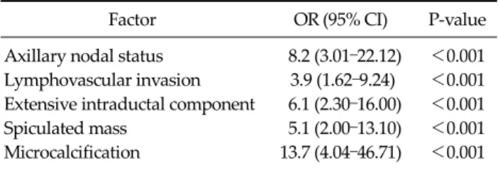
Results: Positive axillary lymph node (odds ratio [OR], 8.2; 95% confidence interval [CI], 3.01 to 22.12), lymphovascular in- vasion (LVI; OR, 3.9; 95% CI, 1.62 to 9.24), extensive intraductal component (EIC; OR, 6.1; 95% CI, 2.30 to 16.00), presence of spiculation (OR, 5.1; 95% CI, 2.00 to 13.10) or presence of microcalcification in the mammography (OR, 13.7; 95% CI, 4.04 to 46.71) have been found to be the independent and adverse factors affecting surgical margin positivity. Conclusion:
Considering decision making for the extent of the excision and for achieving negative surgical margin before BCS, positive ax- illary lymph node, LVI, EIC, spiculation or microcalcification in mammography are related as predictor factors for positive surgical margin.
Key Words: Breast ductal carcinoma, Large-core needle biopsy, Segmental mastectomy
INTRODUCTION
The National Institute of Health (NIH) Consensus Conference in 1990 concluded that breast conservation surgery (BCS) was an appropriate method of treatment for women with early-stage breast cancers (stages I and II).
BCS has been increasingly performed for patients with ear- ly breast cancer since the NIH Consensus Conference [1].
The objective of BCS is to completely remove the breast cancer and achieve negative surgical margins while main- taining the best possible breast cosmesis. Surgical margin positivity is the most important factor for local recurrence
Table 1.Characteristics of the patients
Characteristic
No. of patients (n = 216)
Positive surgical margins
(n = 58)
P-value
Age (yr) 0.333
≤40 42 (19) 14 (24)
>40 174 (81) 44 (76)
Tumor size (cm) 0.002
≤2 120 (56) 22 (38)
>2 96 (44) 36 (62)
Grade 0.170
1 17 (8) 7 (12)
2 137 (63) 37 (64)
3 62 (29) 14 (24)
LVI <0.001
Present 86 (40) 39 (67)
Absent 130 (60) 19 (33)
EIC <0.001
Present 70 (33) 42 (72)
Absent 146 (67) 16 (28)
Molecular subtype 0.749
Luminal A 117 (54) 29 (50)
Luminal B 54 (25) 18 (31)
Her-2 (+) 17 (8) 7 (12)
Triple negative 28 (13) 4 (7)
Axillary nodal status <0.001
Positive 61 (28) 28 (48)
Negative 155 (72) 30 (52)
Breast density 0.574
1 40 (19) 13 (22)
2 59 (27) 11 (19)
3 88 (41) 30 (52)
4 29 (13) 4 (7)
Spiculated mass <0.001
Present 60 (28) 28 (48)
Absent 156 (72) 30 (52)
Microcalcification <0.001
Present 37 (17) 27 (47)
Absent 179 (83) 31 (53)
Multifocality 0.254
Present 9 (4) 4 (7)
Absent 207 (96) 54 (93)
Location in quadrants 0.215
Upper outer 137 (64) 41 (70)
Upper inner 33 (15) 9 (15)
Lower outer 21 (10) 5 (9)
Lower inner 16 (7) 2 (3)
Central 9 (4) 1 (2)
Values are presented as number (%).
LIV, lymphovascular invasion; EIC, extensive intraductal compo- nent; Her-2, human endothelial growth factor receptor 2.
after BCS [2-4]. BCS after tru-cut biopsy is known to be more efficient in providing negative surgical margin com- pared with excisional biopsy [5-7]. We used tru-cut biopsy, except in special cases, as the primary histological diag- nostic method in our clinic. The data on the factors asso- ciated with surgical margin positivity and BCS for in- vasive ductal cancer in the setting of preoperative tru-cut biopsy are limited [8]. The purpose of the present study is to evaluate the factors affecting surgical margin positivity among patients with invasive ductal breast cancer who underwent BCS after preoperative tru-cut biopsy.
METHODS
Two hundred sixteen patients with stage I, II invasive ductal breast carcinoma who had histological diagnosis with preoperative tru-cut biopsy and underwent BCS at the Department of General Surgery in Ondokuz Mayis University School of Medicine between January 2004 and September 2012 were the subject of this retrospective study. All patients underwent sentinel lymph node biopsy with isosulphane blue and patients with metastatic senti- nel lymph node biopsy underwent level I, II axillary node dissection. Injectable sterile solutions of 1% isosulphane blue (monosodium salt of 2,5-disulphonated triphenyl methane) was prepared by the Department of Pharma- ceutical Technology at Istanbul University Faculty of Pharmacy using a stock solution obtained from Sigma company (5 g isosulphan blue, Sigma catalog No: P1888, Sigma-Aldrich chemical Co., Deisenhofen, Germany). The findings of mammography, and breast ultrasonography, and pathological examination have been recorded.
Information regarding surgical margin positivity (nega- tive, positive), age (≤40, >40), tumor size (≤2 cm, >2 cm), tumor grade (1, 2, 3), presence of lymphovascular in- vasion (LVI), presence of extensive intraductal component (EIC), axillary lymph node status (negative, positive), es- trogen receptor (ER) status, progesterone receptor (PR) status, human endothelial growth factor receptor 2 (Her-2) status, molecular subtypes (luminal A, luminal B, Her-2 (+), triple negative), multifocality (present, absent), tu- mor location in breast quadrants (upper outer, upper in- ner, lower outer, lower inner, central), mammographic
density of the breast (Breast Imaging Reporting and Data System density categories 1, 2, 3, 4), and presence of spicu-
Table 2.Logistic regression analysis and independent factors for positive surgical margin
Factor OR (95% CI) P-value
Axillary nodal status 8.2 (3.01–22.12) <0.001 Lymphovascular invasion 3.9 (1.62–9.24) <0.001 Extensive intraductal component 6.1 (2.30–16.00) <0.001 Spiculated mass 5.1 (2.00–13.10) <0.001 Microcalcification 13.7 (4.04–46.71) <0.001 OR, odds ratio; CI, confidence interval.
lation or the presence of microcalcification in the mam- mography have been recorded and analyzed (Table 1).
Positive ER and PR status was defined as ≥5% of tumor cell nuclei showing specific staining. Specific membrane staining for c-erbB2 (Her-2/ neu) was reported as positive, with the intensity of the staining observed on a scale of 0–3.
Intensity of 0 to 1+ was considered negative, whereas 3+
staining was considered positive. An intermediate score of 2+ was confirmed by fluorescence in situ hybridization amplification [9]. Tumors with ER and/or PR positive and Her-2 negative were classified as luminal A, tumors with ER and/or PR positive and Her-2 positive as luminal B, tu- mors with ER and PR negative and Her-2 positive as Her-2 (+), and tumors with ER and PR negative and Her-2 neg- ative as triple negative molecular subtype. We staged the patients using the tumor-node-metastasis stage according to the 7th American Joint Committee on Cancer system [10].
We generally used curvilinear incisions, though radial incisions were sometimes used for tumors located at the three or nine o’clock positions. To achieve a clear surgical margin, we attempted to obtain a margin of 1 cm of grossly normal breast tissue around the tumor. A skin island over- lying the tumor was also excised for very superficial tumors. All the breast tissue under subcutaneous fat tissue was removed for tumors located close to the breast skin.
Underlying breast tissue down to the pectoralis major muscle including the fascia of the muscle was excised for tumors located deep in the breast. Nipple-areola complex (NAC) was removed for tumors located at the central quadrant in case the tumor was close to or invaded the NAC. Preoperative localization of 16 nonpalpable tumors was performed by wire localization, and specimen radiog- raphy was performed for these tumors. Breast magnetic resonance imaging was not used as routinely, but was used for the evaluation of the suspected lesions found on mammography or ultrasonography in 7 patients. Intraop- erative margin excision and frozen section analysis were performed for intraoperative margin assessment in 13 pa- tients for whom the breast surgeon suspected margin pos- itivity during the surgery. The presence of tumor cells at or closer than 1 mm to the inked margins was accepted as positive surgical margin. Variables to be analyzed have
been recorded into the computer by using the SPSS ver.
15.0 (SPSS Inc., Chicago, IL, USA). The categorical data have been identified numerically and by percentage. In univariate analysis, the comparisons of the factors affect- ing surgical margin positivity were made by chi-square test. Logistic regression test was used to detect in- dependent factors affecting surgical margin positivity, and the odds ratios (ORs), and 95% confidence interval (CI) were calculated. P < 0.05 has been accepted as sig- nificance level.
RESULTS
General characteristics of the patients have been given in Table 1. The median age of the patients was 50 (range, 27 to 82). Ninety-six patients were stage IA (45%), 2 were stage IB (1%), 67 (31%) were stage IIA, 37 (17%) were stage IIB, 11 were stage IIIA (5%), and 3 were stage IIIC (1%).
Positive surgical margin was detected in 58 patients (27%).
Ten patients with positive surgical margin had mastec- tomy (17%), and 48 (83%) had re-excisions as the second surgery. At the end of the second surgeries, of the 4 pa- tients that had persistent positive surgical margin, two had re-excision and the other two had mastectomy.
In the univariate analysis (Table 1), tumor size (P = 0.002), positive axillary lymph node (P < 0.001), LVI (P < 0.001), EIC (P < 0.001), presence of spiculation in the mammog- raphy (P < 0.001) and presence of microcalcification in the mammography (P < 0.001) were found to be significantly associated with positive surgical margin. In multivariate analysis (Table 2), positive axillary lymph node (OR, 8.2;
95% CI, 3.01 to 22.12), LVI (OR, 3.9; 95% CI, 1.62 to 9.24), EIC (OR, 6.1; 95% CI, 2.30 to 16.00), presence of spiculation in the
mammography (OR, 5.1; 95% CI, 2.00 to 13.10), and pres- ence of microcalcification in the mammography (OR, 13.7;
95% CI, 4.04 to 46.71) were found to be the independent and adverse factors affecting surgical margin positivity.
DISCUSSION
The objective with patients having BCS is to provide a clean surgical margin with acceptable breast cosmetics.
There is still no consensus regarding the definition of clean surgical margin. Eleven percent of the surgeons attending a study presented by Azu et al. [11] has defined clear surgi- cal margin as the inexistence of tumor cells in the painted margin, 42% defined it as 1–2 mm, 28% as ≥5 mm, and 19% as ≥10 mm. We have accepted clear surgical margin as the inexistence of tumor cells at or closer than 1 mm to the inked margin.
The findings of our study revealed that metastatic axil- lary lymph nodes, LVI, EIC, spiculated mass or micro- calcifications in mammography were associated with pos- itive surgical margin after BCS. Young age [12-14], high grade [15], and high density of the breast [16] have been re- ported as factors affecting positive surgical margin after BCS. Contrary to these studies, in some studies it has been shown that young age [17,18] and grade [6,19] do not have any influence on surgical margin positivity. These factors have also been found not significant in the present study.
In studies where the tumor size was analyzed, it has been reported that it affected surgical margin positivity [20,21].
In the present study; while the tumor size was important in univariate analysis, in multivariate analysis it lost significance. EIC [22-24] and LVI [3,22] were reported as significant factors associated with positive surgical mar- gin in parallel with our study. Park et al. [25] have detected that patients who had 4 or more metastatic axillary lymph nodes had more positive surgical margins. In our study, we have found that metastatic axillary lymph node af- fected positive surgical margin significantly.
There are just a few studies analyzing the affect of the molecular subtypes of breast cancer on surgical margin positivity among patients who underwent BCS. Our find- ings do not agree with the study by Sioshansi et al. [9] who
reported that triple negative breast cancer is associated with an increased risk of residual invasive carcinoma after lumpectomy. Atalay and Irkkan [26] have demonstrated that the presence of tumor in re-excision following BCS is significantly higher in patients with Her-2 (+) subgroup.
However, Sioshani et al. [9] and Atalay et al. [26] analyzed the association of molecular subtypes with residual carci- noma, and with re-excision after lumpectomy, re- spectively, whereas we analyzed the association of sub- types with positive surgical margins.
Preoperative mammography could also have been used for decision making for the extent of excision in patients who are candidates for BCS. We agree with many studies that showed that the presence of microcalcification in mam- mography was an adverse factor affecting positive margin [27-29]. In our study, we have found that surgical margin positivity was significantly higher in patients who had a spicular appearance in mammography. However, a spicu- lated mass, which had been analyzed in only two studies, was not found to affect positive surgical margin [20,30].
Our findings support the findings of the study by Sioshansi et al. [9] who showed that multifocality is not significantly associated with an increased risk of residual invasive breast carcinoma, but contrast with the studies re- porting that multifocality was as an adverse factor affect- ing positive surgical margin [21,27,29]. However, the number of patients with multifocality is very low in our series (3%); therefore, although surgical margin positivity is higher in patients with multifocal disease compared with those without multifocality (44% vs. 26%), this is not significant. Our study could lack the ability to show a dif- ference at this level. Tumor location in breast quadrants was not found to be predictive of positive margins in our series as suggested by previous studies [6,30].
A limited study, found in the literature, analysed the factors affecting positive surgical margin among invasive breast cancer patients who had preoperative core or fine-needle biopsy was performed by Smitt and Horst [8].
In that study they reported that only lobular histology and LVI were significantly related with positive surgical mar- gin among 67 invasive breast cancer patients who under- went preoperative diagnostic biopsy [8].
Consequently, metastatic axillary lymph nodes, EIC,
LVI, presence of spiculation or presence of micro- calcification in mammography have been found as the in- dependent and adverse factors affecting positive surgical margin.
By using the factors affecting positive surgical margin, while communicating with the patient, we can describe more realistically the risks of the margin positivity thereby helping the patient understand better. We could also bene- fit from these factors for defining the extent of the excision size, and we could provide a clear surgical margin with fewer surgeries and reduce the treatment costs and morbidity.
CONFLICTS OF INTEREST
No potential conflict of interest relevant to this article was reported.
REFERENCES
1. Consensus statement: treatment of early-stage breast cancer. National Institutes of Health Consensus Develop- ment Panel. J Natl Cancer Inst Monogr 1992;(11):1-5.
2. Klimberg VS, Harms S, Korourian S. Assessing margin status. Surg Oncol 1999;8:77-84.
3. Singletary SE. Surgical margins in patients with ear- ly-stage breast cancer treated with breast conservation therapy. Am J Surg 2002;184:383-93.
4. Peterson ME, Schultz DJ, Reynolds C, Solin LJ. Outcomes in breast cancer patients relative to margin status after treatment with breast-conserving surgery and radiation therapy: the University of Pennsylvania experience. Int J Radiat Oncol Biol Phys 1999;43:1029-35.
5. White RR, Halperin TJ, Olson JA Jr, Soo MS, Bentley RC, Seigler HF. Impact of core-needle breast biopsy on the sur- gical management of mammographic abnormalities. Ann Surg 2001;233:769-77.
6. Waljee JF, Hu ES, Newman LA, Alderman AK. Predictors of re-excision among women undergoing breast-conserv- ing surgery for cancer. Ann Surg Oncol 2008;15:1297-303.
7. Staradub VL, Rademaker AW, Morrow M. Factors influ- encing outcomes for breast conservation therapy of mam- mographically detected malignancies. J Am Coll Surg 2003;196:518-24.
8. Smitt MC, Horst K. Association of clinical and pathologic variables with lumpectomy surgical margin status after preoperative diagnosis or excisional biopsy of invasive breast cancer. Ann Surg Oncol 2007;14:1040-4.
9. Sioshansi S, Ehdaivand S, Cramer C, Lomme MM, Price LL, Wazer DE. Triple negative breast cancer is associated with an increased risk of residual invasive carcinoma after lumpectomy. Cancer 2012;118:3893-8.
10. Edge SB, Byrd DR, Compton CC, Fritz AG, Greene FL, Trotti A. AJCC cancer staging manual. 7th ed. New York:
Springer; 2010.
11. Azu M, Abrahamse P, Katz SJ, Jagsi R, Morrow M. What is an adequate margin for breast-conserving surgery? Surge- on attitudes and correlates. Ann Surg Oncol 2010;17:
558-63.
12. Lovrics PJ, Cornacchi SD, Farrokhyar F, Garnett A, Chen V, Franic S, et al. Technical factors, surgeon case volume and positive margin rates after breast conservation surgery for early-stage breast cancer. Can J Surg 2010;53:305-12.
13. DiBiase SJ, Komarnicky LT, Schwartz GF, Xie Y, Mansfield CM. The number of positive margins influences the out- come of women treated with breast preservation for early stage breast carcinoma. Cancer 1998;82:2212-20.
14. Mirza NQ, Vlastos G, Meric F, Buchholz TA, Esnaola N, Singletary SE, et al. Predictors of locoregional recurrence among patients with early-stage breast cancer treated with breast-conserving therapy. Ann Surg Oncol 2002;9:256-65.
15. Cellini C, Hollenbeck ST, Christos P, Martins D, Carson J, Kemper S, et al. Factors associated with residual breast cancer after re-excision for close or positive margins. Ann Surg Oncol 2004;11:915-20.
16. Bani MR, Lux MP, Heusinger K, Wenkel E, Magener A, Schulz-Wendtland R, et al. Factors correlating with re- excision after breast-conserving therapy. Eur J Surg Oncol 2009;35:32-7.
17. Keskek M, Kothari M, Ardehali B, Betambeau N, Nasiri N, Gui GP. Factors predisposing to cavity margin positivity following conservation surgery for breast cancer. Eur J Surg Oncol 2004;30:1058-64.
18. Chagpar AB, Martin RC 2nd, Hagendoorn LJ, Chao C, McMasters KM. Lumpectomy margins are affected by tu- mor size and histologic subtype but not by biopsy technique. Am J Surg 2004;188:399-402.
19. Sabel MS, Rogers K, Griffith K, Jagsi R, Kleer CG, Diehl KA, et al. Residual disease after re-excision lumpectomy for close margins. J Surg Oncol 2009;99:99-103.
20. Dillon MF, Hill AD, Fleming FJ, O'Doherty A, Quinn CM, McDermott EW, et al. Identifying patients at risk of com- promised margins following breast conservation for lobu- lar carcinoma. Am J Surg 2006;191:201-5.
21. Cabioglu N, Hunt KK, Sahin AA, Kuerer HM, Babiera GV, Singletary SE, et al. Role for intraoperative margin assess- ment in patients undergoing breast-conserving surgery.
Ann Surg Oncol 2007;14:1458-71.
22. American College of Radiology. Practice guideline for the breast conservation therapy in the management of invasive breast carcinoma. J Am Coll Surg 2007;205:362-76.
23. Newman LA, Washington TA. New trends in breast con- servation therapy. Surg Clin North Am 2003;83:841-83.
24. Smitt MC, Nowels K, Carlson RW, Jeffrey SS. Predictors of reexcision findings and recurrence after breast conser-
vation. Int J Radiat Oncol Biol Phys 2003;57:979-85.
25. Park S, Park HS, Kim SI, Koo JS, Park BW, Lee KS. The im- pact of a focally positive resection margin on the local con- trol in patients treated with breast-conserving therapy. Jpn J Clin Oncol 2011;41:600-8.
26. Atalay C, Irkkan C. Predictive factors for residual disease in re-excision specimens after breast-conserving surgery.
Breast J 2012;18:339-44.
27. Ramanah R, Pivot X, Sautiere JL, Maillet R, Riethmuller D.
Predictors of re-excision for positive or close margins in breast-conservation therapy for pT1 tumors. Am J Surg 2008;195:770-4.
28. Walls J, Knox F, Baildam AD, Asbury DL, Mansel RE, Bundred NJ. Can preoperative factors predict for residual malignancy after breast biopsy for invasive cancer? Ann R Coll Surg Engl 1995;77:248-51.
29. Kurniawan ED, Wong MH, Windle I, Rose A, Mou A, Buchanan M, et al. Predictors of surgical margin status in breast-conserving surgery within a breast screening pro- gram. Ann Surg Oncol 2008;15:2542-9.
30. Moore MM, Borossa G, Imbrie JZ, Fechner RE, Harvey JA, Slingluff CL Jr, et al. Association of infiltrating lobular car- cinoma with positive surgical margins after breast-con- servation therapy. Ann Surg 2000;231:877-82.