Comparative analysis of intraoperative radiofrequency ablation versus non-anatomical hepatic resection for small hepatocellular
carcinoma: short-term result
Yongwoo Yune, Seokwhan Kim, Insang Song, and Kwangsik Chun
Department of surgery, Chungnam National University Hospital, Daejeon, Korea
Backgrounds/Aims: To compare the clinical outcomes of intraoperative radiofrequency ablation (RFA) and non-anatomi- cal hepatic resection (NAHR) for small hepatocellular carcinoma (HCC). Methods: From February 2007 to January 2015, clinical outcomes of thirty four patients with HCC receiving RFA or NAHR were compared, retrospectively.
Results: There was no difference of patient and tumor characteristic between the two groups that received RFA or NAHR. The 1, 2, and 3-year recurrence rates following RFA were 32.2%, 32.2% and 59.3% respectively, and 6.7%, 33.3% and 33.3% following NAHR respectively (p=0.287). The 1, 2 and 3-year overall survival (OS) rates following RFA were 100%, 88.9% and 76.2% respectively, and 100%, 85.6% and 85.6%, respectively, following NAHR (p=0.869).
We did not find a definite statistical difference in recurrence rate and OS rate between the two groups. In the multi- variate analysis, number of tumor was an independent prognostic factor for recurrence and albumin was an in- dependent prognostic factor for OS. Conclusions: We recommend non-anatomical hepatic resection rather than intra- operative RFA in small sized HCC, due to a higher recurrence rate in intraoperative RFA. Intraoperative RFA was inferior to non-anatomical hepatic resection in terms of recurrence rate. We need to select the optimal treatment consid- ering liver function and possibility of recurrence. (Korean J Hepatobiliary Pancreat Surg 2015;19:173-180)
Key Words: Hepatocellular carcinoma; Radiofrequency ablation; Liver resection
Received: September 27, 2015; Revised: November 4, 2015; Accepted: November 8, 2015 Corresponding author: Kwangsik Chun
Deparment of surgery, Chungnam National University Hospital, 282 Munwha-ro, Jung-gu, Daejeon 35015, Korea Tel: +82-42-280-7185, Fax: +82-42-257-8024, E-mail: [email protected]
Copyright Ⓒ 2015 by The Korean Association of Hepato-Biliary-Pancreatic Surgery
This is an Open Access article distributed under the terms of the Creative Commons Attribution Non-Commercial License (http://creativecommons.org/
licenses/by-nc/4.0) which permits unrestricted non-commercial use, distribution, and reproduction in any medium, provided the original work is properly cited.
Korean Journal of Hepato-Biliary-Pancreatic Surgery ∙ pISSN: 1738-6349ㆍeISSN: 2288-9213
INTRODUCTION
Hepatocellular carcinoma (HCC) is the most frequent primary hepatic malignancy.1 Hepatic resection and liver transplantation is recommended by the latest guidelines for early HCC, meeting the Milan criteria with the 5-year survival rate potentially reaching 50 to 75%.2,3 However, a limited number of patients can be treated with liver transplantation due to its strict indication, high cost and limited donor liver availabiity.4
Generally, anatomical hepatic resection is preferred when treating HCC because HCC has a tendency to in- vade the portal veins and spread along intrasegmental branches.5 Few patients however are suitable for liver re- section because of poor liver function6 such as cirrhosis, chronic liver disease and the difficulty in predicting post- operative liver failure. Non-anatomical hepatic resection
(NAHR) is an attractive alternative treatment option for patients with cirrhotic liver limiting resectability of liver.
It has been previously reported that non-anatomical re- section is equal to anatomical resection, and in some cases non-anatomical hepatic resection is recommended over anatomical resection.7,8 Radiofrequency ablation (RFA), which is one of the local ablative techniques, was reported to be effective in achieving complete tumor necrosis. RFA is currently used for treating resectable small HCC.9 Although the effectiveness is less well established than hepatic resection, RFA has been widely accepted for treat- ing patients with unresectable HCC.7 RFA is an attractive treatment due to its advantages over liver resection, in- cluding a reduced destruction of normal liver tissue, lower cost, lower complication rate, and shorter hospital stay.10,11 In spite of this, there is still debate over whether RFA or Hepatic resection is the most suitable therapy for
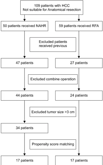
Fig. 1. Patient selection process.
small HCC. Many previous studies have compared hepatic resection and RFA, however, there is a lack of studies comparing the efficacy of NAHR and RFA.
This study aims to compare the effectiveness of RFA and NAHR, especially the short term results, for small sized HCC retrospectively.
MATERIALS AND METHODS
Procedure and indication of RFA
The indications of intraoperative RFA are as follows:
tumors which were superficially located, but the patient was not indicated for liver resection due to advanced liver cirrhosis which means that the percutaneous RFA ap- proach is not possible.; tumors which were deeply located or located at posterior surface segment 6, 7, 8 of the liver and liver resection was not indicated due to advanced liv- er disease and/or advanced variceal formation.
The RFA operation methods have been previously reported.9 The Cool-tipTM (18Gauge) Radiofrequency Ablation System and the EvidentTM Microwave Ablation System (Covidien, CO, USA) were used. Dynamic liver Magnetic resonance imaging (MRI) was performed, pre- operatively, to determine exact tumor localization, size and number. After careful liver mobilization to prevent tumor injury, the whole liver was scanned by intraoperative so- nography and tumor localization performed. In the laparo- scopic approach, patient position was important. A supine position was useful for anteriorly located tumors and whole liver scanning, but if the tumor was located in seg- ment 7 or the posterior surface of segment 6 of liver, 45 degree right side up position was more useful for liver mobilization and traction during ablation. Ablation size was selected according to tumor size, was the electrode being larger than the tumor. A radiofrequency electrode was carefully inserted into the tumor and ablation was per- formed by the operator. In the superficially located tumor, the radiofrequency electrode was inserted into normal liver tissue to prevent tumor popup during ablation. If tumor size was larger than 2 cm, two or more ablations were performed for a larger safety zone of at least 1 cm. Single radiofrequency current was emitted for 12 minutes.
Patients
From February 2007 to January 2015, we performed in-
traoperative RFA on 59 patients and NAHR on 50 patients with HCC at Chungnam National University Hospital.
These patients were not suitable for anatomical resection because of unfavorable ICG test results, or were candidates for liver transplantation. We excluded patients who had received any previous treatment for HCC, or liver trans- plantation during the follow up period. We also excluded patients who received combined operations for other disease. Small HCC was defined as a tumor size less than 3 cm whether it was single or multiple on preoperative hepatic imaging. 24 patients of intraoperative RFA and 34 patients of NAHR were enrolled in this study. Initially in- traoperative RFA was indicated for patients with the worst liver function, more advanced liver cirrhosis and deep loca- tion of the tumor, when compared to those put forward for NAHR, meaning that baseline patient characteristics would be different. To overcome this, 17 pairs of matched
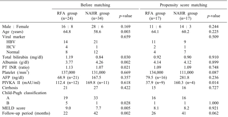
Table 1. Demographics and clinical characteristics of patients
Before matching Propensity score matching
RFA group (n=24)
NAHR group
(n=34) p-value RFA group
(n=17)
NAHR group
(n=17) p-value Male : Female
Age (years) Viral marker HBV HCV Normal
Total bilirubin (mg/dl) Albumin (g/dl) PT INR (ratio) Platelet (/mm3) AFP (ng/dl)
PIVKA II (mAU/ml) Cirrhosis
Child-Pugh classification A
B MELD score
Follow-up period (months)
16 : 8 64.8
14 4 8 1.19 3.77 1.13 137,000 68.9 (n=21) 112.4 (n=12)
21
19 5 9.0 22
28 : 6 58.6
21 1 12 0.84 4.26 1.07 131,000
167.5 169.8 (n=11)
27
33 1 7.7 42
0.169 0.003 0.659
0.030 0.002 0.021 0.669 0.357 0.618 0.422
0.028 0.005 0.002
11 : 6 64.1
11 2 4 0.92 4.14 1.09 134,000 79.5 (n=16)
37.9 (n=9) 15
16 1 8.1 26
14 : 3 60.2
9 1 7 0.90 4.12 1.09 111,000
281.8 160.3 (n=4)
16
16 1 8.2 41
0.244 0.225 0.509
0.910 0.899 0.748 0.087 0.236 0.014 0.727
1.000 0.921 0.062 One patient receiving RFA was co-infection of HBV & HCV. RFA, radiofrequency ablation; HAHR, non-anatomical hepatic resection; HBV, hepatitis B virus HCV, hepatitis C virus
patients were enrolled in this study by propensity score matching method (Fig. 1).
The diagnosis of HCC was based on imaging modality, including enhanced computed tomography (CT), MRI, and tumor markers. Considering cancer cell seeding dur- ing liver biopsy, preoperative liver biopsy was not sug- gested for all patients. Diagnosis of HCC mainly de- pended on typical findings; early-phase enhancement or late-phase contrast washout. Elevation of alpha-fetoprotein (AFP), history of hepatic viral infection or heavy alcohol consumption were also considered supplemental. Prior to treatment, all patients underwent basal laboratory tests in- cluding bilirubin, albumin and prothrombin activity. In terms of tumor location, a subcapsular lesion was defined as a tumor located within 2 cm from the liver capsule and segment was defined according to the Couinaud segments.
Written informed consent was obtained from all patients before treatment.
Follow-up
Follow-up contrast enhanced computed tomography was performed immediately after RFA, and every three to four months in the first 2 years for every patient. For each follow up, blood tests including liver function tests
and tests of serum AFP, were conducted. If HCC recurred we performed proper treatment for patients considering health status.
Statistics
All analysis was performed using the statistical soft- ware SPSS version 19.0 (SPSS Inc., Chicago, IL).
Comparisons between the two groups were done using the Student’s t-test for continuous data and the chi-square test for categorical data. Propensity score matching method was used for matching baseline characteristics of the two groups. The relative prognostic significance of the varia- bles in predicting recurrence-free survival was analyzed using multivariate Cox proportional hazards regression analysis. The recurrence rate was calculated using the Kaplan-Meier method. Significant difference was consid- ered when p<0.05.
RESULTS
Patient & tumor characteristics
The demographics and clinical characteristic are sum- marized in Tables 1 and 2. The NAHR group was young- er in mean ages (58.6 vs. 64.8 years; p=0.003) and had
Table 2. Tumor characteristics
Before matching Propensity score matching
RFA group (n=24)
NAHR group
(n=34) p-value RFA group
(n=17)
NAHR group
(n=17) p-value
Size (mean) Number (mean) Location (tumor) Superficial Deep
Segmental distribution Segment VI Segment VII Segment VIII Other
1.9 1.2 (n=28)
16 12 (n=28)
3 5 5 15
2.1 1.1 (n=36)
31 5 (n=36)
10 6 6 14
0.139 0.184 0.009
0.338
1.8 1.2 (n=20)
12 8 (n=20)
3 4 3 10
2.2 1.1 (n=19)
15 4 (n=19)
7 2 1 9
0.060 0.641 0.200
0.563
Fig. 2. Comparison of the recurrence rates after propensity matching analysis.
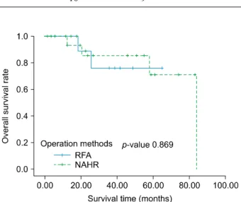
Fig. 3. Comparison of the overall survival rates after propen- sity matching analysis.
better PT INR result (1.07 vs. 1.13; p=0.021), albumin (4.26 vs 3.77 g/dl; p=0.002), and model for end-stage liv- er disease (MELD) score (7.7 vs. 9.0; p=0.005) than the RFA group. There was more Child-Pugh classification A in the NAHR group. There was no significant difference in sex, viral marker, cirrhosis, platelet count between the two groups. More tumors were located in the subcapsular area in the NAHR group (p=0.009) but segmental dis- tribution were not significantly different. Mean tumor size and tumor number were not different between two group.
Follow-up periods were significantly longer in the NAHR group (42 months vs 22 months, p=0.002). Baseline char- acteristics, in particular liver function, were different be- tween two groups. 17 pairs of patient were selected by propensity core matching method for matching baseline characteristics between two groups. The baseline patient and tumor characteristics were not statistically different
between two groups (Tables 1, 2).
Comparison of the recurrence and overall survival rates
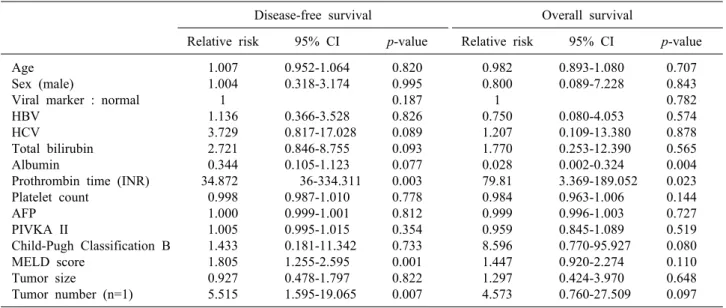
The recurrence rate at 1, 2 and 3 years were 32.2%, 32.2% and 59.3% respectively in the RFA group, and 6.7%, 33.3% and 33.3% respectively in the NAHR group (p=0.287) (Fig. 2). In univariate analysis, PT INR, MELD score, and tumor number were predictors of tumor re- currence (p=0.003, p=0.001, p=0.007, respectively) (Table 3). In multivariate analysis, tumor number was a predictor of recurrence (p=0.019) (Table 4). The OS rates at 1, 2 and 3 years were 100%, 88.9% and 76.2% respectively in the RFA group, and 100%, 85.6% and 85.6% re- spectively in the NAHR group (p=0.869) (Fig. 3). In uni- variate analysis, albumin and PT INR were predictors of overall survival (OS) (p=0.004 and p=0.023, respectively)
Table 4. Results of the multivariate analysis of factors related to the disease-free survival and overall survival after propensity matching analysis
Disease-free survival Overall survival
Relative risk 95% CI p-value Relative risk 95% CI p-value Albumin
Prothrombin time (INR) Number of tumor (n=2) MELD score
- 14.70 5.18 1.71
- 0.1-218.6 1.30-20.60 0.97-3.02
- 0.583 0.019 0.063
0.05 11.51
- -
0.003-0.62 0.01-165.9
- -
0.020 0.336
- - Table 3. Results of the univariate analysis of factors related to the disease-free survival and overall survival
Disease-free survival Overall survival
Relative risk 95% CI p-value Relative risk 95% CI p-value Age
Sex (male)
Viral marker : normal HBV
HCV
Total bilirubin Albumin
Prothrombin time (INR) Platelet count
AFP PIVKA II
Child-Pugh Classification B MELD score
Tumor size
Tumor number (n=1)
1.007 1.004 1 1.136 3.729 2.721 0.344 34.872 0.998 1.000 1.005 1.433 1.805 0.927 5.515
0.952-1.064 0.318-3.174
0.366-3.528 0.817-17.028
0.846-8.755 0.105-1.123 36-334.311
0.987-1.010 0.999-1.001 0.995-1.015 0.181-11.342
1.255-2.595 0.478-1.797 1.595-19.065
0.820 0.995 0.187 0.826 0.089 0.093 0.077 0.003 0.778 0.812 0.354 0.733 0.001 0.822 0.007
0.982 0.800 1 0.750 1.207 1.770 0.028 79.81 0.984 0.999 0.959 8.596 1.447 1.297 4.573
0.893-1.080 0.089-7.228
0.080-4.053 0.109-13.380 0.253-12.390 0.002-0.324 3.369-189.052
0.963-1.006 0.996-1.003 0.845-1.089 0.770-95.927
0.920-2.274 0.424-3.970 0.760-27.509
0.707 0.843 0.782 0.574 0.878 0.565 0.004 0.023 0.144 0.727 0.519 0.080 0.110 0.648 0.097
(Table 3). In multivariate analysis, albumin was a pre- dictor of recurrence rate (p=0.020) (Table 4).
DISCUSSION
HCC is the most frequent primary hepatic malignancy and, with the advance of diagnostic imaging modality, the diagnosis of small HCC has improved considerably. It has allowed patients to have the opportunity to cure HCC by hepatic resection or local ablation, for example using RFA.1,12,13 However, many clinicians find it difficult to se- lect the optimal therapy and treatment for individual pa- tients with HCC. Although hepatic resection is considered the treatment of choice for HCC, many factors, including liver dysfunction, general condition of the patient and tu- mor location, and portal vein invasion of tumor, often lim- it the indication for hepatic resection and extent of treatment.14,15 Under such circumstances, NAHR or RFA could be appropriate treatments for small sized HCC.
To date there has been no study comparing NAHR and
RFA to determine which is the better treatment for pa- tients with small HCCs. Several randomized controlled trials (RCT) have compared the efficacy of hepatic re- section and RFA. Imai et al. concluded that in 2-3 cm HCC, hepatic resection would be the first-line therapy be- cause of an observed higher survival rate and lower re- currence rate.16 Wang et al.17 reported that for HCC pa- tients in the BCLC very early/early stage, surgical re- section yielded better disease-free survival rate than RFA.
However, in these RCTs, non-anatomical hepatic resection and anatomical hepatic resection, and percutaneous RFA and intraoperative RFA, were not distinguished. In our study, we compared the survival rates between NAHR and intraoperative RFA in patients with HCC less than 3 cm.
Of course these patients were treated with the intension to treat.
In this situation, the different recurrence rates between the two groups could be related to differences in back- ground characteristics, especially liver function related factors. The result of high recurrence and low OS rate in
the NAHR may be the result of liver function, as liver function is related to liver fibrosis, and liver fibrosis is associated with a high risk of multi-centric carcinogenesis.18 As such prothrombin activity, albumin, total bilirubin, and Child-Pugh class could be the strongest predictors.19-26 To equalize these variables of the two groups, we used the propensity score matching method. In this study, OS and recurrence rates of NAHR surpassed those of intra- operative RFA group, except after 2-years when OS and recurrence rates were not significantly different between the two groups. This result may be explained by initial small scale cohorts.
Tumor recurrence post-RFA differed from post-liver re- section that makes sufficient safety margin. In immediate postoperative state, RFA area could be confirmed via a comparison of before and after CT scan, but this imaging may not be sufficient for defining tumor necrosis and suf- ficient margin27 because of mobilization during intra- operative manipulation. As such, some RFA results could cause misinterpretation of complete ablation. Tumor pop-up is another key problem. Superficially located tu- mors that are directly punctured can pop-up though the needle hole. This complication could be a source of peri- toneal drop metastasis but can be easily prevented by puncturing though normal tissue and capping the tumor with surgical gauze. These tumor pop-ups could also cause tumor spreading to small intrahepatic vessels and bile ducts because of high pressure gradients that occur during ablation.28 These could be the main cause of high rate of recurrence in the initial first year post treatment. In our study, the cumulative rate of tumor recurrence in the first year was much higher than that following NAHR.
Tanaka et al.29 compared the survival impact of anatomic versus non anatomic resection in patients with solitary HCC. The 1, 3, and 5-year disease-free survival rates in the non-anatomic group were 21%, 52%, and 76%
respectively. The 1, 3, and 5-year OS rates in the non-ana- tomic group were 97%, 91%, and 61% respectively.
Okamura et al.30 compared the disease-free survival and OS rates using propensity matching analysis in the non-ana- tomic resection group and found that the cumulative 1, 3 and 5-year recurrence-free survival rates were 69.7, 46.5 and 31.9% respectively, and the cumulative 1, 3, and 5-year OS rates were 96.4%, 90.1% and 79.7% respectively. Wang et al.17 compared overall survival and disease-free survival
between surgical resection and RFA. They found that for patients who underwent RFA the 1, 3, and 5-year cumu- lative OS rates were 98.1%, 82.8, and 82.8% respectively, and the 1, 3, and 5-year cumulative disease-free survival rates were 67.1%, 46.4%, and 38.0%. Our study found sim- ilar results of recurrence and OS rates.
Goh et al.31 reported that the number of tumors (>3 nodules) was an independent negative predictor of dis- ease-free survival and OS. Increasing number of tumors means multiplicity of liver fibrosis, as liver fibrosis in- crease the risk of carcinogenesis. Tumors recurred simul- taneously at remnant liver tissue, even though hepatic re- section or RFA were carried out. In this study, we per- formed univariate analysis and multivariate cox propor- tional hazard regression analysis. Only tumor number af- fected recurrence rate in the multivariate analysis.
As aforementioned, liver function is related to liver fib- rosis, and liver fibrosis is associated with carcinogenesis of the liver. In this study, albumin, number of tumors, and MELD score, statistically affected recurrence rate, whilst Albumin and INR statistically affected OS rate in uni- variate analysis. The other liver function related factors tested were not related to recurrence and OS rates with statistical significance. We think this is most likely due to small sample size, and short-term follow-up study.
Although, without statistically significant differences, liver function-related factors, including PT INR, albumin, total bilirubin, and grading systems (Child-Pugh class and MELD score) also seems to be a prognostic factor of over- all survival rate and recurrence rate. This suggests that the severity of the underlying liver disease may be a risk fac- tor of HCC recurrence and overall survival. It also sup- ports the importance of liver status in carcinogenesis.
HCC patients with low levels of AFP and PIVKA-II had more favorable clinical characteristics and showed a better prognosis than those with elevated levels of AFP or PIVKA-II.32 We found that tumor markers were not re- lated to tumor recurrence or OS. We attempted to evaluate the tumor markers for each patient before operation, but some patients were missing. Additionally, we checked PIVKA-II for preoperative patients but could only gather a small data set.
Our study had several limitations. First of all, our study included a small sample size and short follow-up period.
We think that this accounts for the absence of statistically
significant differences. Secondly, it was a retrospective study. Thus, our study has fundamental flaws by a se- lection bias. We overcame selection bias by using propen- sity score matching methods, but it made a result from a smaller sample size.
In this study, the tumor number is a predictor of disease free survival and albumin is a predictor of overall survival in small sized HCC patients. We recommend non-anatom- ical hepatic resection over intraoperative RFA in small sized HCC, because of high recurrence rate in intra- operative RFA. We did not find intraoperative RFA to be inferior to NAHR in any other way, except recurrence rate. Clinicians need to select the optimal treatment con- sidering liver function and possibility of recurrence. Our study highlights the requirement for a large volume long-term follow-up study to further analyze these treat- ments and when they are best applied.
REFERENCES
1. Martin P. Hepatocellular carcinoma: risk factors and natural history. Liver Transpl Surg 1998;4(5 Suppl 1):S87-S91.
2. European Association For The Study Of The Liver, European Organisation For Research And Treatment Of Cancer. EASL-EORTC clinical practice guidelines: management of hepatocellular carcinoma.
J Hepatol 2012;56:908-943.
3. Bruix J, Sherman M; American Association for the Study of Liver Diseases. Management of hepatocellular carcinoma: an update. Hepatology 2011;53:1020-1022.
4. Wang Y, Luo Q, Li Y, Deng S, Wei S, Li X. Radiofrequency ablation versus hepatic resection for small hepatocellular carci- nomas: a meta-analysis of randomized and nonrandomized con- trolled trials. PLoS One 2014;9:e84484.
5. Makuuchi M, Imamura H, Sugawara Y, Takayama T. Progress in surgical treatment of hepatocellular carcinoma. Oncology 2002;62 Suppl 1:74-81.
6. Kobayashi M, Ikeda K, Kawamura Y, Yatsuji H, Hosaka T, Sezaki H, et al. High serum des-gamma-carboxy prothrombin level predicts poor prognosis after radiofrequency ablation of hepatocellular carcinoma. Cancer 2009;115:571-580.
7. Marubashi S, Gotoh K, Akita H, Takahashi H, Ito Y, Yano M, et al. Anatomical versus non-anatomical resection for hep- atocellular carcinoma. Br J Surg 2015;102:776-784.
8. Tomimaru Y, Eguchi H, Marubashi S, Wada H, Kobayashi S, Tanemura M, et al. Equivalent outcomes after anatomical and non-anatomical resection of small hepatocellular carcinoma in patients with preserved liver function. Dig Dis Sci 2012;57:
1942-1948.
9. Yun D, Kim S, Song I, Chun K. Comparative analysis of Laparoscopic versus open surgical radiofrequency ablation for malignant liver tumors. Korean J Hepatobiliary Pancreat Surg 2014;18:122-128.
10. Livraghi T, Meloni F, Di Stasi M, Rolle E, Solbiati L, Tinelli C, et al. Sustained complete response and complications rates af- ter radiofrequency ablation of very early hepatocellular carcino-
ma in cirrhosis: Is resection still the treatment of choice?
Hepatology 2008;47:82-89.
11. Majno PE, Mentha G, Mazzaferro V. Partial hepatectomy versus radiofrequency ablation for hepatocellular carcinoma: confirming the trial that will never be, and some comments on the in- dications for liver resection. Hepatology 2010;51:1116-1118.
12. Livraghi T, Meloni F, Morabito A, Vettori C. Multimodal im- age-guided tailored therapy of early and intermediate hep- atocellular carcinoma: long-term survival in the experience of a single radiologic referral center. Liver Transpl 2004;10(2 Suppl 1):S98-S106.
13. Takayama T, Makuuchi M, Kojiro M, Lauwers GY, Adams RB, Wilson SR, et al. Early hepatocellular carcinoma: pathology, imaging, and therapy. Ann Surg Oncol 2008;15:972-978.
14. Kagawa T, Koizumi J, Kojima S, Nagata N, Numata M, Watanabe N, et al; Tokai RFA Study Group. Transcatheter arte- rial chemoembolization plus radiofrequency ablation therapy for early stage hepatocellular carcinoma: comparison with surgical resection. Cancer 2010;116:3638-3644.
15. Liu Z, Zhou Y, Zhang P, Qin H. Meta-analysis of the therapeutic effect of hepatectomy versus radiofrequency ablation for the treatment of hepatocellular carcinoma. Surg Laparosc Endosc Percutan Tech 2010;20:130-140.
16. Imai K, Beppu T, Chikamoto A, Doi K, Okabe H, Hayashi H, et al. Comparison between hepatic resection and radiofrequency ablation as first-line treatment for solitary small-sized hep- atocellular carcinoma of 3 cm or less. Hepatol Res 2013;43:
853-864.
17. Wang JH, Wang CC, Hung CH, Chen CL, Lu SN. Survival com- parison between surgical resection and radiofrequency ablation for patients in BCLC very early/early stage hepatocellular carcinoma. J Hepatol 2012;56:412-418.
18. Koike Y, Shiratori Y, Sato S, Obi S, Teratani T, Imamura M, et al. Risk factors for recurring hepatocellular carcinoma differ according to infected hepatitis virus-an analysis of 236 consecutive patients with a single lesion. Hepatology 2000;32:1216-1223.
19. Choi D, Lim HK, Rhim H, Kim YS, Lee WJ, Paik SW, et al.
Percutaneous radiofrequency ablation for early-stage hep- atocellular carcinoma as a first-line treatment: long-term results and prognostic factors in a large single-institution series. Eur Radiol 2007;17:684-692.
20. Lam VW, Ng KK, Chok KS, Cheung TT, Yuen J, Tung H, et al. Risk factors and prognostic factors of local recurrence after radiofrequency ablation of hepatocellular carcinoma. J Am Coll Surg 2008;207:20-29.
21. Lencioni R, Cioni D, Crocetti L, Franchini C, Pina CD, Lera J, et al. Early-stage hepatocellular carcinoma in patients with cir- rhosis: long-term results of percutaneous image-guided radio- frequency ablation. Radiology 2005;234:961-967.
22. Tateishi R, Shiina S, Teratani T, Obi S, Sato S, Koike Y, et al.
Percutaneous radiofrequency ablation for hepatocellular carcinoma.
An analysis of 1000 cases. Cancer 2005;103:1201-1209.
23. Guglielmi A, Ruzzenente A, Battocchia A, Tonon A, Fracastoro G, Cordiano C. Radiofrequency ablation of hepatocellular carcino- ma in cirrhotic patients. Hepatogastroenterology 2003;50:480-484.
24. Raut CP, Izzo F, Marra P, Ellis LM, Vauthey JN, Cremona F, et al. Significant long-term survival after radiofrequency ablation of unresectable hepatocellular carcinoma in patients with cirrhosis.
Ann Surg Oncol 2005;12:616-628.
25. Xu HX, Lu MD, Xie XY, Yin XY, Kuang M, Chen JW, et al.
Prognostic factors for long-term outcome after percutaneous thermal ablation for hepatocellular carcinoma: a survival analysis of 137 consecutive patients. Clin Radiol 2005;60:1018-1025.
26. N'Kontchou G, Mahamoudi A, Aout M, Ganne-Carrié N, Grando
V, Coderc E, et al. Radiofrequency ablation of hepatocellular carcinoma: long-term results and prognostic factors in 235 Western patients with cirrhosis. Hepatology 2009;50:1475-1483.
27. Mazzaferro V, Battiston C, Perrone S, Pulvirenti A, Regalia E, Romito R, et al. Radiofrequency ablation of small hepatocellular carcinoma in cirrhotic patients awaiting liver transplantation: a prospective study. Ann Surg 2004;240:900-909.
28. Iida H, Aihara T, Ikuta S, Yamanaka N. Effectiveness of im- pedance monitoring during radiofrequency ablation for predicting popping. World J Gastroenterol 2012;18:5870-5878.
29. Tanaka K, Shimada H, Matsumoto C, Matsuo K, Nagano Y, Endo I, et al. Anatomic versus limited nonanatomic resection for solitary hepatocellular carcinoma. Surgery 2008;143:607-615.
30. Okamura Y, Ito T, Sugiura T, Mori K, Uesaka K. Anatomic ver- sus nonanatomic hepatectomy for a solitary hepatocellular carci- noma: a case-controlled study with propensity score matching.
J Gastrointest Surg 2014;18:1994-2002.
31. Goh BK, Chow PK, Teo JY, Wong JS, Chan CY, Cheow PC, et al. Number of nodules, Child-Pugh status, margin positivity, and microvascular invasion, but not tumor size, are prognostic factors of survival after liver resection for multifocal hep- atocellular carcinoma. J Gastrointest Surg 2014;18:1477-1485.
32. Kang SH, Kim DY, Jeon SM, Ahn SH, Park JY, Kim SU, et al. Clinical characteristics and prognosis of hepatocellular carci- noma with different sets of serum AFP and PIVKA-II levels. Eur J Gastroenterol Hepatol 2012;24:849-856.