Letter to the Editor
500 Ann Dermatol
Received October 19, 2012, Revised December 1, 2012, Accepted for publication December 5, 2012
Corresponding author: Dong-Youn Lee, Department of Dermatology, Samsung Medical Center, 81 Irwon-ro, Gangnam-gu, Seoul 135-710, Korea. Tel:
82-2-3410-3543, Fax: 82-2-3410-3869, E-mail: dylee@ skku.edu
This is an Open Access article distributed under the terms of the Creative Commons Attribution Non-Commercial License (http://
creativecommons.org/licenses/by-nc/3.0) which permits unrestricted non-commercial use, distribution, and reproduction in any medium, provided the original work is properly cited.
sed in specific types of cancer, KAI1 is over expressed in MM. Interestingly, KAI1 was not expressed in cutaneous BCCs, SCCs and normal tissues. More work is needed to characterize the pathway through which KAI1 is over- expressed in MM skin tumor.
ACKNOWLEDGMENT
This work was supported by the Soonchunhyang Univer- sity Research Fund.
REFERENCES
1. Dong JT, Isaacs WB, Barrett JC, Isaacs JT. Genomic organi- zation of the human KAI1 metastasis-suppressor gene. Geno- mics 1997;41:25-32.
2. Chen Z, Mustafa T, Trojanowicz B, Brauckhoff M, Gimm O, Schmutzler C, et al. CD82, and CD63 in thyroid cancer. Int J Mol Med 2004;14:517-527.
3. Hinoda Y, Adachi Y, Takaoka A, Mitsuuchi H, Satoh Y, Itoh F, et al. Decreased expression of the metastasis suppressor
gene KAI1 in gastric cancer. Cancer Lett 1998;129:229-234.
4. Guo XZ, Friess H, Maurer C, Berberat P, Tang WH, Zim- mermann A, et al. KAI1 is unchanged in metastatic and nonmetastatic esophageal and gastric cancers. Cancer Res 1998;58:753-758.
5. Yusenko MV, Kovacs G. Identifying CD82 (KAI1) as a marker for human chromophobe renal cell carcinoma. Histopatho- logy 2009;55:687-695.
6. Geradts J, Maynard R, Birrer MJ, Hendricks D, Abbondanzo SL, Fong KM, et al. Frequent loss of KAI1 expression in squamous and lymphoid neoplasms. An immunohistoche- mical study of archival tissues. Am J Pathol 1999;154:1665- 1671.
7. Farhadieh RD, Smee R, Ow K, Yang JL, Russell PJ, Crouch R, et al. Down-regulation of KAI1/CD82 protein expression in oral cancer correlates with reduced disease free survival and overall patient survival. Cancer Lett 2004;213:91-98.
8. Okochi H, Kato M, Nashiro K, Yoshie O, Miyazono K, Furue M. Expression of tetra-spans transmembrane family (CD9, CD37, CD53, CD63, CD81 and CD82) in normal and neoplastic human keratinocytes: an association of CD9 with alpha 3 beta 1 integrin. Br J Dermatol 1997;137:856-863.
http://dx.doi.org/10.5021/ad.2013.25.4.500
Lichenoid Drug Eruption after Low-Dose Imatinib Mesylate Treatment
Jae-Hyung Lee, Jong-Yoon Chung, Mi-Young Jung, Cho Rok Kim, Ji-Ho Park, Ji-Hye Park
1, Jong-Hee Lee, Joo-Heung Lee, Jun-Mo Yang, Dong-Youn Lee
Department of Dermatology, Samsung Medical Center, Sungkyunkwan University School of Medicine,
1Department of Dermatology, Kangbuk Samsung Hospital, Sungkyunkwan University School of Medicine, Seoul, Korea
Dear Editor:
Imatinib mesylate, a selective antitumor tyrosine kinase inhibitor, has been approved as the first-line therapy for gastrointestinal stromal cell tumor and chronic myeloid leukemia. Lichenoid drug eruption (LDE) is known to
cause uncommon adverse cutaneous reactions of imatinib mesylate, usually appearing with 400 mg or more of imatinib mesylate per day1,2. Here, we present a case of severe LDE after low-dose imatinib mesylate treatment for gastrointestinal stromal cell tumor.
Letter to the Editor
Vol. 25 No. 4, 2013 501 Fig. 2. Histopathological findings. (A) Dense band like lymphocytic infiltration in the upper dermis and deep dermal lymphocytic infiltration (H&E, ×40). (B) Lichenoid lymphocytic infiltration with ‘saw-toothed’ rete ridges and vacuolar degeneration of the cells in the basal layer. Cytoid bodies in the granular layer (H&E, ×100).
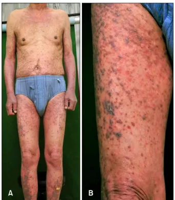
Fig. 1. (A) Symmetrically involved erythematous to violaceous papules and plaques, covered extensive body surface area. (B) Slightly elevated violaceous papules and plaque on the right thigh.
A 77-year-old Korean male patient came to our derma- tology clinic in March 2011 for skin rash over the entire body. He was diagnosed with gastrointestinal stromal cell tumor with multiple liver metastasis, and began imatinib
with 400 mg per day on September 2007. Two months later, violaceous pruritic papules developed symmetrically on both legs. The skin lesion progressed to the whole body despite a dose reduction to 300 mg per day. His skin lesion was markedly improved after a medication change to sunitinib. However, his kidney function gradually worsened and consequently, sunitinib was changed to imatinib again with a reduced dose to 300 mg per day from December 2010. Extensive skin lesion reappeared soon and a dose reduction to 200 mg per day had no effect on improving skin lesion. On physical examination, violaceous, slightly elevated papules symmetrically co- vered the extensive body surface (Fig. 1A). The distri- bution was mainly on both extremities (Fig. 1B). Skin bio- psy was performed on his left lower leg. Specimen show- ed lichenoid lymphocytic infiltration on the upper dermis and deep dermal perivascular lymphocytic infiltration (Fig.
2A). In the high-magnification, ‘saw-toothed’ rete-ridges were similar to those of lichen planus; however, the find- ing of cytoid bodies in the granular layer was favorable to LDE (Fig. 2B). Based on the clinical and histologic findings, a diagnosis of LDE due to imatinib was made.
Adverse cutaneous reactions after imatinib are quite com- mon and various2. Among them, imatinib-induced LDE is a relatively uncommon adverse skin reaction and has been reported in 15 cases to date2. In the PubMed online research, all 15 affected patients took more than 400 mg per day of imatinib, except 1 case which showed a lasting LDE despite the dose reduction from 400 mg per day to 200 mg per day3. One case of a Korean patient with LDE
Letter to the Editor
502 Ann Dermatol
Received November 22, 2012, Accepted for publication December 5, 2012
Corresponding author: Bark-Lynn Lew, Department of Dermatology, Kyung Hee University Hospital at Gangdong, 892 Dongnam-ro, Gangdong-gu, Seoul 134-727, Korea. Tel: 82-2-440-7329, Fax: 82-2-440- 7336, E-mail: bellotte@hanmail.net
This is an Open Access article distributed under the terms of the Creative Commons Attribution Non-Commercial License (http://
creativecommons.org/licenses/by-nc/3.0) which permits unrestricted non-commercial use, distribution, and reproduction in any medium, provided the original work is properly cited.
after imatinib was reported to date; yet, the case differs with our case in that the eruption was well-controlled with a dose reduction to 300 mg per day4. Ugurel et al.5 repor- ted that imatinib acts as a dose dependent inducer of the development of LDE and may show mild reactivity to low or intermediate dose (200 to 600 mg/day). However, in this case, the patient suffered from extensive drug eruption in spite of the low dose of 200 mg per day. We present here LDE after imatinib treatment of 400 mg per day to a low dose of 200 mg per day. This case presented that a low dose of imatinib does not always prevent LDE and can induce severe eruption in some patients. We suggest clinicians to keep in mind that all patients taking imatinib mesylate have the possibility of extensive LDE even in a low dose and therefore should carry a close observation.
REFERENCES
1. Kawakami T, Kawanabe T, Soma Y. Cutaneous lichenoid eruption caused by imatinib mesylate in a Japanese patient with chronic myeloid leukaemia. Acta Derm Venereol 2009;
89:325-326.
2. Amitay-Laish I, Stemmer SM, Lacouture ME. Adverse cutane- ous reactions secondary to tyrosine kinase inhibitors inclu- ding imatinib mesylate, nilotinib, and dasatinib. Dermatol Ther 2011;24:386-395.
3. Dalmau J, Peramiquel L, Puig L, Fernández-Figueras MT, Roé E, Alomar A. Imatinib-associated lichenoid eruption: acitretin treatment allows maintained antineoplastic effect. Br J Der- matol 2006;154:1213-1216.
4. Yang JH, Shin JW, Kim HD, Park YL, Lee SY, Whang KU.
Imatinib mesylate (Gleevec(TM))-induced lichenoid drug eruption improved by tentative dose-reduction and topical steroid. Korean J Dermatol 2011;49:155-158.
5. Ugurel S, Hildenbrand R, Dippel E, Hochhaus A, Schadendorf D. Dose-dependent severe cutaneous reactions to imatinib. Br J Cancer 2003;88:1157-1159.
http://dx.doi.org/10.5021/ad.2013.25.4.502
Minor Salivary Gland Sialolithiasis of the Upper Lip
Dong-Woo Suh, Eun-Ju Lee, Bark-Lynn Lew, Woo-Young Sim
Department of Dermatology, Kyung Hee University School of Medicine, Seoul, Korea
Dear Editor:
Sialolithiasis is a common disease of the salivary glands.
Most calculi occur in the major salivary glands such as the submandibular glands (80% to 92%) and parotid glands (16% to 19%), while minor salivary glands are rarely affected (2%)1. Minor salivary gland sialolithiasis is charac- terized by a small, solitary submucosal nodule, which is hard and in some cases can be movable in the surround- ing tissue2. Since it is rare and its clinical features are not
always typical, clinical misdiagnosis is possible3. Most otolaryngologists and dentists are relatively familiar with sialolithiasis, but many dermatologists are not. In order to heighten the awareness of this disease and to facilitate diagnosis, we report a case of minor salivary gland sialoli- thiasis that was initially misdiagnosed clinically.
A 56-year-old man presented with a six-year history of an asymptomatic solitary submucosal nodule on the upper lip. He reported no history of trauma to the lip. Physical