JCS
Journal of Chest SurgeryClinical Research Modus Operandi: Irrigation of the Modified Eloesser Flap in
Heterogeneous Suppurative Lung Pathologies
Rajkamal Vishnu, M.Ch.1, Guruprasad D. Rai, M.Ch.1, Ganesh Sevagur Kamath, M.Ch.1, Vijaya Kumara, M.D.2
Departments of 1Cardiovascular and Thoracic Surgery and 2Anesthesia, Kasturba Medical College, Manipal Academy of Higher Education, Manipal, India
ARTICLE INFO
Received November 5, 2020 Revised December 24, 2020 Accepted January 25, 2021 Corresponding author Guruprasad D. Rai Tel 91-9886730726 Fax 91-9886730726 E-mail [email protected] ORCID
https://orcid.org/0000-0002-8785-0803
Background: Refractory empyemas with collapsed lung and persistent bronchopleural fistulas pose significant problems to thoracic surgeons and impose a substantial burden in terms of morbidity and mortality. The modified Eloesser flap procedure is a useful palli- ative option for clearing infections. Herein, we present our experiences with the modified Eloesser flap procedure in mixed suppurative lung pathologies with a new technique of irrigation for persistent infection.
Methods: A retrospective review was carried out of 56 patients who underwent the modified Eloesser flap with continuous irrigation at Katurba Medical College. These pa- tients had severe morbidities and were not suitable for major thoracic resection surgery, and electively underwent modified Eloesser flap surgery. Regular follow-up was done at 1, 3, 6, and 12 months. Patients with persistent infections were treated with our continuous irrigation technique.
Results: The most important finding was that all patients with active sputum acid-fast bacilli–positive findings became sputum smear–negative during the first month of fol- low-up. Half (50%) of the patients had a patent stoma. Eleven patients had persistent infec- tions, necessitating continuous irrigation. The infection was fully cleared after 1 month in 9 patients, while 2 patients required second irrigation and continued to receive follow-up. In the remaining 50% of the patients, the stoma closed completely, and the lung expanded fully.
Conclusion: The modified Eloesser flap is a simple procedure. In suppurative patholo- gies, infections were well controlled and the general condition of the patients improved.
Our continuous irrigation method showed promising results in patients with persistent purulent discharge.
Keywords: Modified Eloesser flap, Lung disease, Therapeutic irrigation, Thoracic empy- emas tuberculosis, Povidone-iodine
Copyright©The Korean Society for Thoracic and Cardiovascular Surgery. 2021. All right reserved.
This is an Open Access article distributed under the terms of the Creative Commons Attribution Non-Commercial License (http://creativecommons.org/licenses/
Introduction
The management of refractory empyemas with collapsed lung and persistent bronchopleural fistulas (BPF) poses a significant problem to thoracic surgeons. In India, tuber- culosis (TB) is the most prevalent cause of empyema and causes considerable morbidity and mortality. Parapneu- monic effusions are the commonest cause of refractory empyema, while other causes (e.g., post–lung resection sur- gery, trauma, etc.) are less frequently identified [1]. These patients have poor lung function, chronic infection, and malnutrition, which make them unfit for major thoracic
surgery. Furthermore, these patients are initially treated with antibiotics, tube thoracostomy, and thrombolysis, but the infection sometimes does not resolve. For these pa- tients, Eloesser developed a procedure that is a useful palli- ative option for clearing the infection [2]. Later, the modi- fied Eloesser flap (MEF) developed by Symbas et al. [3]
emerged as a successful alternative for treatment of empy- ema, BPF [4], and post-surgical space infection. Many oth- er techniques for pleural obliteration once the infection is under control, after the MEF procedure, have been de- scribed [5,6]. Herein, we describe our experiences of the MEF procedure in mixed suppurative lung pathologies
https://doi.org/10.5090/jcs.20.144 pISSN: 2765-1606 eISSN: 2765-1614 J Chest Surg. 2021;54(2):137-142
https://doi.org/10.5090/jcs.20.144
JCS
with a new technique of irrigation for persistent infections and present the results.
Methods
Data collection was carried out through a retrospective review of 56 consecutive patients undergoing a MEF at Kasturba Medical College, Manipal, India. The observa- tional study period was 5 years, from January 2012 to Jan- uary 2017. Institutional ethical approval was obtained, and the study was conducted according to the institutional eth- ical protocol. Patients undergoing MEF in our study had various suppurative lung pathologies such as chronic em- pyema, BPF, and post–lung resection space infection. There were 44 (78.5%) male patients and 12 (21.4%) female pa- tients. Their mean age was 42.3 years (range, 21–69 years).
Our study subjects were routinely investigated for per- sistent lung pathologies and underwent conventional chest roentgenograms and standard laboratory evaluations. Spu- tum was obtained for acid-fast bacilli (AFB) staining in all the patients, of whom 9 (17.85%) had multi-drug-resistant pulmonary tuberculosis (MDR-TB) and underwent the conventional MDR-TB regimen as per the Revised Nation- al TB Control Programme, India prior to surgery (Table 1).
Preoperative pulmonary function testing, chest computed tomography (CT), and fiber-optic bronchoscopy scanning were performed prior to surgery. A screening of patients’
history for diabetes, prior pulmonary TB, and cardiac ill- ness was done. Most of our patients were referred from the Department of Pulmonary Medicine after multiple rounds of failed medical treatment, and others were old postopera-
tive lung resection patients (these patients are previously operated long time ago) requiring MEF for space infection.
Common therapeutic modalities that were used prior to MEF included image-guided catheter placement, thrombo- lytic administration, intercostal drain (ICD) placement, and decortication.
In our study, all the patients electively underwent MEF.
The surgical technique of MEF followed the description of Symbas et al. [3]. The patients were put in the right or left lateral position depending on the side of MEF to be done.
All the patients underwent a uniform U-shaped inverted skin incision which was decided after needle localization on the table, scars of a previous intercostal tube, or thora- cotomy. The flap was fashioned at the basal region of the thorax. A window was placed near the diaphragm for max- imum gravity-dependent drainage when the patient sat in an upright or semi-upright position. After fashioning the skin and subcutaneous tissue, deeper planes were dissected using electrocautery in the same inverted U-shaped flap until adequate width and length were achieved. Together, it had a tongue-like appearance, and was later used to create the window. The rib to be resected depended on the extent of drainage required. Once the pleura was opened, surgical debridement of non-viable tissue was done and tissue sam- ples were collected for cultures. A thorough wash was per- formed to clear all the infective foci. Once again, the ade- quacy of the window was checked before creating the stoma. The soft tissue flap was now re-fashioned inward against the diaphragm on the most dependent position and the skin edges of the upper region were not sutured to the pleural surface. The wound was inspected for hemostasis and a moist dressing with paraffin gauge was placed. Most of the patients required 2–3 rounds of dressing with low thoracic suction to remove all the retained collection with saline irrigation (except in a patient with BPF) for faster re- covery [4]. All patients were educated about wound care at home and regular dressing.
Patients with persistent purulent discharge were treated with a continuous irrigation technique. These patients were admitted, and routine cultures and blood investiga- tions were performed. For the irrigation process, a 12F pig- tail catheter was placed at the second anterior intercoastal space at midclavicular line with ultrasound-guided cathe- ter insertion by the Seldinger technique under local anes- thesia. To avoid fluid spillage and to allow easy mainte- nance of the window flap, a colostomy bag was placed at the window site. Continuous irrigation was carried out twice daily for 5 days. Irrigation was first performed with 200 mL of 1% povidone-iodine solution over a period of 60 Table 1. Preoperative patient status, tuberculosis category, and
treatment
Variable No. (%)
Total no. of patients in the study group 56 (100.00) Total no. of pulmonary tuberculosis patients 20 (35.71) Patients on antitubercular drugs prior to MEFa),b) 20 (35.71) Sputumpositive at the time of MEF 9 (16.07) Sputumnegative at the time of MEF 11 (19.64)
Total no. of MDRTB patients 9 (16.07)
MEF, modified Eloesser flap; MDR, multidrugresistant; TB, tuberculosis;
RNTCP, Revised National TB Control Programme.
a)For new cases of pulmonary tuberculosis, the treatment regimen as per RNTCP included 2 months of isoniazid, rifampicin, ethambutol, and pyrazinamide in the intensive phase and 4 months of isoniazid, rifampicin, and ethambutol in the continuation phase. b)The MDRTB treatment regimen as per RNTCP included 6–9 months of kanamycin, levofloxacin, ethionamide, cycloserine, pyrazinamide, and ethambutol in the intensive phase and 18 months of levofloxacin, ethionamide, cycloserine, and ethambutol in the continuation phase.
Rajkamal Vishnu, et al. Continuous Irrigation Method in Modified Eloesser Flap
JCS
minutes, followed by a 1-L saline flush over a period of 20 minutes. Patients were then encouraged to change posi- tions frequently to maintain a uniform distribution. The collected fluid was disposed along with the colostomy bag and a moist dressing with a paraffin gauge was applied.
The same procedure was repeated. Meanwhile, antibiotics were started according to the culture results and sensitivi- ty.From the collected data, information was extracted on patients’ age, sex, the indications for surgery, past medical history, the presence of active TB, preoperative investiga- tions, intraoperative procedures, length of postoperative stay, microorganism culture reports, status of stoma paten- cy, complications of the procedure, and postoperative fol- low-up.
Observational data were entered from patients’ charts and analyzed using SPSS ver. 15.0 (SPSS Inc., Chicago, IL, USA).
The study was approved by the Institutional Review Board of Kasturba Medical College (IRB approval no., IEC 415-2020). Informed consent was obtained from all indi- vidual participants included in the study.
Results
In our study, 44 men and 12 women were admitted with mixed suppurative lung pathology. The most common conditions requiring the MEF that we encountered were chronic empyema (30 patients, 53.5%), post–lung resection space infection (10 patients, 17.8%), BPFs after lung surgery (6 patients, 10.7%), trapped lung (4 patients, 7.14%), and chronic empyema with BPF (6 patients, 10.7%), as detailed in (Fig. 1). All the patients were routinely investigated with basic blood analyses, sputum samples for AFB, chest roent-
genograms, chest CT, and intraoperative fluid and tissue cultures before undergoing the MEF procedure. The most common organisms from intraoperative fluid and tissue cultures were Gram-positive organisms (mainly Staphylo- coccus and Streptococcus) and Gram-negative organisms (mainly Pseudomonas) (Fig. 2). Eighteen patients were di- agnosed with diabetes mellitus, but fortunately had well-controlled blood sugar levels. Every patient had ICD in situ for a relatively long period with failure of lung ex- pansion and other medical management. Fifty-six patients electively underwent MEF. The mean time period from disease onset to MEF was 8±4.42 weeks. The operative technique was the same for all the patients, using U-shaped inverted flap. The intraoperative and in-hospital outcomes are presented in Table 2. Adequate intraoperative drainage was achieved in patients with space infection and empy- ema, and there were no perioperative complications or deaths. In all patients with diabetes mellitus, the MEF
30 53.5
10 17.8
6 10.7
4 7.14
Number Percentage
Chronic empyema
Post lung resection
space infection
BPF post-surgical
Trapped lung
Chronic empyema
with BPF 6
10.7
Fig. 1. Heterogeneity of infections. BPF, bronchopleural fistula.
50 45 40 35 30 25 20 15 10
%ofthenumberofpatients 5
Gram-positiv e
Gram-negativ e
Mycobacteriu m
Fungal
Anaerobes 0
46.4
26.7
17.85
5.34 3.54
Fig. 2. Types of microorganisms. Values are presented as % of the number of patients.
Table 2. Intraoperative and postoperative outcomes of patients undergoing the modified Eloesser flap and our irrigation method
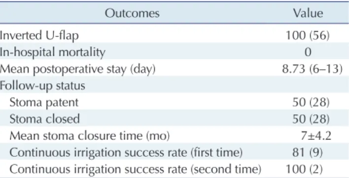
Outcomes Value
Inverted Uflap 100 (56)
Inhospital mortality 0
Mean postoperative stay (day) 8.73 (6–13)
Followup status
Stoma patent 50 (28)
Stoma closed 50 (28)
Mean stoma closure time (mo) 7±4.2
Continuous irrigation success rate (first time) 81 (9) Continuous irrigation success rate (second time) 100 (2) Values are presented as % (number), mean (range), or mean±standard deviation.
https://doi.org/10.5090/jcs.20.144
JCS
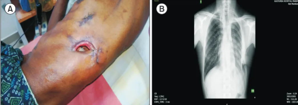
showed favorable granulation and healing, which was achieved with adequate blood glucose control (Fig. 3A, B).
Regular follow-up was done at 1, 3, 6, and 12 months post- operatively, and 2 deaths occurred during follow-up due to respiratory arrest with sepsis and cachexia. Most of our patients clinically improved with weight gain and in- creased appetite after MEF. Due to continuous gravity-de- pendent drainage and the absence of stasis of purulent flu- id, the infection was controlled well in most of the patients during the first month of follow-up. In 28 patients (50%) who underwent MEF, the window was completely closed, of whom 92.9% (26) had complete lung expansion and 2 (7.1%) required enlargement of the window for drainage as the air leak persisted. The mean closure period was 7±4.20 months. In the remaining 28 patients (50%), the window was patent; of these patients, 17 (60.7%) had complete lung ex- pansion and 11 (39.2%) had persistent infections and con- tinued to receive follow-up. The 11 patients with persistent infections (of whom 9 patients had MDR-TB and 2 patients had Gram-positive bacterial infections) were treated with our continuous irrigation technique described above (Fig.
4A, B). During the first month of follow-up, out of 11 pa- tients who underwent continuous irrigation with a patent window, 9 patients showed no infective discharge and their lungs had fully expanded (7 patients with MDR-TB con- verted to sputum smear–negative and 2 patients with Gram- positive bacteria showed negative culture results) and con- tinued to receive regular follow-up. Two patients with MDR- TB with persistent infections were treated for a second time with continuous irrigation after 1 month, and contin- ued to receive follow-up with no further purulent dis- charge and negative sputum smear findings.
Discussion
Many different techniques for window drainage have been developed with modifications for a few decades. The
original window drainage procedure was introduced by Eloesser [2] was developed for draining tuberculous empy- emas and pleural space infections. His original technique involved a superior U-shaped flap over the most dependent position with resection of the underlying ribs. His drainage method acted as a 1-way valve where purulent content exits and air is prevented from re-entering the pleural space.
The valve also provides negative pressure, which encourag- es lung expansion. This method was further modified by Symbas et al. [3] in 1971 for the treatment of thoracic em- pyema. The modified technique consists of an inverted U-shaped flap at the basal region of the thorax with resec- tion of the ribs. Once the cavity is totally drained and the underlying ribs resected, the tongue flap is then tacked to the inferior-most aspect of the drained space. In recent years, the MEF has been a very effective drainage tech- nique for the treatment of thoracic empyema [7].
In our current study, we adopted the MEF using an in- verted U-shaped flap in all patients [3]. Our indications for surgery extended beyond chronic empyema to include BPF and post-resection space infections. Prior to surgery, all patients underwent nonsurgical treatments such as antibi- otics, multiple thoracentesis, and intercostal tube place-
Fig. 3. (A) Postoperative followup after 1 month, showing a well
healed stoma. (B) Postoperative chest Xray with an expanded lung on the left side.
A B
Fig. 4. (A, B) Continuous irrigation method with a pigtail catheter and colostomy bag for collection of drainage.
A B
Rajkamal Vishnu, et al. Continuous Irrigation Method in Modified Eloesser Flap
JCS
ment [1,7]. The reasons for performing this procedure in- cluded anticipation of prolonged intercostal tube placement and non-compliance with intercostal tube placement and position. In this study, we observed that an active pulmo- nary TB patient improved with MEF, and as a result of the continuous irrigation method with povidone-iodine, all MDR-TB patients converted to sputum smear AFB-nega- tive status during follow-up [8]. Overall, in our patients, who had serious morbidities and were not suitable for ma- jor thoracic resection surgery, MEF served as a palliative procedure improving quality of life [7]. To highlight the advantages of MEF in our patients: (1) it is a minor proce- dure well tolerated by patients with severe morbidities; (2) aggressive diabetes control promoted a faster recovery with healthy granulation tissue; (3) infection control was achieved in most of our patients with symptomatic improvement (reduction in purulent discharge, increase in appetite, and weight gain) and enhanced overall quality of life; (4) BPF recovered quickly with lung expansion, changing into a bronchocutaneous fistula [4]; and (5) the chest deformity after spontaneous stoma closure was very minimal and the residual defect was a small indentation on the skin at the site of the stoma. For patients with a persistent stomal opening, thoracoplasty has been advised as an option.
During follow-up, 11 patients had persistent infections that were treated with continuous irrigation. This technique was previously used for post-pneumonectomy space infec- tions.
The advantages of our continuous irrigation method are that: (1) a thorough cleaning of the purulent substance is possible; (2) it is less painful, as a 12F pigtail catheter is used under local anesthesia; (3) lung injury is avoided, as the catheter is inserted using ultrasound guidance; (4) us- ing a colostomy bag has additional benefits for avoiding spillage of contents and easy maintenance of the MEF sto- ma; (5) the use of only 1% povidone-iodine is non-toxic to the lung and leads to better healing [9,10]; and (6) all MDR- TB patients showed negative sputum smear results during follow-up after undergoing our continuous irrigation tech- nique [11,12].
In conclusion, the modified Eloesser type of drainage is a simple and safe procedure to perform. Prompt clearance of infection is seen after the procedure. In most cases, it is sufficient for the closure of BPF. In cases where fistula clo- sure did not occur, the infection was well controlled. Addi- tionally, our continuous irrigation technique had the fol- lowing advantages: (1) it helped to resolve persistent infections; (2) it was very convenient for both surgeons and patients; and (3) adequate infection control was observed
in this irrigation technique, as it was a continuous method.
The general condition of patients improved, providing the opportunity to undertake a more extensive procedure at a later date at a considerably lower risk. This technique is highly valuable for patients who do not understand their disease and who are not compliant with instructions per- taining to the care of a long-standing ICD. The residual chest deformity after window closure is acceptable. This study shows how an alternative, less-invasive method of ir- rigation can prevent recurrent lung infection and improve quality of life.
Our study is probably the first one to describe this tech- nique and hence has limitations as a retrospective observa- tional study with no prior studies to compare. We also ac- knowledge that the small sample size may not provide adequate statistical power to the study, thereby limiting the extrapolation of the study results to the general population.
Conflict of interest
No potential conflict of interest relevant to this article was reported.
ORCID
Rajkamal Vishnu: https://orcid.org/0000-0002-0294-6196 Guruprasad D. Rai: https://orcid.org/0000-0002-8785-0803 Ganesh Sevagur Kamath: https://orcid.org/0000-0003-1020-2717 Vijaya Kumara: https://orcid.org/0000-0003-4642-7213
References
1. Magovern CJ, Rusch VW. Parapneumonic and post-traumatic pleural space infections. Chest Surg Clin N Am 1994;4:561-82.
2. Eloesser L. Of an operation for tuberculous empyema. Ann Thorac Surg 1969;8:355-7.
3. Symbas PN, Nugent JT, Abbott OA, Logan WD Jr, Hatcher CR Jr.
Nontuberculous pleural empyema in adults: the role of a modified Eloesser procedure in its management. Ann Thorac Surg 1971;12:69- 78.
4. Galvin IF, Gibbons JR, Maghout MH. Bronchopleural fistula: a nov- el type of window thoracostomy. J Thorac Cardiovasc Surg 1988;96:
433-5.
5. Clagett OT, Geraci JE. A procedure for the management of postpneu- monectomy empyema. J Thorac Cardiovasc Surg 1963;45:141-5.
6. Gharagozloo F, Margolis M, Facktor M, Tempesta B, Najam F. Post- pneumonectomy and postlobectomy empyema. Thorac Surg Clin 2006;16:215-22.
7. Thourani VH, Lancaster RT, Mansour KA, Miller JI Jr. Twenty-six
https://doi.org/10.5090/jcs.20.144
JCS
years of experience with the modified Eloesser flap. Ann Thorac Surg 2003;76:401-5.
8. Subotic D, Yablonskiy P, Sulis G, et al. Surgery and pleuro-pulmo- nary tuberculosis: a scientific literature review. J Thorac Dis 2016;8:
E474-85.
9. Gharagozloo F, Trachiotis G, Wolfe A, DuBree KJ, Cox JL. Pleural space irrigation and modified Clagett procedure for the treatment of early postpneumonectomy empyema. J Thorac Cardiovasc Surg 1998;116:943-8.
10. Banerjee A, Subbarao KS. Use of povidone iodine for the manage- ment of post-pneumonectomy empyema. Chest 1983;84:507-8.
11. Rikimaru T, Kondo M, Kajimura K, et al. Efficacy of common anti- septics against multidrug-resistant mycobacterium tuberculosis. Int J Tuberc Lung Dis 2002;6:763-70.
12. Rikimaru T, Kondo M, Kajimura K, et al. Bactericidal activities of commonly used antiseptics against multidrug-resistant mycobacteri- um tuberculosis. Dermatology 2002;204 Suppl 1:15-20.